. 2021 Mar;160(4):1240-1255.e3.
doi: 10.1053/j.gastro.2020.11.008. Epub 2020 Nov 13.

A Novel Role of SLC26A3 in the Maintenance of Intestinal Epithelial Barrier Integrity

Affiliations

A Novel Role of SLC26A3 in the Maintenance of Intestinal Epithelial Barrier Integrity

Anoop Kumar et al. Gastroenterology. 2021 Mar.

Abstract

Background & aims: The down-regulated in adenoma (DRA) protein, encoded by SLC26A3, a key intestinal chloride anion exchanger, has recently been identified as a novel susceptibility gene for inflammatory bowel disease (IBD). However, the mechanisms underlying the increased susceptibility to inflammation induced by the loss of DRA remain elusive. Compromised barrier is a key event in IBD pathogenesis. The current studies were undertaken to elucidate the impact of DRA deficiency on epithelial barrier integrity and to define underlying mechanisms.

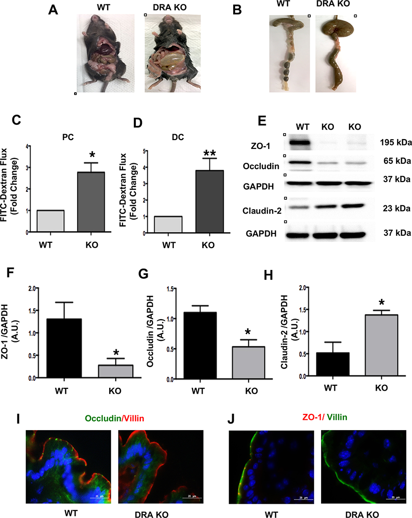
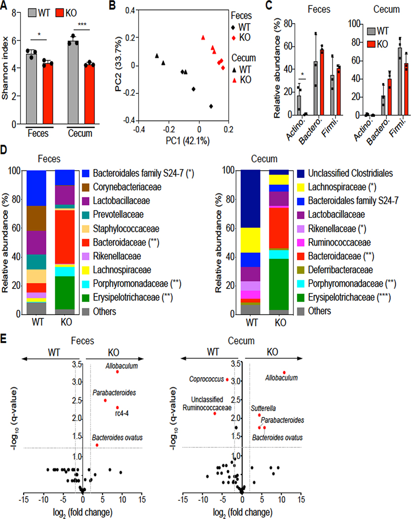
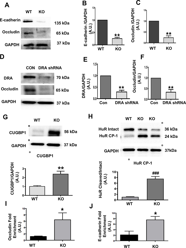
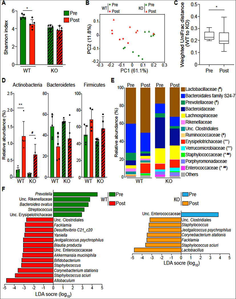
Methods: Wild-type and DRA-knockout (KO) mice and crypt-derived colonoids were used as models for intestinal epithelial response. Paracellular permeability was measured by using fluorescein isothiocyanate-dextran flux. Immunoblotting, immunofluorescence, immunohistochemistry, and ribonucleoprotein immunoprecipitation assays were performed. Gut microbiome analysis was conducted to investigate the impact of DRA deficiency on gut microbial communities.

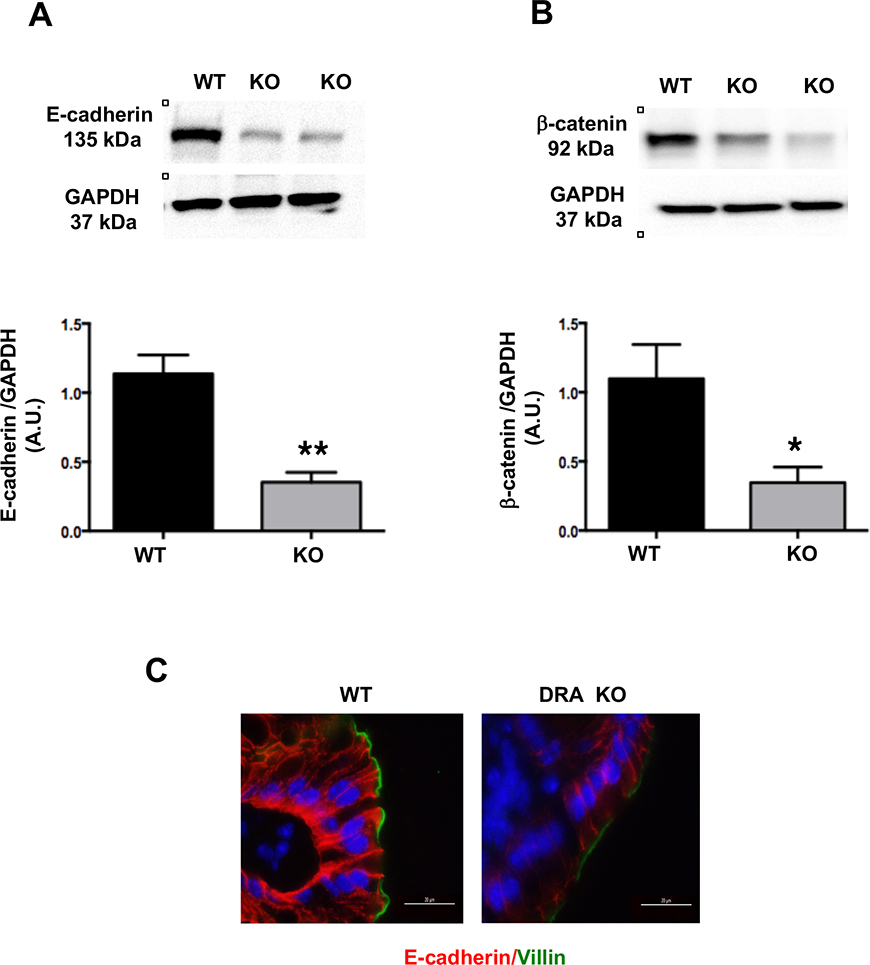
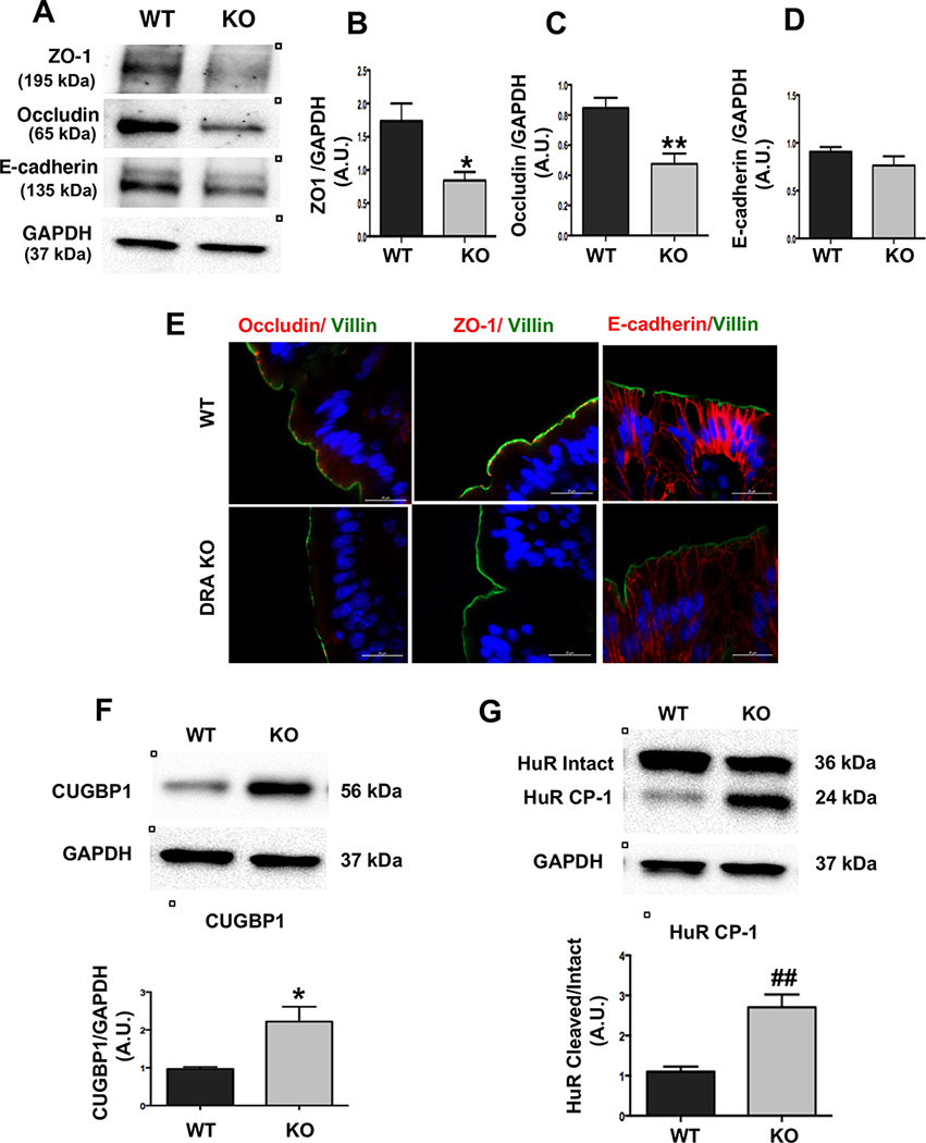
Results: DRA-KO mice exhibited an increased colonic paracellular permeability with significantly decreased levels of tight junction/adherens junction proteins, including ZO-1, occludin, and E-cadherin. A similar expression pattern of occludin and E-cadherin was observed in colonoids derived from DRA-KO mice and short hairpin RNA-mediated DRA knockdown in Caco-2 cells. Microbial analysis showed gut dysbiosis in DRA-KO mice. However, cohousing studies showed that dysbiosis played only a partial role in maintaining tight junction protein expression. Furthermore, our results showed increased binding of RNA-binding protein CUGBP1 with occludin and E-cadherin genes in DRA-KO mouse colon, suggesting that posttranscriptional mechanisms play a key role in gut barrier dysfunction.

Conclusions: To our knowledge, our studies demonstrate a novel role of DRA in maintaining the intestinal epithelial barrier function and potential implications of its dysregulation in IBD pathogenesis.

Keywords: Down-regulated in Adenoma; Gut Microbiota; Inflammatory Bowel Disease; Intestinal Chloride Transporter.


Conflict of interest statement

Conflict of interest: none

Figures

Figure 1:
Figure 1:. Loss of DRA results in increased paracellular permeability and decreased TJ protein expression in mouse colon:
DRA KO mice exhibit severe diarrhea, a distended abdomen and enlarged cecum (Figures A and B). DRA KO mice showed increased paracellular permeability in proximal colon (PC) and distal colon (DC) (Figures C and D). Intestinal permeability was determined by comparing mucosal-to-serosal flux of 4 kDa-FITC dextran (FD4) fluorescence over 120 minutes (n=3–4). Effects of DRA knockdown on protein levels of TJ components in wild type and DRA KO mice distal colon by immunoblotting are shown (Figure E) (n=3–6). GAPDH was used as the internal control. Densitometric analyses of band intensities are shown (Figures F, G, H). Values are means ± SEM. Data was analyzed by unpaired t-test (*p <0.05 vs. wild type; **p <0.01 vs. wild type). Immunofluorescence staining of distal colon sections of wild type and DRA KO mice for occludin (green), villin (red), and DAPI (blue) (Figure I) and ZO-1 (red), villin (green), and DAPI (blue) (Figure J). Scale bar: 20 μm.
Figure 2:
Figure 2:. Loss of DRA results in decreased AJ protein expression in mouse colon:
Distal colonic mucosal lysates of wild type and DRA KO mice were subjected to SDS-PAGE and probed with E-cadherin and β-catenin antibodies. (Figures A and B): Densitometric analysis was performed of relative band intensities with GAPDH as internal control. Values are means ± SEM and representative blots of 3–6 independent experiments are shown. Data was analyzed by unpaired t-test (*p <0.05 vs. wild type; **p <0.01 vs. wild type). (C) Immunofluorescence staining of distal colonic sections of wild type and DRA KO mice for E-cadherin (red), villin (green), and DAPI (blue) is shown. Scale bar: 20 μm.
Figure 3.
Figure 3.. Loss of DRA alters gut microbial composition.
(Figure A) Shannon index measuring bacterial diversity by 16S rRNA sequencing in the feces and cecal contents of DRA KO and WT mice (n=3). (Figure B) Weighted Unifrac principal coordinate analysis (PCoA) plot separating fecal and cecal microbiota composition between DRA KO and WT mice. (Figure C) Top phylum abundance in the feces and cecal contents. (Figure D) Top family abundance of fecal and cecal microbes. Asterisks (*) indicate statistical significance between the groups of mice. (Figure E) Significantly different taxa between DRA KO and WT mice in fecal and cecal samples in red. The dashed lines represent thresholds (log2 fold change > 1, FDR P <0.05). *p <0.05, **p <0.01, ***p <0.001.
Figure 4:
Figure 4:. Loss of DRA results in reduced TJ/AJ protein expression in colonoids and Caco-2 cells and increased CUGBP1 expression, HuR protein cleavage, and binding of CUBGP1 to 3’ UTR of occludin and E-cadherin in DRA KO mice:
Protein expression of E-cadherin and occludin in colonoids from wild type and DRA KO mice by western blotting (Figure A). Densitometric analyses of band intensities (GAPDH was used as the internal control) are shown (Figures B and C). DRA and occludin immunoblotting in lentiviral shRNA mediated knockdown of DRA in Caco-2 cells are shown (Figure D). Densitometric analyses of band intensities (GAPDH was used as the internal control) are shown (Figures E and F). Values (for figures 4B,4C,4E,4F) are means ± SEM from blots from 3–4 independent experiments. Data was analyzed by unpaired t-test (**p <0.01 vs. wild type). Western blot and densitometric analysis of CUGBP1 and HuR protein levels in distal colonic mucosal lysates of wild type or DRA KO mice (Figures G and H). Results represent means ± SEM of 3–4 mice (**p <0.01 compared to wild type, ###p <0.001, cleaved compared with intact HuR). CUGBP1 binding (over IgG) to 3’UTR of occludin and E-cadherin as measured by RNP-IP Assay (Figures I and J). Values are means ± SEM of blots of 4–5 independent experiments. Data was analyzed by unpaired t-test (*p <0.05 compared to wild type).
Figures 5:
Figures 5:. DSS-colitis results in decreased protein levels of DRA, occludin, and increased protein levels of CUGBP1 and cleaved-HuR:
Representative western blots and densitometric analysis of DRA and occludin protein levels in distal colonic mucosal tissue lysates of DSS or control mice (Figures A and B). Results represent means ± SEM of 4 mice. (*p <0.05 and **p <0.01 compared to controls). Representative western blots and densitometric analysis of CUGBP1 and HuR protein (intact and cleaved HuR) levels in distal colonic mucosal tissue lysates of wild type or DRA KO mice (Figures C and D). Results represent means ± SEM of 3 mice. Data was analyzed by unpaired t-test (*p <0.05 CUGBP1 in DSS mice compared to control mice. #p <0.05, cleaved HuR/intact HuR in DSS mice compared to control mice).
Figure 6.
Figure 6.. Cohousing contributes to partial exchange of fecal microbiota in DRA KO and WT mice.
Shannon index of fecal microbiota in post-cohoused DRA KO and WT mice compared to their pre-cohoused counterparts (n=5) (Figure A). Weighted Unifrac PCoA plot showing community variation in indicated mouse groups (Figure B). Weighted Unifrac distances between cohoused mice and their pre-cohoused controls showing the reduced similarity between DRA KO and WT mice after cohousing (Figure C). Averaged relative abundances of top phyla (Figure D) and families (Figure E) in pre- and post-cohoused DRA KO and WT mice. LEfSe plots showing differentiating taxa (species level) between post-cohoused mice and their pre-cohoused controls (Figure F). *p <0.05, **p <0.01, pre-cohoused WT vs. post-cohoused WT; #p <0.05, ##P <0.01, pre-cohoused KO vs. post-cohoused KO.
Figure 7:
Figure 7:. TJ/AJ protein and RBPs expression in cohoused wild type and DRA KO mice:
Protein expression of ZO-1, occludin and E-cadherin in distal colon of wild type and DRA KO co-housed mice (Figure A). Densitometric analyses (GAPDH was used as the internal control) of band intensities are shown (Figures B, C and D). Values are means ± SEM of blots from 4 wild type and 5 KO co-housed mice. Data was analyzed by unpaired t-test (*p <0.05 vs wild type, **p <0.01 vs. wild type). Immunostaining for ZO-1, occludin, and E-cadherin is shown (Figure E). Scale bar: 20 μm. Protein expression of CUGBP-1 and HuR in distal colon of wild type and DRA KO co-housed mice by western analysis and densitometric analyses (GAPDH-internal control) of band intensities is shown (Figures F and G). Values are means ± SEM of blots from 4 wild type and 5 KO mice. Data was analyzed by unpaired t-test (*p <0.05 vs wild type, ## p <0.01 vs. wild type).

References

    1. Priyamvada S, Gomes R, Gill RK, et al. Mechanisms Underlying Dysregulation of Electrolyte Absorption in Inflammatory Bowel Disease-Associated Diarrhea. Inflamm Bowel Dis 2015;21:2926–35. - PMC - PubMed
    1. Anbazhagan AN, Priyamvada S, Alrefai WA, et al. Pathophysiology of IBD associated diarrhea. Tissue Barriers 2018:e1463897. - PMC - PubMed
    1. Shen L, Su L, Turner JR. Mechanisms and functional implications of intestinal barrier defects. Dig Dis 2009;27:443–9. - PMC - PubMed
    1. Al-Sadi R, Guo S, Ye D, et al. TNF-alpha modulation of intestinal epithelial tight junction barrier is regulated by ERK1/2 activation of Elk-1. Am J Pathol 2013;183:1871–1884. - PMC - PubMed
    1. Al-Sadi R, Guo S, Ye D, et al. Mechanism of IL-1beta modulation of intestinal epithelial barrier involves p38 kinase and activating transcription factor-2 activation. J Immunol 2013;190:6596–606. - PMC - PubMed

Publication types

MeSH terms