Randomized Controlled Trial
. 2021 Mar:230:106-111.e6.
doi: 10.1016/j.jpeds.2020.11.015. Epub 2020 Nov 13.

Limitations of Conventional Magnetic Resonance Imaging as a Predictor of Death or Disability Following Neonatal Hypoxic-Ischemic Encephalopathy in the Late Hypothermia Trial

Collaborators, Affiliations
Randomized Controlled Trial

Limitations of Conventional Magnetic Resonance Imaging as a Predictor of Death or Disability Following Neonatal Hypoxic-Ischemic Encephalopathy in the Late Hypothermia Trial

Abbot R Laptook et al. J Pediatr. 2021 Mar.

Abstract

Objective: To investigate if magnetic resonance imaging (MRI) is an accurate predictor for death or moderate-severe disability at 18-22 months of age among infants with neonatal encephalopathy in a trial of cooling initiated at 6-24 hours.

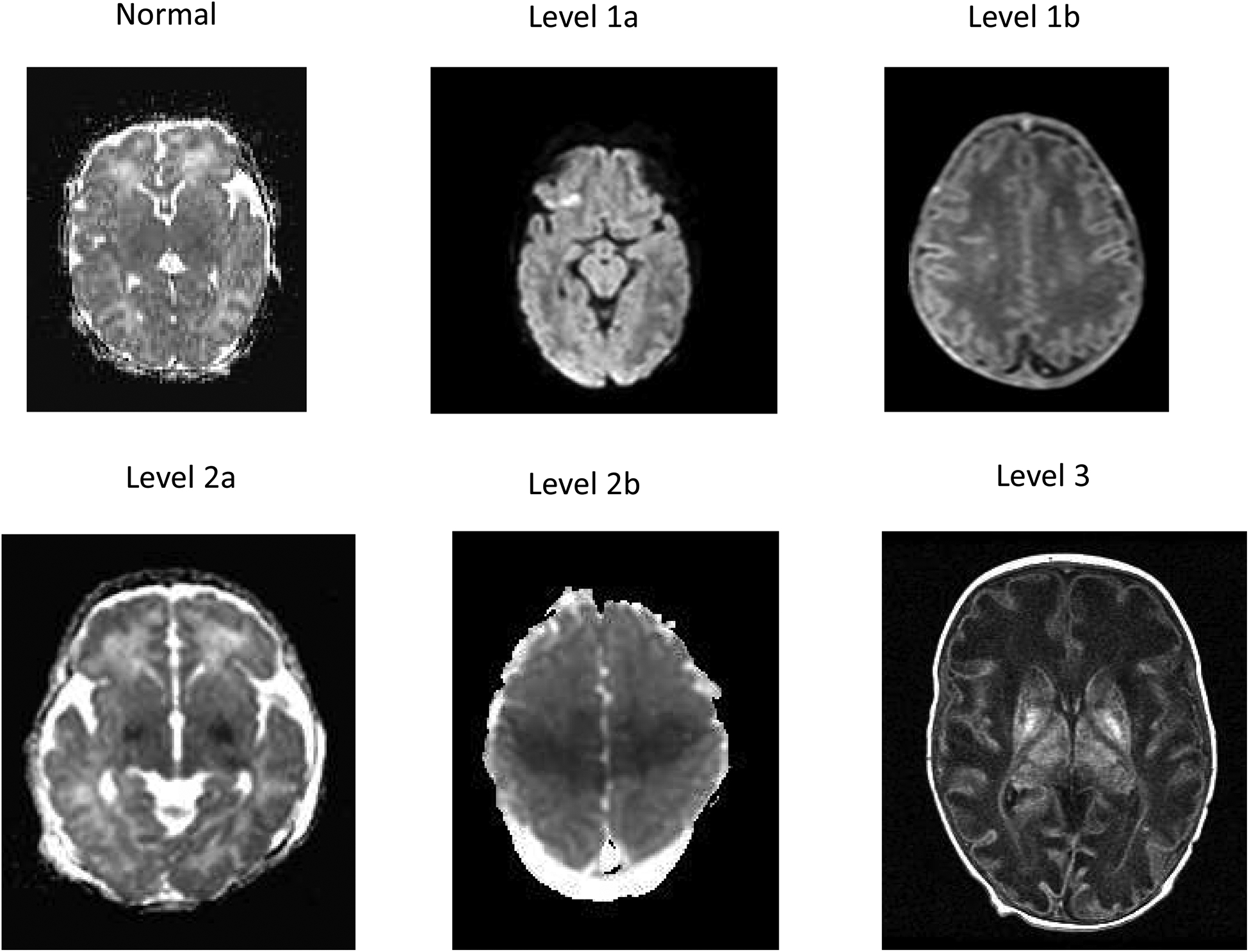
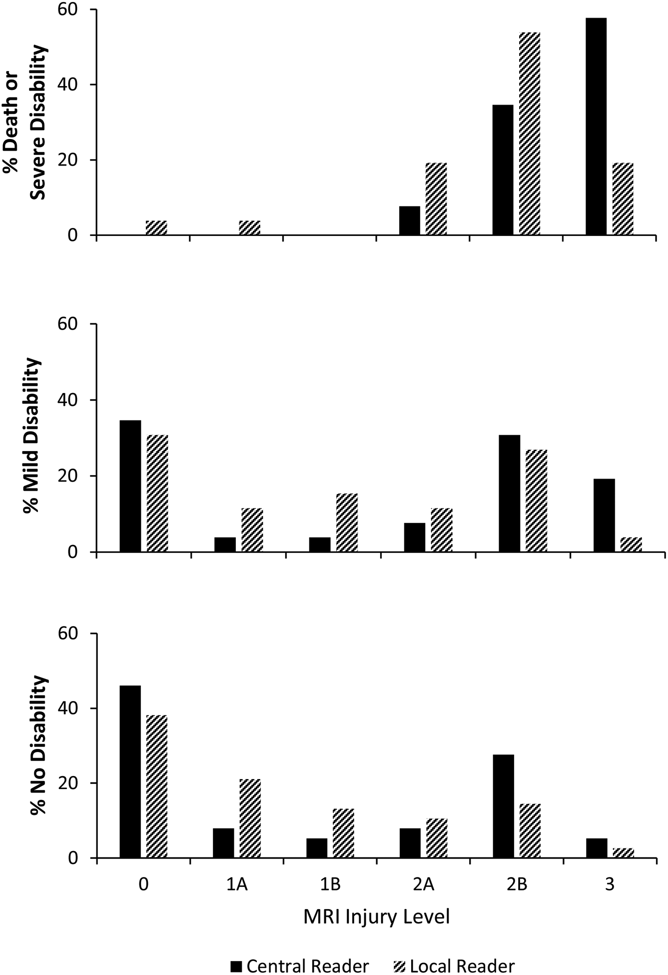
Study design: Subgroup analysis of infants ≥36 weeks of gestation with moderate-severe neonatal encephalopathy randomized at 6-24 postnatal hours to hypothermia or usual care in a multicenter trial of late hypothermia. MRI scans were performed per each center's practice and interpreted by 2 central readers using the Eunice Kennedy Shriver National Institute of Child Health and Human Development injury score (6 levels, normal to hemispheric devastation). Neurodevelopmental outcomes were assessed at 18-22 months of age.

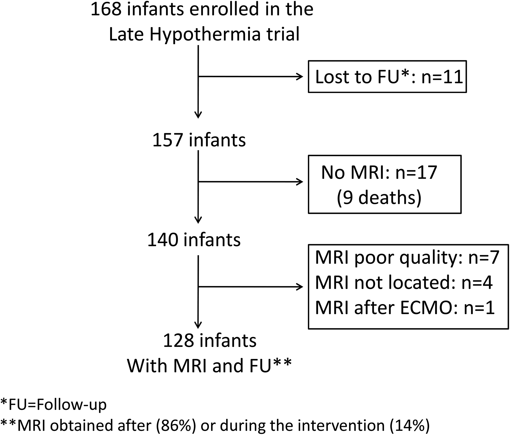
Results: Of 168 enrollees, 128 had an interpretable MRI and were seen in follow-up (n = 119) or died (n = 9). MRI findings were predominantly acute injury and did not differ by cooling treatment. At 18-22 months, death or severe disability occurred in 20.3%. No infant had moderate disability. Agreement between central readers was moderate (weighted kappa 0.56, 95% CI 0.45-0.67). The adjusted odds of death or severe disability increased 3.7-fold (95% CI 1.8-7.9) for each increment of injury score. The area under the curve for severe MRI patterns to predict death or severe disability was 0.77 and the positive and negative predictive values were 36% and 100%, respectively.

Conclusions: MRI injury scores were associated with neurodevelopmental outcome at 18-22 months among infants in the Late Hypothermia Trial. However, the results suggest caution when using qualitative interpretations of MRI images to provide prognostic information to families following perinatal hypoxia-ischemia.

Trial registration: Clinicaltrials.gov: NCT00614744.

Keywords: brain cooling; hypoxic-ischemic encephalopathy; imaging.


Conflict of interest statement

The National Institutes of Health, the Eunice Kennedy Shriver National Institute of Child Health and Human Development (NICHD), the National Center for Research Resources, and the National Center for Advancing Translational Sciences provided grant support for the Neonatal Research Network’s Late Hypothermia Trial (recruitment 4/17/2008 – 7/13/2014 and follow-up from 10/17/2009 – 9/12/2016) through cooperative agreements. Participating NRN sites collected data and transmitted it to RTI International, the data coordinating center (DCC) for the network, which stored, managed, and analyzed the data for this study. While NICHD staff did have input into the study design, conduct, analysis, and manuscript drafting, the content is solely the responsibility of the authors and does not necessarily represent the official views of the National Institutes of Health. On behalf of the NRN, A.D. (DCC Principal Investigator), Breda Munoz, PhD, and B.D. (DCC Statistician) had full access to all of the data in the study, and with the NRN Center Principal Investigators, take responsibility for the integrity of the data and accuracy of the data analysis. The authors declare no conflicts of interest.

Figures

Figure 1:
Figure 1:. MRI Levels of Injury after Hypoxic-Ischemic Encephalopathy and 18–22 Month Outcome
Representative images of the MRI levels of injury based on the NICHD pattern of signal abnormalities are presented and defined as follows: Level 0: Normal signal throughout the brain on a diffusion image Level 1a: Minimal cerebral lesions only without involvement of basal ganglia (BG), or thalamus (T), or anterior/posterior limb of the internal capsule (ALIC/PLIC respectively) and no areas of watershed infarction. A diffusion weighted image reveals a punctate lesion in the frontal region. Level 1b: More extensive cerebral lesions not corresponding to a watershed or vascular distribution without BGT, PLIC, ALIC, involvement. A T1 weighted image indicates multiple high intensity punctate lesions in the white matter bilaterally. Level 2a: Any BGT, ALIC, PLIC involvement or watershed infarction noted without any other cerebral lesions. A diffusion weighted image indicates abnormal signal intensity in the medial BGT and decreased signal intensity in the posterior limb of the internal capsule bilaterally. Level 2b: Involvement of either BGT, ALIC, PLIC or areas of watershed/vascular distribution and additional cerebral lesions. A diffusion weighted image indicates restricted diffusion in the BGT and in the peri-rolandic and posterior parasagittal regions bilaterally. Level 3: Cerebral hemispheric devastation. A T1 weighted image indicates global involvement of the white matter with attenuated signal intensity, a simplified cortical gray matter pattern and increased signal intensity in the BGT with loss of signal intensity of the PLIC.
Figure 2:
Figure 2:
Flow diagram of infants enrolled in the Late Hypothermia trial who were analyzed for the secondary study.
Figure 3:
Figure 3:. MRI Injury Scores after Hypoxic-Ischemic Encephalopathy and 18–22 Month Outcome
The 18–22 month outcome is plotted as function of the NICHD injury score for infants with death or severe disability (top panel), and for infants with mild disability (middle panel) or without disability (bottom panel). The injury score was determined by the central readers and included adjudicated interpretations. Black columns represent central readers and hashed columns represent local readers.

References

    1. American College of Obstetricians and Gynecologists, American Academy of Pediatrics. Neonatal Encephalopathy and Neurologic Outcome, 2nd Ed. Washington, DC: American College of Obstetricians and Gynecologists; 2014.
    1. Barkovich AJ, Hajnal BL, Vigneron D, Sola A, Partridge JC, Allen F, et al. Prediction of neuromotor outcome in perinatal asphyxia: evaluation of MR scoring systems. AJNR Am J Neuroradiol. 1998;19:143–9. - PMC - PubMed
    1. Miller SP, Ramaswamy V, Michelson D, Barkovich AJ, Holshouser B, Wycliffe N, et al. Patterns of brain injury in term neonatal encephalopathy. J Pediatr. 2005;146:453–60. - PubMed
    1. Myers RE. Two patterns of perinatal brain damage and their conditions of occurrence. Am J Obstet Gynecol. 1972;112:246–76. - PubMed
    1. Rutherford M, Ramenghi LA, Edwards AD, Brocklehurst P, Halliday H, Levene M, et al. Assessment of brain tissue injury after moderate hypothermia in neonates with hypoxic-ischaemic encephalopathy: a nested substudy of a randomised controlled trial. Lancet Neurol. 2010;9:39–45. - PMC - PubMed

Publication types

Associated data