Review
. 2021 Jan;41(1):20-32.
doi: 10.1111/liv.14730. Epub 2020 Nov 29.

Pathophysiological mechanisms of liver injury in COVID-19

Affiliations
Review

Pathophysiological mechanisms of liver injury in COVID-19

Alexander D Nardo et al. Liver Int. 2021 Jan.

Abstract

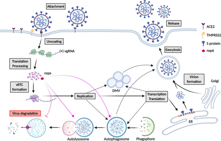
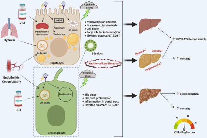
The recent outbreak of coronavirus disease 2019 (COVID-19), caused by the Severe Acute Respiratory Syndrome Coronavirus-2 (SARS-CoV-2) has resulted in a world-wide pandemic. Disseminated lung injury with the development of acute respiratory distress syndrome (ARDS) is the main cause of mortality in COVID-19. Although liver failure does not seem to occur in the absence of pre-existing liver disease, hepatic involvement in COVID-19 may correlate with overall disease severity and serve as a prognostic factor for the development of ARDS. The spectrum of liver injury in COVID-19 may range from direct infection by SARS-CoV-2, indirect involvement by systemic inflammation, hypoxic changes, iatrogenic causes such as drugs and ventilation to exacerbation of underlying liver disease. This concise review discusses the potential pathophysiological mechanisms for SARS-CoV-2 hepatic tropism as well as acute and possibly long-term liver injury in COVID-19.


Conflict of interest statement

The Medical Universities of Graz and Vienna have filed patents for the medical use of norUDCA and MT is listed as co‐inventor. MT has served as a speaker for Falk Foundation, Gilead, Intercept and MSD; he has advised for Albireo, BiomX, Boehringer Ingelheim, Falk Pharma GmbH, Genfit, Gilead, Intercept, Jannsen, MSD, Novartis, Phenex, Regulus and Shire. He further received travel grants from Abbvie, Falk, Gilead and Intercept and research grants from Albireo, CymaBay, Falk, Gilead, Intercept, MSD and Takeda. SL has received personal fees from Roche, AstraZeneca, Novartis and Biogena outside the submitted work, Authors not named here have disclosed no conflicts of interest.

Figures

Figure 1
Figure 1
SARS‐CoV‐2 life cycle in host cells. SARS‐CoV‐2 attachment to host cells in liver (eg hepatocytes) may be mediated by the interaction of Spike (S) protein with ACE2. S protein is cleaved by the transmembrane serine protease 2 (TMPRSS2), allowing the cellular entry of the virus. Once uncoated, the viral genome ((+) vgRNA) is released and translated by the ribosome into pp1a and pp1ab (not shown), that are further cleaved into 16 non‐structural proteins (nsps). Following the viral replication/transcription complex (vRTC) assembly, nsp6 (in red) induces autophagosome formation, where viral replication might take place (purple dashed lines). Viral replication might also occur in double‐membrane vesicles (DMV) (black dashed lines). nsp6‐mediated inhibition of autophagosome/lysosome expansion might prevent viral degradation (purple dashed inhibitory line). Newly synthesized viral structural and accessory proteins assemble to form the nucleocapsid and viral envelope at the ER–Golgi intermediate compartment (lower right). Mature virions are then released through the exploitation of the host vesicular system (upper right). DMV and autophagosomes might also be used by the virus for exocytosis and release of mature virions (black dashed lines)
Figure 2
Figure 2
Proposed pathophysiology for liver injury upon SARS‐CoV‐2 infection. COVID‐19‐associated hepatocellular damage is mainly characterized by moderate steatosis, lobular and portal inflammation, apoptotic/necrotic foci and elevation of plasma ALT and AST (upper left panel). Preliminary observations suggest that the injury might be caused by hepatocellular infection with direct cytopathic effects of SARS‐CoV‐2, which could induce mitochondrial dysfunction and ER stress contributing to steatosis. Furthermore, SARS‐CoV‐2 infection might also activate mTOR, which eventually inhibits autophagy (as a mechanism of viral degradation) and facilitates viral escape from the immune system. In addition, cytokine storm, hypoxic conditions due to ARDS and drug‐induced liver injury (DILI) may contribute. COVID‐19‐associated cholangiocellular injury has also been observed and is mainly characterized by bile duct proliferation, occasionally bile plug formation and elevation of plasma γGT and ALP (lower left panel). From a hepatological perspective, COVID‐19‐positive patients may be divided into three categories: patients without pre‐existing chronic liver disease, patients with early stage chronic liver disease and patients with advanced chronic liver disease/cirrhosis. COVID‐19‐associated liver injury may have a more severe outcome in patients with pre‐existing liver disease, such as non‐alcoholic fatty liver disease (NAFLD) and associated metabolic comorbidity. Moreover, COVID‐19 may induce hepatic decompensation with increased mortality in cirrhotic patients (right panel)

References

    1. John Hopkins University and Medicine . COVID‐19 Map ‐ Johns Hopkins Coronavirus Resource Center. John Hopkins Coronavirus Resource Center 1 https://coronavirus.jhu.edu/map.html (2020)
    1. Xie M, Chen Q. Insight into 2019 novel coronavirus — An updated interim review and lessons from SARS‐CoV and MERS‐CoV. Int J Infect Dis. 2020;94:119‐124. - PMC - PubMed
    1. Zhang C, Shi L, Wang FS. Liver injury in COVID‐19: management and challenges. The Lancet Gastroenterol Hepatol. 2020;5:428‐430. - PMC - PubMed
    1. Xu L, Liu J, Lu M, Yang D, Zheng X. Liver injury during highly pathogenic human coronavirus infections. Liver Int. 2020;40:liv.14435. - PMC - PubMed
    1. Sun J, Aghemo A, Forner A, Valenti L. COVID‐19 and liver disease. Liver Int. 2020. 10.1111/liv.14470 - DOI - PubMed

Publication types

Substances