

Extreme Diversity of the Human Vascular Mesenchymal Cell Landscape
- PMID: 33190596
- PMCID: PMC7763765
- DOI: 10.1161/JAHA.120.017094
Extreme Diversity of the Human Vascular Mesenchymal Cell Landscape
Erratum in
-
Correction to: Extreme Diversity of the Human Vascular Mesenchymal Cell Landscape.J Am Heart Assoc. 2021 Jan 5;10(1):e014643. doi: 10.1161/JAHA.119.014643. Epub 2020 Dec 25. J Am Heart Assoc. 2021. PMID: 33356377 Free PMC article. No abstract available.
Abstract
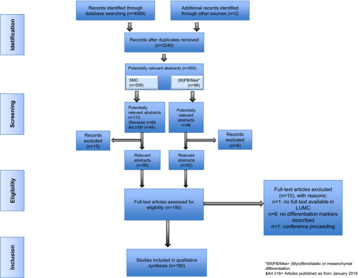
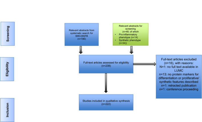
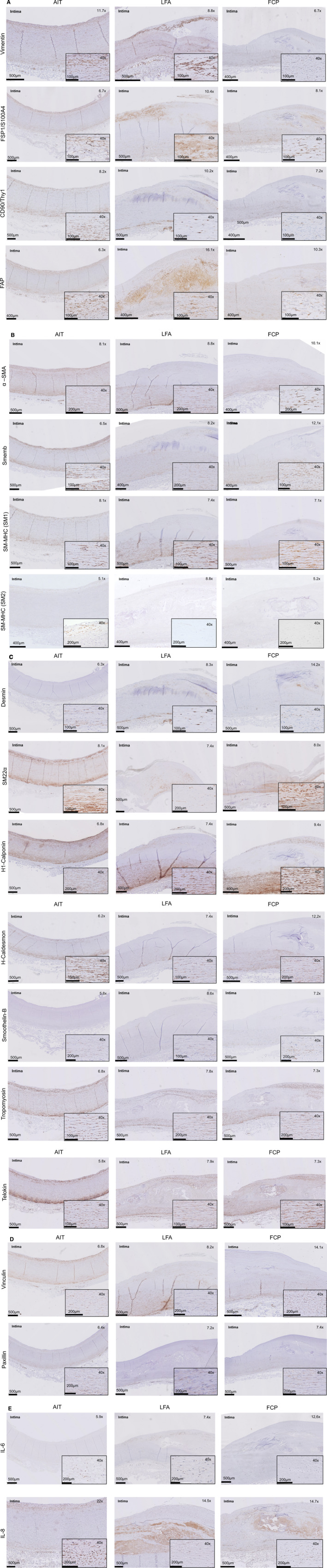
Background Human mesenchymal cells are culprit factors in vascular (patho)physiology and are hallmarked by phenotypic and functional heterogeneity. At present, they are subdivided by classic umbrella terms, such as "fibroblasts," "myofibroblasts," "smooth muscle cells," "fibrocytes," "mesangial cells," and "pericytes." However, a discriminative marker-based subclassification has to date not been established. Methods and Results As a first effort toward a classification scheme, a systematic literature search was performed to identify the most commonly used phenotypical and functional protein markers for characterizing and classifying vascular mesenchymal cell subpopulation(s). We next applied immunohistochemistry and immunofluorescence to inventory the expression pattern of identified markers on human aorta specimens representing early, intermediate, and end stages of human atherosclerotic disease. Included markers comprise markers for mesenchymal lineage (vimentin, FSP-1 [fibroblast-specific protein-1]/S100A4, cluster of differentiation (CD) 90/thymocyte differentiation antigen 1, and FAP [fibroblast activation protein]), contractile/non-contractile phenotype (α-smooth muscle actin, smooth muscle myosin heavy chain, and nonmuscle myosin heavy chain), and auxiliary contractile markers (h1-Calponin, h-Caldesmon, Desmin, SM22α [smooth muscle protein 22α], non-muscle myosin heavy chain, smooth muscle myosin heavy chain, Smoothelin-B, α-Tropomyosin, and Telokin) or adhesion proteins (Paxillin and Vinculin). Vimentin classified as the most inclusive lineage marker. Subset markers did not separate along classic lines of smooth muscle cell, myofibroblast, or fibroblast, but showed clear temporal and spatial diversity. Strong indications were found for presence of stem cells/Endothelial-to-Mesenchymal cell Transition and fibrocytes in specific aspects of the human atherosclerotic process. Conclusions This systematic evaluation shows a highly diverse and dynamic landscape for the human vascular mesenchymal cell population that is not captured by the classic nomenclature. Our observations stress the need for a consensus multiparameter subclass designation along the lines of the cluster of differentiation classification for leucocytes.
Keywords: atherosclerosis; fibroblasts; myofibroblasts; vascular smooth muscle cells.
Conflict of interest statement
None.
Figures

References
-
- Topouzis S, Majesky M. Smooth muscle lineage diversity in the chick embryo. Dev Biol. 1996;178:430–445. - PubMed
-
- Lacolley P, Regnault V, Nicoletti A, Li Z, Michel J. The vascular smooth muscle cell in arterial pathology: a cell that can take on multiple roles. Cardiovasc Res. 2012;95:194–204. - PubMed
-
- Riches K, Clark E, Helliwell RJ, Angelini TG, Hemmings KE, Bailey MA, Bridge KI, Scott DJA, Porter KE. Progressive development of aberrant smooth muscle cell phenotype in abdominal aortic aneurysm disease. J Vasc Res. 2018;55:35–46. - PubMed
-
- Bellini A, Mattoli S. The role of the fibrocyte, a bone marrow‐derived mesenchymal progenitor, in reactive and reparative fibroses. Lab Invest. 2007;87:858–870. - PubMed
Publication types
MeSH terms
LinkOut - more resources
Full Text Sources
Medical
Research Materials
Miscellaneous
