Review
. 2021 Jun;39(6):613-623.
doi: 10.1016/j.tibtech.2020.10.006. Epub 2020 Nov 13.

Image-Based Live Cell Sorting

Affiliations
Review

Image-Based Live Cell Sorting

Cody A LaBelle et al. Trends Biotechnol. 2021 Jun.

Abstract

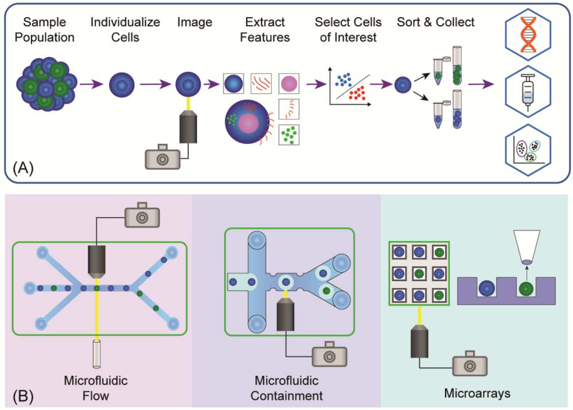
Technologies capable of cell separation based on cell images provide powerful tools enabling cell selection criteria that rely on spatially or temporally varying properties. Image-based cell sorting (IBCS) systems utilize microfluidic or microarray platforms, each having unique characteristics and applications. The advent of IBCS marks a new paradigm in which cell phenotype and behavior can be explored with high resolution and tied to cellular physiological and omics data, providing a deeper understanding of single-cell physiology and the creation of cell lines with unique properties. Cell sorting guided by high-content image information has far-reaching implications in biomedical research, clinical medicine, and pharmaceutical development.

Keywords: cell sorting; cytometry; imaging; microarrays; microdevice; microfluidics.


Conflict of interest statement

Conflict of Interest

N.L.A. and C.E.S. disclose a financial interest in Cell Microsystems, Inc. All other authors declare no conflicts.

Figures

Figure 1 (Key Figure):
Figure 1 (Key Figure):. Overview of Image Based Cell Sorting and Main Technologies.
(A) IBCS platforms typically follow a process in which a sample of cells is collected, the sample is separated into either single cells or small colonies, each cell or colony is imaged, features are extracted using ML or traditional image analysis techniques, cells of interest are selected for isolation, and the cells are sorted into collection containers. The cells are often then used in additional assays, sequencing pipelines, or therapies. (B) The three principle strategies used in IBCS are microfluidic flow devices, microfluidic containment devices, and microarrays. In microfluidic flow (left), cells rapidly move within a fluid stream and are imaged with specialized imaging techniques, and then sorted into collection containers in a high-throughput manner. Using microfluidic containment (center), cells are individualized and captured within droplets or stationary traps for conventional imaging and medium-throughput sorting using fluid flow. Alternatively, elements in microarrays (right) are loaded with single or multiple cells, imaged with conventional imaging techniques—often temporally—and cells are isolated using low-throughput mechanical or optical retrieval methods.
Figure 2:
Figure 2:. Microfluidic Flow IBCS Platforms.
(A) These IBCS platforms often use spatial and temporal excitation waveforms to form images, enabling information-rich signal collection. Machine learning strategies are then applied to the images to classify cells and the individual cells are sorted in a high-throughput manner. (B) Intelligent image-activated cell sorting system and images of Jurkat cells captured by the system. Reproduced from [21] with permission. (C) Machine learning based real-time image-guided cell sorting and classification system, using a spatial filter to produce excitation waveforms that are transformed into 2-D images for analysis. Reproduced from [19] with permission. (D) Ghost cytometry cell sorting platform which employs an optical filter to produce 1-D waveforms used for cell classification. Reprinted from [18] with permission.
Figure 3.
Figure 3.. Microfluidic Containment IBCS.
(A) Schematic of the major steps or common components of containment IBCS. (B) Use of droplet microfluidics to separate interacting cells. Reproduced from [31] with permission. (C) Example of a cell containment strategy that relies on cell docking in a restricted channel location. A bead-based secretion assay is also integrated into this sorting platform. Reprinted from [37]. (D) IBCS device that relies on valve closure to entrap cells within a small region of a microchannel. Adapted from [35] under a Creative Commons license (http://creativecommons.org/licenses/by/4.0/).
Figure 4.
Figure 4.. Overview of Microarray-Based IBCS.
(A) Workflow for microarray platforms used in IBCS. (B) Sorting using confocal imaging on microraft arrays. Adapted from [58] with permission. (C) Microchip for DEP arraying and sorting of cells. Adapted from [51] with permission. (D) Sorting cells based on temporal imaging after placement into arrayed docking sites. Adapted from [54] with permission.

References

    1. Bossel Ben-Moshe N et al. (2019) Predicting bacterial infection outcomes using single cell RNA-sequencing analysis of human immune cells. Nat. Commun 10, 3266. - PMC - PubMed
    1. Buenrostro JD et al. (2018) Integrated Single-Cell Analysis Maps the Continuous Regulatory Landscape of Human Hematopoietic Differentiation. Cell 173, 1535–1548.e16 - PMC - PubMed
    1. Kim C et al. (2018) Chemoresistance Evolution in Triple-Negative Breast Cancer Delineated by Single-Cell Sequencing. Cell 173, 879–893.e13 - PMC - PubMed
    1. Mitra AK et al. (2016) Single-cell analysis of targeted transcriptome predicts drug sensitivity of single cells within human myeloma tumors. Leukemia 30, 1094–1102 - PubMed
    1. Krieg C et al. (2018) High-dimensional single-cell analysis predicts response to anti-PD-1 immunotherapy. Nat. Med 24, 144–153 - PubMed

Publication types

MeSH terms