Review
. 2020 Nov-Dec;11(6):1031-1038.
doi: 10.1016/j.jcot.2020.08.027. Epub 2020 Sep 12.

Associated both-column acetabular fracture: An overview of operative steps and surgical technique

Affiliations
Review

Associated both-column acetabular fracture: An overview of operative steps and surgical technique

Vincenzo Giordano et al. J Clin Orthop Trauma. 2020 Nov-Dec.

Abstract

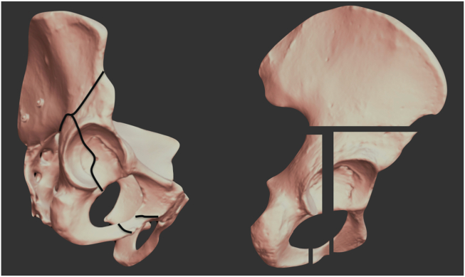
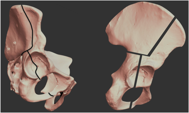
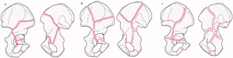
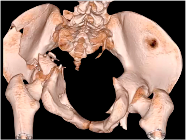
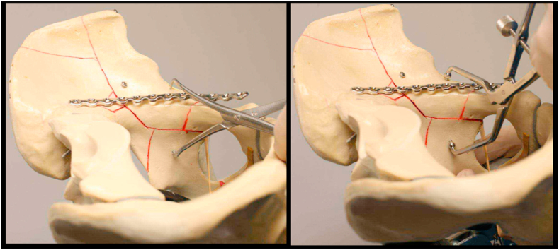
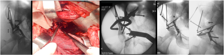
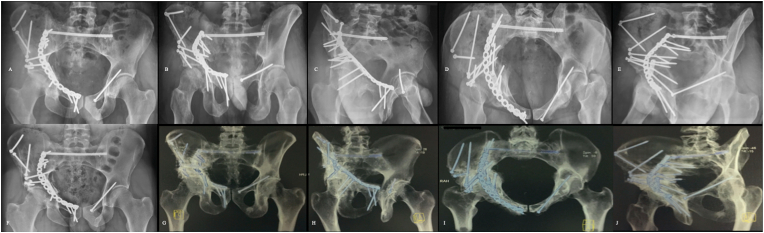
The management of both-column fractures of the acetabulum is challenging for the orthopaedic surgeon. Operative treatment is usually recommended in this particular fracture pattern, as residual joint surface displacement has been shown to increase local contact stress, drastically leading to rapid cartilage destruction. In this review, we present an overview of operative steps and surgical technique for both-column acetabular fracture reconstruction. Therefore, we demonstrate how correct understanding of fracture morphology and displacement, preoperative preparation, including choice of approach and patient positioning, reduction strategies, and programmed sequential fixation, starting from superior fracture lines on the anterior acetabular column and ending on the posterior components of this fracture type, may provide satisfactory outcomes in this difficult acetabular fracture pattern.

Keywords: Acetabular fracture; Approach to the acetabulum; Both-column acetabular fracture; Hip fracture.


Conflict of interest statement

None.

Figures

Fig. 1
Fig. 1
Pelvic plastic model. A, T-shaped pattern. In this fracture pattern, the proximal vertical fracture line generally exits between the ASIS∗ and the AIIS∗∗; B, Y-shaped pattern. In this fracture pattern, the proximal vertical fracture line can exit either between the ASIS and the iliac tuberosity (low anterior column fracture) or up in the iliac crest (high anterior column fracture). ∗ASIS (anterior superior iliac spine); ∗∗AIIS (anterior inferior iliac spine).
Fig. 1
Fig. 1
Pelvic plastic model. A, T-shaped pattern. In this fracture pattern, the proximal vertical fracture line generally exits between the ASIS∗ and the AIIS∗∗; B, Y-shaped pattern. In this fracture pattern, the proximal vertical fracture line can exit either between the ASIS and the iliac tuberosity (low anterior column fracture) or up in the iliac crest (high anterior column fracture). ∗ASIS (anterior superior iliac spine); ∗∗AIIS (anterior inferior iliac spine).
Fig. 2
Fig. 2
Y-shaped fracture types. A, Low anterior column fracture – the vertical fracture line exits between the ASIS and the iliac tuberosity; B, High anterior column fracture – the vertical fracture line exits up in the iliac crest. Note that there is also a secondary fracture line exiting between the ASIS and the iliac tuberosity. This pattern is also called H-shaped type; C, High anterior column fracture – the vertical fracture line exits up in the iliac crest. Observe that a secondary fracture line extends into the SI joint. We call this pattern X-shaped type.
Fig. 3
Fig. 3
3-D pelvic image demonstrating the posterior aspect of pelvis. Associated posterior wall (white arrowheads) is often a large, noncomminuted, nondisplaced or minimally anteriorly displaced fragment. It occurs in nearly two fifths of cases and is generally created by a pull-type mechanism. (courtesy Adrién Roa-Zoppi, MD)
Fig. 4
Fig. 4
AP right hip radiograph. A lateral compressive force applied to the greater trochanter with the hip in slight abduction and external rotation displaces the acetabulum columns medially. The femoral head acts as a hammer, directly striking on the anterior medial wall of the acetabulum and quadrilateral plate.
Fig. 5
Fig. 5
The spur sign is considered pathognomonic of both-column fracture. It is better observed in the obturator oblique radiographic view and the CT scan.
Fig. 6
Fig. 6
AP pelvis radiograph and 3D-CT reconstruction of the pelvis of a 35-year-old male who fell from approximately 8 m height. Images show a high anterior column pattern extending into the SI joint (X-type) associated with a displaced posterior wall acetabular fracture. Observe that posterior column component is minimally displaced.
Fig. 7
Fig. 7
Axial tomographic cuts from the same patient revealing a complex fracture of the anterior component and an associated unique displaced posterior wall acetabular fracture. Note the low-anterior column acetabular fracture on the contralateral side.
Fig. 8
Fig. 8
A constant triangular bone piece is always present in the iliac fossa close to the inferior part of the SI joint (left image, black arrows). Reduction of the superior part of the fracture should start with this key fragment as this restores the upper medial portion of the pelvic brim (right image).
Fig. 9
Fig. 9
Pelvic plastic model for demonstration purposes. Reduction should begin proximally in the iliac wing through the 1st window of the ilioinguinal approach. The key fragment is reduced first and temporarily secured with a 1.6-mm K-wire, then the iliac crest is reduced and fixed with multiple 3.5-mm long cortical lag-screws placed between the inner and outer tables of the ilium.
Fig. 10
Fig. 10
Intraoperative fluoroscopic images from the same patient presented in Fig. 6, Fig. 7. Reduction and fixation of the iliac wing using multiple 3.5-mm cortical screws. Additionally, a suprapectineal plate was used to fix the medial roof fragment.
Fig. 11
Fig. 11
Pelvic plastic model for demonstration purposes. Anterior column is usually externally rotated and medialized. A Farabeuf clamp helps manipulate and handling the anterior column through the 1st and 2nd windows of the ilioinguinal approach. Final reduction can be accomplished using a tong clamp and a Schanz pin. It is imperative that the femoral head is reduced under the acetabular roof.
Fig. 12
Fig. 12
Pelvic plastic model for demonstration purposes. Posterior column and quadrilateral plate are reduced through both the 1st and 2nd windows of the ilioinguinal approach Weber pointed clamps and asymmetrical Matta clamps are of special value to aid reduction. Fixation is accomplished using long cortical lag-screws placed through or outside the pelvic brim buttress plate.
Fig. 13
Fig. 13
Reduction of the posterior wall fragment is achieved through additional dissection on the outer table of the ilium. Intraoperative images from the same patient presented in Fig. 6, Fig. 7, Fig. 10. Note the posterior wall fragment completely displaced (black arrows). The use of an asymmetric long-arm pelvic clamp through the 2nd window of the ilioinguinal approach allows anatomic reduction of the posterior wall fragment. Fixation is done with 3.5-mm long cortical lag-screws placed from the inner iliac fossa to the posterior wall.
Fig. 14
Fig. 14
Radiographic and tomographic images from the same patient presented in Fig. 6, Fig. 7, Fig. 10, Fig. 13. A to E: Immediate post-operative AP pelvis, outlet view, obturator oblique, inlet view, and iliac oblique radiographs. Observe the anatomic reduction of the right hemipelvis and the acetabulum. SI joint was fixed with a 7.0-mm cannulated screw in S1. The left acetabulum fracture (infra-tectal simple transverse fracture) was fixed with a percutaneous retrograde 3.5-mm long cortical screw; F to J: AP pelvis and 3D reconstruction tomographic outlet, obturator oblique, inlet, and iliac oblique views after 3-year follow-up. No signs of osteoarthritis, heterotopic ossification, or vascular damage to the femoral heads.

Similar articles

Cited by

References

    1. Giannoudis P.V., Grotz M.R.W., Papakostidis C., Dinopoulos H. Operative treatment of displaced fractures of the acetabulum. A meta-analysis. J Bone Joint Surg Br. 2005;87(1):2–9. - PubMed
    1. Kelly J., Ladurner A., Rickman M. Surgical management of acetabular fractures – a contemporary literature review. Injury. 2020;S0020–1383(20):30525–30528. doi: 10.1016/j.injury.2020.06.016. - DOI - PubMed
    1. Letournel E. Acetabulum fractures: classification and management. Clin Orthop Relat Res. 1980;(151):81–106. - PubMed
    1. Gänsslen A., Frink M., Hildebrand F., Krettek C. Both column fractures of the acetabulum: epidemiology, operative management and long-term-results. Acta Chir Orthop Traumatol Cech. 2012;79(2):107–113. - PubMed
    1. Pierannunzii L., Fischer F., Tagliabue L., Calori G.M., D’Imporzano M. Acetabular both-column fractures: essential of operative management. Injury. 2010;41(11):1145–1149. doi: 10.1016/j.injury.2010.08.011. - DOI - PubMed