. 2020 Nov 6:14:4765-4774.
doi: 10.2147/DDDT.S270443. eCollection 2020.

Effects of Snake-Derived Phospholipase A2 Inhibitors on Acute Pancreatitis: In vitro and in vivo Characterization

Affiliations

Effects of Snake-Derived Phospholipase A2 Inhibitors on Acute Pancreatitis: In vitro and in vivo Characterization

Yanping Wu et al. Drug Des Devel Ther. .

Abstract

Objective: We aimed to investigate the effects of snake-derived phospholipase A2 inhibitor (PLA2) from Sinonatrix percarinata and Bungarus multicinctus on acute pancreatitis in vivo and in vitro and assess the mechanisms.

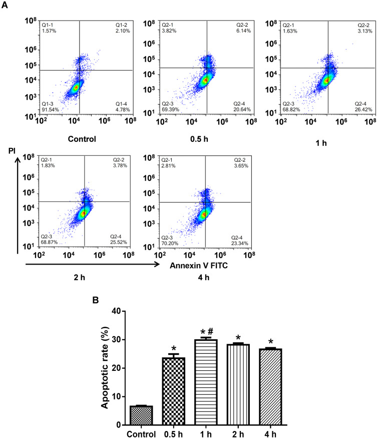
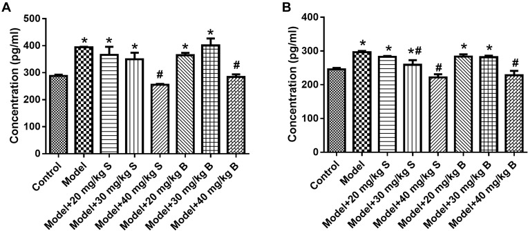
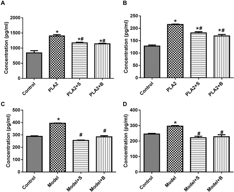
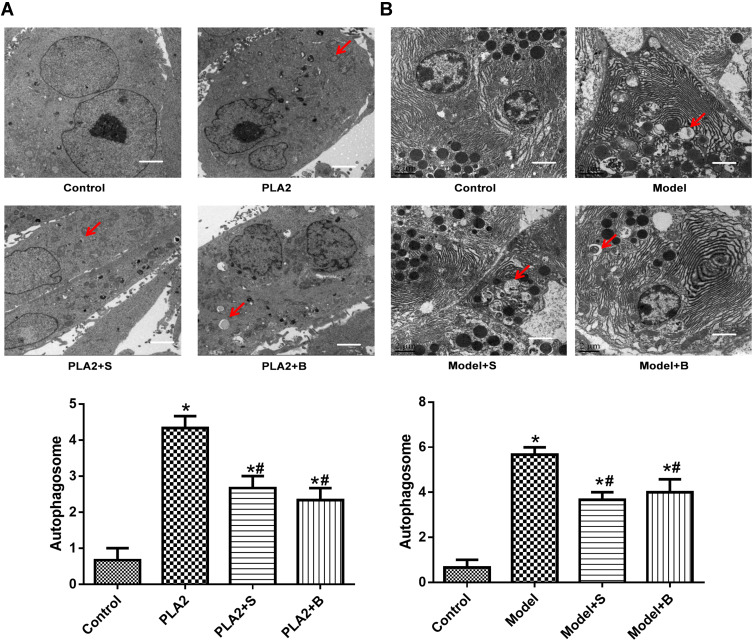
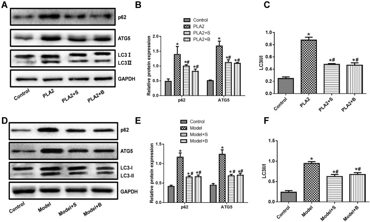
Methods: The levels of platelet-activating factor (PAF) and tumor necrosis factor (TNF)-α were detected by ELISA, and the characteristics of autophagy were detected by transmission electron microscopy and Western blotting (LC3, p62, and ATG5).

Results: In vitro experiments showed that PLA2 treatment caused obvious formation of autophagic bodies. By contrast, Sinonatrix and Bungarus peptides reduced the number of autophagic bodies. The concentrations of PAF and TNF-α, and the expressions of p62, autophagy-related 5 (ATG5), and microtubule-associated protein 1A/1B-light chain 3 (LC3)II/LC3I in the PLA2-treated group were significantly higher than in the control group (P<0.05). The concentrations of PAF and TNF-α, and the expressions of p62, ATG5, and LC3II/LC3I in the Sinonatrix or Bungarus peptide treatment groups were significantly lower than in the PLA2-treated cells (P<0.05). In the pancreatic tissue, autophagic bodies were observed in the model group; autophagic bodies were remarkably reduced in Sinonatrix or Bungarus peptide-treated groups compared with the model group. In vivo experiments also showed that the levels of PAF and TNF-α, and the expressions of p62, ATG5, and LC3II/LC3I were significantly higher in the model group than in the control group (P<0.05). The levels of PAF and TNF-α in the model group, and the expressions of p62, ATG5, and LC3II/LC3I in Sinonatrix or Bungarus peptide-treated groups were significantly lower than in the model group (P<0.05).

Conclusion: Sinonatrix or Bungarus peptide could ameliorate the features of acute pancreatitis, likely through regulating autophagy.

Keywords: PLA2; acute pancreatitis; autophagy; snake-derived PLI.


Conflict of interest statement

All authors declare no personal, financial or non-financial conflicts of interest for this work.

Figures

Figure 1
Figure 1
Determining the effect of PLA2 treatment by flow cytometry. (A) Representative images of flow cytometry; (B) Quantitative apoptosis data. Data were expressed as mean and standard deviation. N=6 in each group (Bonferroni test). *P<0.05: compared with the control group; #P<0.05: compared with 0.5 h.
Figure 2
Figure 2
Effects of snake-derived PLIs on PAF and TNF-α levels in pancreatic tissue. (A) PAF level; (B) TNF-α level. Data were expressed as mean and standard deviation. N=6 in each group (Bonferroni test). *P<0.05: compared with the control group; #P<0.05: compared with the model group.
Figure 3
Figure 3
Snake-derived PLI reduced PAF and TNF-α levels in PLA 2-treated cells and AP mice model. (A, B) The level of PAF and TNF-α in the pancreatic acinar cells; (C, D) The level of PAF and TNF-α in vivo. Data were expressed as mean and standard deviation. N=6 in each group (Bonferroni test). *P<0.05: compared with the control group; #P<0.05: compared with the model group.
Figure 4
Figure 4
Snake-derived PLI reduced autophagic body formation in PLA 2-treated cells and AP mice model. (A) TEM results of cells; (B) TEM results of pancreatic tissue. Arrows indicate the autophagic bodies. *P<0.05: compared with the control group; #P<0.05: compared with the model group.
Figure 5
Figure 5
Snake-derived PLI reduced the expression of LC3, p62, and ATG5 in PLA 2-treated cells and AP mice model. (AC) LC3, p62, and ATG5 in PLA 2-treated cells; (DF) LC3, p62, and ATG5 in AP mice model. The original blots are shown in Supplementary Figure S1. Data were expressed as mean and standard deviation. N=6 in each group (Bonferroni test). *P<0.05: compared with the control group; #P<0.05: compared with the model group.

Similar articles

Cited by

References

    1. Sun H, Zuo HD, Lin Q, et al. MR imaging for acute pancreatitis: the current status of clinical applications. Ann Transl Med. 2019;7(12):269. doi:10.21037/atm.2019.05.37 - DOI - PMC - PubMed
    1. Singh VK, Yadav D, Garg PK. Diagnosis and management of chronic pancreatitis: a review. JAMA. 2019;322(24):2422–2434. doi:10.1001/jama.2019.19411 - DOI - PubMed
    1. Gravante G, Garcea G, Ong SL, et al. Prediction of mortality in acute pancreatitis: a systematic review of the published evidence. Pancreatology. 2009;9(5):601–614. doi:10.1159/000212097 - DOI - PubMed
    1. Hietaranta A, Kemppainen E, Puolakkainen P, et al. Extracellular phospholipases A2 in relation to systemic inflammatory response syndrome (SIRS) and systemic complications in severe acute pancreatitis. Pancreas. 1999;18(4):385–391. doi:10.1097/00006676-199905000-00009 - DOI - PubMed
    1. Xiao H, Pan H, Liao K, Yang M, Huang C. Snake venom PLA2, a promising target for broad-spectrum antivenom drug development. Biomed Res Int. 2017;2017:6592820. doi:10.1155/2017/6592820 - DOI - PMC - PubMed