. 2020 Nov 6:13:4209-4219.
doi: 10.2147/DMSO.S275660. eCollection 2020.

High-Intensity Interval Training Attenuates Ketogenic Diet-Induced Liver Fibrosis in Type 2 Diabetic Mice by Ameliorating TGF-β1/Smad Signaling

Affiliations

High-Intensity Interval Training Attenuates Ketogenic Diet-Induced Liver Fibrosis in Type 2 Diabetic Mice by Ameliorating TGF-β1/Smad Signaling

Qiang Zhang et al. Diabetes Metab Syndr Obes. .

Abstract

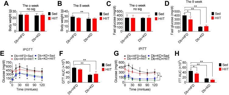
Objective: Ketogenic diet (KD) and high-intensity interval training (HIIT) have preclinical benefits for type 2 diabetes (Db). However, the health risks of long-term KD use in diabetes should be ascertained and prevented. We hypothesized that KD-induced liver fibrosis in type 2 diabetic mice could be ameliorated by HIIT.

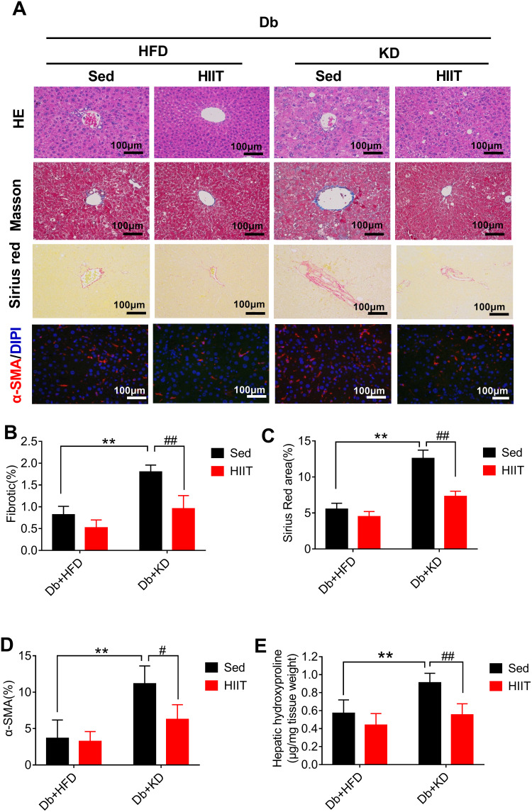
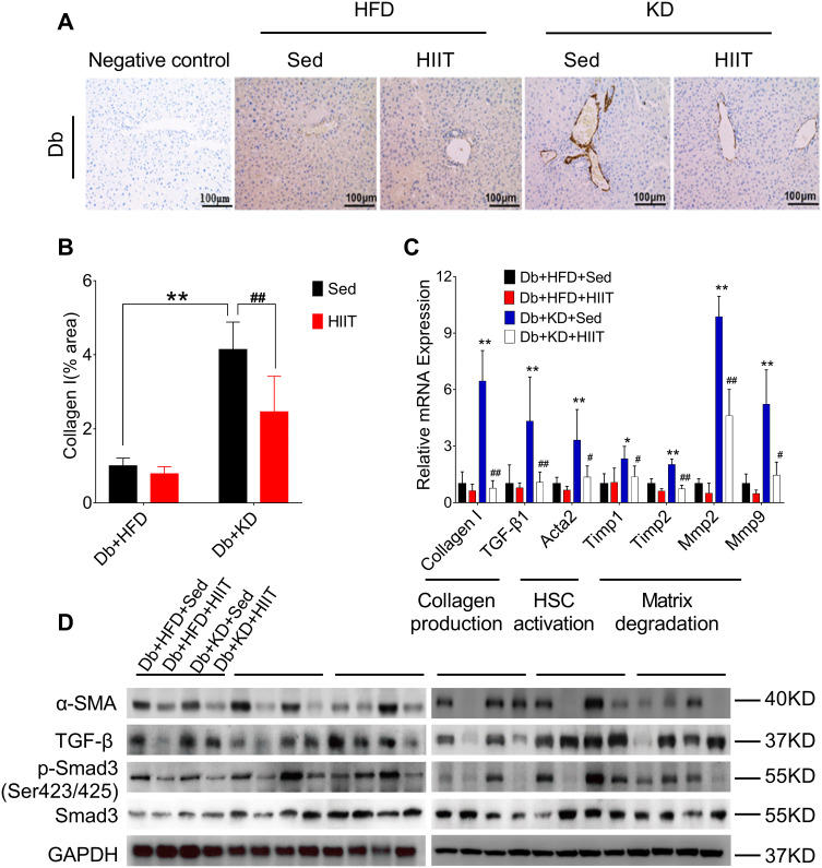
Methods: Streptozotocin-induced type 2 diabetic mice were divided into high-fat diet (HFD) control (Db+HFD+Sed), KD control (Db+KD+Sed), HFD coupled with HIIT (Db+HFD+HIIT), and KD coupled with HIIT (Db+KD+HIIT) groups (n=6, per group). Control mice were kept in sedentary (Sed), while HIIT group mice underwent 40-minute high-intensity interval training three alternate days per week. After 8-week intervention, the indicators of body weight and insulin resistance, oxidative stress markers, hepatic fibrosis, genetic and protein expression of related pathways were tested.

Results: We found that fasting blood glucose level was reduced in the Db+HFD+HIIT, Db+KD+Sed, and Db+KD+HIIT groups. Insulin sensitivity was increased in diabetic mice of these groups, whereas ROS levels were decreased in mice that underwent HIIT. The immunohistochemical staining of liver, serum index, and hepatic parameters of diabetic mice in the KD group revealed liver fibrosis, which was significantly attenuated by HIIT. Besides, these effects of HIIT were the outcome of hepatic stellate cell's inactivation, reduced protein expression of matrix metalloproteinases and tissue inhibitor of metalloproteinases, and the inhibition of TGF-β1/Smad signaling.

Conclusion: KD had a profound fibrotic effect on the liver of type 2 diabetic mice, whereas HIIT ameliorated this effect. KD did not show any apparent benefit as far as glucose tolerance and homeostasis were concerned. Concisely, our results demonstrated that KD should be coupled with HIIT for the prevention and preclinical mitigation of type 2 diabetes.

Keywords: ROS; TGF-β1/Smad signal; diabetes; hepatic fibrosis; high-intensity interval training; ketogenic diet.


Conflict of interest statement

Dr. Yi Sun and Dr. ShuZhe Ding report grants from the National Natural Science Foundation of China, during the conduct of the study. The authors report no other potential conflicts of interest for this work.

Figures

Figure 1
Figure 1
Effect of the ketogenic diet and the ketogenic diet coupled high-intensity interval on body weight and glucose homeostasis in diabetic mice. The analysis of body weight (A) before the intervention, (B) after 8-week HIIT intervention; fasting blood glucose (C) before the intervention, (D) after HIIT intervention; (E, F) glucose tolerance test, and (G, H) insulin tolerance test, after HIIT intervention. Data were presented as means ± SD (n=6 each group) and *P <0.05, **P <0.01, compared to Db+HFD+Sed group, no sig. indicates no significance.
Figure 2
Figure 2
Ketogenic diet treatment-induced oxidative stress in diabetic mice. The diabetic mice were exposed to HFD or KD with or without HIIT for 8-week. Analysis of (A) serum H2O2, (B) serum MDA, (C) serum NO, (D) serum GSH-PX and (E) serum SOD in the mice of Db+HFD+Sed, Db+KD+Sed, Db+HFD+HIIT, and Db+KD+HIIT groups. Data were presented as means ± SD (n=6 each group). Groups were statistically compared using two-way ANOVA and Bonferroni post hoc tests. *P <0.05, **P <0.01, compared to Db+HFD+Sed group, ##P <0.01, compared to Db+KD+Sed group.
Figure 3
Figure 3
High-intensity interval training attenuated ketogenic diet-induced hepatic fibrosis and histological changes in livers of diabetic mice. Analysis of mice liver (A), fibrotic area (%) (B), Sirius Red area (%) (C), α-SMA (%) (D) and hepatic hydroxyproline levels (E). Immunostaining was performed to determine the α-SMA expression (red, 200×), and nuclear counterstaining was done using DAPI (blue). Scale bars: 100 μm. Data were presented as means ± SD (n=6 each group). Groups were statistically compared using two-way ANOVA and Bonferroni post hoc tests. **P <0.01, compared to Db+HFD+Sed group, #P <0.05, ##P <0.01 compared to Db+KD+Sed group.
Figure 4
Figure 4
High-intensity interval training attenuated ketogenic diet-induced liver fibrosis. (A) Collagen production was compared between different groups by collagen I antibody staining. (B) Percentages of immunoreactive (collagen (I) areas in liver tissue sections were measured and expressed as relative values obtained by comparing the Db+HFD+Sed group of mice. (C) Gene expression analysis of collagen production, HSCs activation, and matrix degradation in the liver of diabetic mice of different groups. (D) Western blot analysis of mouse liver lysates by using specific antibodies against α-SMA, TGF-β, p-Smad3 (ser 423/425), or Smad3. Data were presented as means ± SD (n=6 each group). Groups were statistically compared using two-way ANOVA and Bonferroni post hoc test. Scale bars: 100 μm. *P <0.05, **P <0.01, compared to Db+HFD+Sed group, #P <0.05, ##P <0.01 compared to the Db+KD+Sed group.

Similar articles

Cited by

References

    1. Organisation IDF. IDF Diabetes Atlas Ninth Edition. IDF. 2019;1(9):34–36. doi:10.1016/j.diabres.2018.05.033 - DOI
    1. American Diabetes Association. Classification and diagnosis of diabetes. Sec. 2. in standards of medical care in diabetes-2016. Diabetes Care. 2016;39(Suppl.1):S13–S22. - PubMed
    1. American Diabetes Association. Diagnosis and Classification of Diabetes Mellitus. Diabetes Care. 2009;32(Suppl 1):S62–S67. doi:10.2337/dc13-S067 - DOI - PMC - PubMed
    1. Lancet T. The diabetes pandemic. Lancet. 2011;378(9786):99. doi:10.1016/S0140-6736(11)61068-4 - DOI - PubMed
    1. Watson JD. Type 2 Diabetes As A Redox Disease. Lancet. 2014;383(9919):841–843. doi:10.1016/S0140-6736(13)62365-X - DOI - PubMed