. 2020 Nov 6:12:11359-11369.
doi: 10.2147/CMAR.S267355. eCollection 2020.

LncRNA MALAT1 Promotes Survival of Epithelial Ovarian Cancer Cells by Downregulating miR-145-5p

Affiliations

LncRNA MALAT1 Promotes Survival of Epithelial Ovarian Cancer Cells by Downregulating miR-145-5p

Ke Wang et al. Cancer Manag Res. .

Retraction in

Abstract

Purpose: This paper was aimed at investigating the regulatory mechanism of long non-coding RNA metastasis-associated lung adenocarcinoma transcript-1 (MALAT1) in epithelial ovarian cancer (EOC).

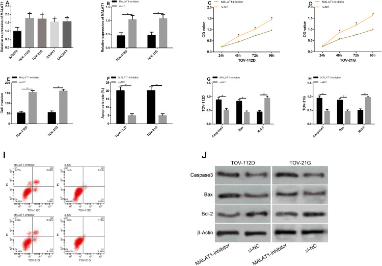
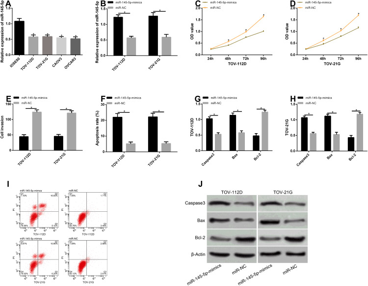
Materials and methods: MALAT1 and miR-145-5p expression in the tissues, serum, and EOC cell lines (TOV-112D, TOV-21G) of patients with EOC were detected. The two genes were transfected into the cells via upregulating or downregulating their expression. Levels of apoptosis-related proteins (Caspase-3, Bax, Bcl-2) were analyzed. Mechanisms of cell proliferation, invasion, and apoptosis were studied.

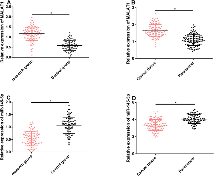
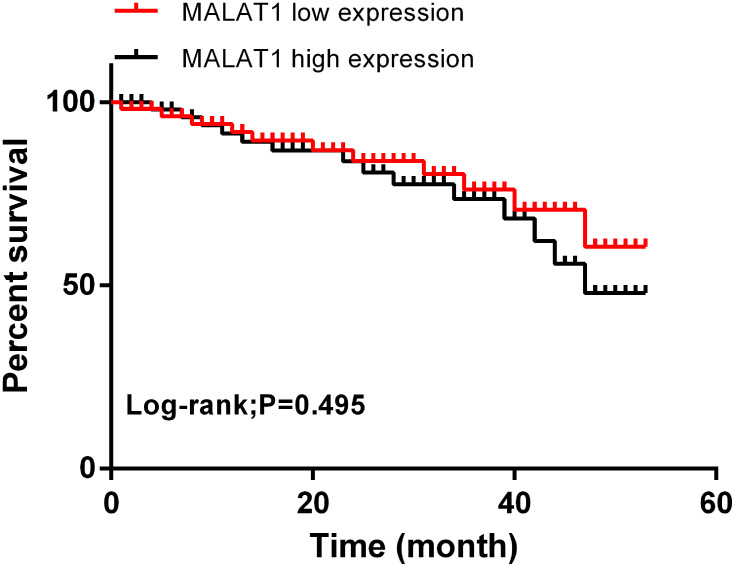
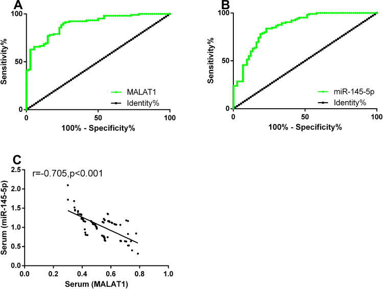
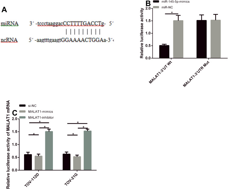
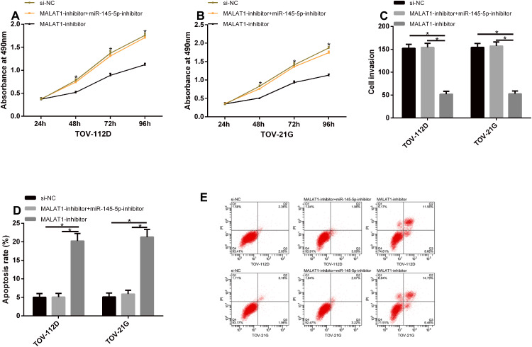
Results: MALAT1 was high expressed in EOC tissues, while miR-145-5p was poorly expressed in them. The areas under the curves (AUCs) of the two genes for diagnosing EOC were greater than 0.850, and the two had a significantly negative correlation. According to multivariate Cox regression analysis, high MALAT1 expression, tumor size, degree of differentiation, case staging, and lymph node metastasis were the independent risk factors affecting prognosis. The 5-year overall survival rate (OSR) of patients with low MALAT1 expression was remarkably higher than that of those with high expression. Overexpressing miR-145-5p and silencing MALAT1 could inhibit EOC cells from proliferating and invading, increase their apoptotic rate, and improve levels of the apoptosis-related proteins. After co-transfection with MALAT1-inhibitor + miR-145-5p-inhibitor, the proliferation and invasion of TOV-112D and TOV-21G cells were inhibited and the apoptotic rate rose more obviously. Inhibiting MALAT1 could increase miR-145-5p expression, thus inhibiting EOC cells from proliferating and invading and thereby increasing their apoptotic rate.

Conclusion: MALAT1 promotes EOC cells' survival by downregulating miR-145-5p so it may become a new direction for EOC diagnosis and gene therapy.

Keywords: EOC; MALAT1; cell survival; miR-145-5p.


Conflict of interest statement

The authors report no conflicts of interest for this work. Ye Zhao and Yi-Min Wang are co-corresponding author.

Figures

Figure 1
Figure 1
MALAT1 and miR-145-5p expression in serum and tissues. (A and B) MALAT1 expression remarkably rose in the serum and tissues of patients with EOC. (C and D) miR-145-5p expression remarkably rose in the serum and tissues of patients with EOC.
Figure 2
Figure 2
Correlation of MALAT1 with prognostic survival. The correlation of high and low MALAT1 expression with the 5-year OSR of patients with EOC.
Figure 3
Figure 3
Diagnostic value of MALAT1 and miR-145-5p expression. (A) The sensitivity and specificity of serum MALAT1 for diagnosing EOC were 90.47% and 74.32%, respectively. (B) The sensitivity and specificity of serum miR-145-5p for diagnosing EOC were 74.79% and 89.79%, respectively. (C) Serum MALAT1 expression was negatively correlated with miR-145-5p expression in the study group (r=0.618, P<0.001).
Figure 4
Figure 4
Effects of MALAT1 expression on biological functions of transfected cells. (A) MALAT1 expression in the cell lines of patients with EOC. (B) MALAT1 expression in TOV-112D and TOV-21G cells after transfection. (C) The proliferation of TOV-112D cells after transfection. (D) The proliferation of TOV-21G cells after transfection. (E) The invasion of TOV-112D and TOV-21G cells after transfection. (F) The apoptosis of TOV-112D and TOV-21G cells after transfection. (G and H) The effects of inhibiting MALAT1 on apoptosis-related proteins in TOV-112D and TOV-21G cells. (I) Flow cytometry maps. (J) Apoptosis-related protein maps.
Figure 5
Figure 5
Effects of miR-145-5p expression on biological functions of transfected cells. (A) miR-145-5p expression in the cell lines of patients with EOC. (B) miR-145-5p expression in TOV-112D and TOV-21G cells after transfection. (C) The proliferation of TOV-112D cells after transfection. (D) The proliferation of TOV-21G cells after transfection. (E) The invasion of TOV-112D and TOV-21G cells after transfection. (F) The apoptosis of TOV-112D and TOV-21G cells after transfection. (G and H) The effects of overexpressing miR-145-5p on apoptosis-related proteins in TOV-112D and TOV-21G cells. (I) Flow cytometry maps. (J) Apoptosis-related protein maps.
Figure 6
Figure 6
Gene identification of MALAT1. (A) The binding site between MALAT1 and miR-145-5p. (B) Results of dual luciferase reporter gene assay. (C) miR-145-5p expression in TOV-112D and TOV-21G cells after transfection.
Figure 7
Figure 7
Rescue experiment. (A and B) The cells in the MALAT1-inhibitor + miR-145-5p-inhibitor group were not different from those in the si-NC group with respect to their proliferation, but they had remarkably increasing proliferation when compared with those in the MALAT1-inhibitor group. (C) The cells in the MALAT1-inhibitor + miR-145-5p-inhibitor group were not different from those in the si-NC group with respect to their invasion, but they had remarkably increasing invasion when compared with those in the MALAT1-inhibitor group. (D) The cells in the MALAT1-inhibitor + miR-145-5p-inhibitor group were not different from those in the si-NC group with respect to their apoptosis, but they had remarkably reducing apoptosis when compared with those in the MALAT1-inhibitor group. (E) Flow cytometry maps.

Similar articles

Cited by

References

    1. Lheureux S, Gourley C, Vergote I, Oza AM. Epithelial ovarian cancer. Lancet. 2019;393:1240–1253. doi:10.1016/S0140-6736(18)32552-2 - DOI - PubMed
    1. Rojas V, Hirshfield KM, Ganesan S, Rodriguez-Rodriguez L. Molecular characterization of epithelial ovarian cancer: implications for diagnosis and treatment. Int J Mol Sci. 2016;17:2113. doi:10.3390/ijms17122113 - DOI - PMC - PubMed
    1. Al Habyan S, Kalos C, Szymborski J, McCaffrey L. Multicellular detachment generates metastatic spheroids during intra-abdominal dissemination in epithelial ovarian cancer. Oncogene. 2018;37:5127–5135. doi:10.1038/s41388-018-0317-x - DOI - PMC - PubMed
    1. Zhao L, Kong H, Sun H, Chen Z, Chen B, Zhou M. LncRNA-PVT1 promotes pancreatic cancer cells proliferation and migration through acting as a molecular sponge to regulate miR-448. J Cell Physiol. 2018;233:4044–4055. - PubMed
    1. Zou C, Wang Q, Lu C, et al. Transcriptome analysis reveals long noncoding RNAs involved in fiber development in cotton (Gossypium arboreum). Sci China Life Sci. 2016;59:164–171. doi:10.1007/s11427-016-5000-2 - DOI - PubMed

Publication types