. 2020 Oct 26:2020:7948095.
doi: 10.1155/2020/7948095. eCollection 2020.

Increased Levels of Soluble CD206 Associated with Rapidly Progressive Interstitial Lung Disease in Patients with Dermatomyositis

Affiliations

Increased Levels of Soluble CD206 Associated with Rapidly Progressive Interstitial Lung Disease in Patients with Dermatomyositis

Ya-Wen Shen et al. Mediators Inflamm. .

Abstract

Objective: Soluble CD206 (sCD206) is considered a macrophage activation marker, and a previous study proved it as a potential biomarker to predict the severity of anti-melanoma differentiation-associated gene 5- (anti-MDA-5-) positive dermatomyositis- (DM-) associated interstitial lung disease (ILD). To investigate the role of sCD206 in various subtypes of DM, we evaluated the serum level of sCD206 in patients with different myositis-specific autoantibodies besides anti-MDA-5 and clarified its clinical significance.

Methods: Commercial enzyme-linked immunosorbent assay kits were used to detect serum concentrations of sCD206 in 150 patients with DM and 52 healthy controls (HCs). Correlations between sCD206 levels and clinical features, laboratory examinations, and pulmonary function test parameters were analysed.

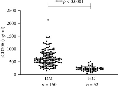
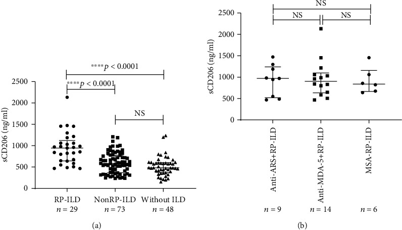
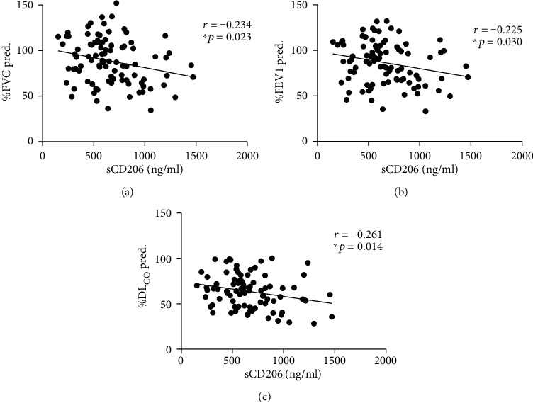
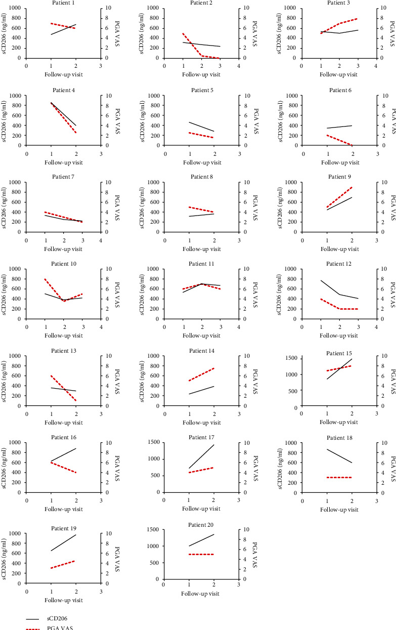
Results: The median concentrations of serum sCD206 in DM patients were significantly higher than those in HCs (p < 0.0001). Furthermore, median sCD206 levels were elevated in patients with ILD (p = 0.001), especially in those with rapidly progressive ILD (RP-ILD) (p < 0.0001). In addition, sCD206 levels were negatively correlated with the pulmonary function test results, including the percent predicted forced vital capacity (r = -0.234, p = 0.023), percent predicted forced expiratory volume in one second (r = -0.225, p = 0.030), and percent predicted carbon monoxide diffusion capacity (r = -0.261, p = 0.014). Age- and gender-adjusted multivariable analysis showed that sCD206 was an independent prognostic factor for RP-ILD in patients with DM. A longitudinal study showed that sCD206 levels were positively correlated with the physician global assessment visual analog scale scores (β = 54.201, p = 0.001).

Conclusion: Serum sCD206 levels were significantly increased in patients with DM and significantly associated with RP-ILD, suggesting that sCD206 is an important biological predictor of RP-ILD in patients with DM.


Conflict of interest statement

The authors declare that they have no conflict of interest.

Figures

Figure 1
Figure 1
The serum levels of sCD206 in patients with DM and HC samples. The serum levels of sCD206 in DM patients were significantly more elevated than those in HCs. sCD206: soluble CD206; HC: healthy control; DM: dermatomyositis. The error bars represent the interquartile range.
Figure 2
Figure 2
The sCD206 levels in all DM patients and in those with RP-ILD with different MSAs. (a) The serum levels of sCD206 in DM patients with RP-ILD, nonRP-ILD, and those without ILD. (b) The serum levels of sCD206 in DM patients with RP-ILD classified based on different MSAs. sCD206: soluble CD206; DM: dermatomyositis; ILD: interstitial lung disease; RP-ILD: rapidly progressive interstitial lung disease; ARS: aminoacyl-tRNA synthetases; MDA-5: melanoma differentiation-associated gene-5; MSA: myositis-specific autoantibodies. The Kruskal-Wallis H test was applied for comparisons between three groups. Error bars represent the interquartile range. NS indicates no significant difference.
Figure 3
Figure 3
The correlations of sCD206 levels with laboratory parameters in patients with DM. (a) sCD206 levels were positively correlated with ferritin levels in patients with DM. (b) sCD206 levels were positively correlated with CRP levels in patients with DM. (c) sCD206 levels were positively correlated with LDH levels in patients with DM. (d) sCD206 levels were positively correlated with TG levels in patients with DM. (e) sCD206 levels were positively correlated with CD19+CD5- B cell percentage in patients with DM. (f) sCD206 levels were negatively correlated with CD4+T cell percentage in patients with DM. sCD206: soluble CD206; DM: dermatomyositis; CRP: C-reactive protein; LDH: lactate dehydrogenase; TG: triglyceride.
Figure 4
Figure 4
The correlation between sCD206 levels and PFTs in patients with DM. (a) sCD206 levels were negatively correlated with %FVC in patients with DM. (b) sCD206 levels were negatively correlated with %FEV1 in patients with DM. (c) sCD206 levels were negatively correlated with %DLCO in patients with DM. sCD206: soluble CD206; DM: dermatomyositis; PFTs: pulmonary function tests; %FVC: percent predicted forced vital capacity; %FEV1: percent predicted forced expiratory volume in one second; %DLCO: percent predicted carbon monoxide diffusion capacity.
Figure 5
Figure 5
ROC curve for the RP-ILD risk prediction model. ROC curve analysis was used to assess the diagnostic value of sCD206 levels in the prediction of RP-ILD. The area under the curve was 0.811 (p < 0.0001), and the optional cut-off value was 792.75 ng/ml with a sensibility of 0.690 and a specificity of 0.835. ROC: receiver operating characteristic; RP-ILD: rapidly progressive interstitial lung disease; sCD206: soluble CD206.
Figure 6
Figure 6
The longitudinal analysis of sCD206 levels and PGA VAS in 20 patients with DM, respectively. sCD20: soluble CD206; DM: dermatomyositis; VAS: visual analog scale; PGA: physician global assessment.

References

    1. Dalakas M. C. Inflammatory muscle diseases. The New England Journal of Medicine. 2015;372(18):1734–1747. doi: 10.1056/NEJMra1402225. - DOI - PubMed
    1. Marie I., Hachulla E., Hatron P. Y., et al. O97 Polymyositis and dermatomyositis: short-term and long-term outcome, and predictive factors of prognosis. The Journal of Rheumatology. 2003;14, article S28 doi: 10.1016/S0953-6205(03)91246-5. - DOI - PubMed
    1. Connors G. R., Christopher-Stine L., Oddis C. V., Danoff S. K. Interstitial lung disease associated with the idiopathic inflammatory myopathies: what progress has been made in the past 35 years? Chest. 2010;138(6):1464–1474. doi: 10.1378/chest.10-0180. - DOI - PubMed
    1. McHugh N. J., Tansley S. L. Autoantibodies in myositis. Nature Reviews Rheumatology. 2018;14(5):290–302. doi: 10.1038/nrrheum.2018.56. - DOI - PubMed
    1. Cavagna L., on Behalf of AENEAS (American and European NEtwork of Antisynthetase Syndrome) Collaborative Group, Nuño L., et al. Serum Jo-1 Autoantibody and isolated arthritis in the antisynthetase syndrome: review of the literature and report of the experience of AENEAS Collaborative Group. Clinical Reviews in Allergy & Immunology. 2017;52(1):71–80. doi: 10.1007/s12016-016-8528-9. - DOI - PubMed

MeSH terms