. 2020 Oct 22:13:527543.
doi: 10.3389/fnmol.2020.527543. eCollection 2020.

Object Recognition Memory: Distinct Yet Complementary Roles of the Mouse CA1 and Perirhinal Cortex

Affiliations

Object Recognition Memory: Distinct Yet Complementary Roles of the Mouse CA1 and Perirhinal Cortex

David A Cinalli Jr et al. Front Mol Neurosci. .

Abstract

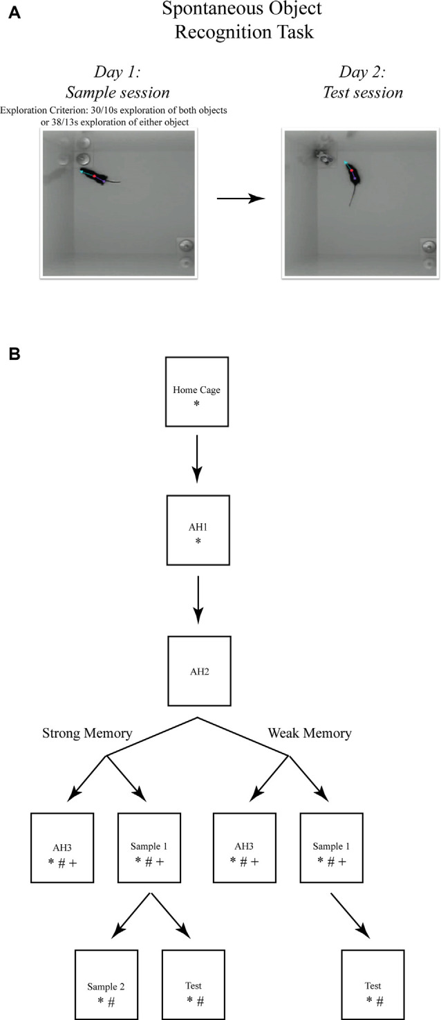
While the essential contribution of the hippocampus to spatial memory is well established, object recognition memory has been traditionally attributed to the perirhinal cortex (PRh). However, the results of several studies indicate that under specific procedural conditions, temporary or permanent lesions of the hippocampus affect object memory processes as measured in the Spontaneous Object Recognition (SOR) task. The PRh and hippocampus are considered to contribute distinctly to object recognition memory based on memory strength. Allowing mice more, or less, exploration of novel objects during the encoding phase of the task (i.e., sample session), yields stronger, or weaker, object memory, respectively. The current studies employed temporary local inactivation and immunohistochemistry to determine the differential contributions of neuronal activity in PRh and the CA1 region of the hippocampus to strong and weak object memory. Temporary inactivation of the CA1 immediately after the SOR sample session impaired strong object memory but spared weak object memory; while temporary inactivation of PRh post-sample impaired weak object memory but spared strong object memory. Furthermore, mRNA transcription and de novo protein synthesis are required for the consolidation of episodic memory, and activation patterns of immediate early genes (IEGs), such as c-Fos and Arc, are linked to behaviorally triggered neuronal activation and synaptic plasticity. Analyses of c-Fos and Arc protein expression in PRh and CA1 neurons by immunohistochemistry, and of Arc mRNA by qPCR after distinct stages of SOR, provide additional support that strong object memory is dependent on CA1 neuronal activity, while weak object memory is dependent on PRh neuronal activity. Taken together, the results support the view that both PRh and CA1 are required for object memory under distinct conditions. Specifically, our results are consistent with a model that as the mouse begins to explore a novel object, information about it accumulates within PRh, and a weak memory of the object is encoded. If object exploration continues beyond some threshold, strong memory for the event of object exploration is encoded; the consolidation of which is CA1-dependent. These data serve to reconcile the dissension in the literature by demonstrating functional and complementary roles for CA1 and PRh neurons in rodent object memory.

Keywords: Arc; hippocampus; muscimol; object recognition; qRT-PCR.


Figures

Figure 1
Figure 1
The spontaneous object recognition (SOR) task. (A) The object recognition memory task protocol consisted of a sample session (left) and a test session (right) conducted within a familiar rectangular arena. During the sample session, the mouse was placed into the familiar arena to freely explore two presented objects until a prescribed sample session exploration criterion was reached. The criteria for strong object memory training was 38 s of exploration of one sample session object, or 30 s of exploration of both objects. The criteria for weak object memory training was 13 s of exploration on one object or 10 s of exploration of both objects. After a 24 h delay, the mouse was returned to the familiar arena for a 5 min test session in which one of the sample objects was replaced with a novel object. Object memory was inferred from an analysis of the differences in time spent exploring both test session objects. These photographs depict an example of the arena configuration and the objects our lab has used to test object memory in mice using the SOR task (Cohen et al., 2013). (B) The SOR task protocols used for immunohistochemical staining of immediate early genes (IEGs) and quantification of mRNA after strong or weak object memory training. Each box represents a behavioral session and each arrow represents a 24 h delay between sessions. *, signifies a group of mice euthanized following that session for Fos protein quantification; #, signifies a group of mice euthanized following that session for Arc protein quantification; and +, signifies a group of mice euthanized following that session for Arc mRNA quantification. Boxes located next to one another, and connected by a common arrow, indicate that the groups were matched for the time in the testing arena.
Figure 2
Figure 2
Representative photomicrographs of tissue sections analyzed for Fos and Arc protein expression and the representative locations where tissue punches were taken for analysis of Arc mRNA. locations. (A) CA1 (top), and PRh (bottom) photomicrographs of guide cannulae placement (scale bars: 200 μm). Data for mice with improper placement were removed from further analyses. (B) Representative photomicrographs of the regions where IEG proteins were counted. Neurons are stained blue, while IEG-positive protein is stained dark brown (scale bars: 100 μm). (C) Representative distribution of muscimol in perirhinal cortex (PRh). This figure depicts a representative example of drug distribution in mice that received bilateral infusions of fluorophore-conjugated muscimol (red fluorescence, BODIPY TMR-X, Molecular Probes). DAPI Fluoromount (Thermo-Fisher) was used to improve visualization of the locally infused fluorophore-conjugated muscimol within the tissue (scale bar: 500 μm). Analysis of images revealed that the fluorophore-conjugated muscimol diffused within the rhinal cortex, but largely remained within the PRh. For representative CA1 spread, see Cohen et al. (2013) and Stackman et al. (2016). (D,E) Shaded circles indicate tissue punch isolation regions for qRT-PCR against respective coronal plates from the mouse stereotaxic atlas (numbers refer to millimeter from Bregma; Franklin and Paxinos, 2008).
Figure 3
Figure 3
Influence of post-sample session local muscimol-induced inactivation of the CA1 or PRh on test session object discrimination depends on strength of object memory training. (A) Mice that achieved the strong object memory training criterion and then received an intra-CA1 infusion of muscimol exhibited significant impairment of object discrimination relative to the intra-CA1 saline-treated mice during the test session. Mice that achieved the strong object memory training criterion and then received an intra-PRh infusion of muscimol exhibited test session object discrimination that was comparable to that of intra-PRh saline-treated mice. (B) Mice that achieved the weak object memory training criterion and then received intra-CA1 muscimol exhibited object discrimination that was comparable to that of intra-CA1 saline-treated mice. Mice that achieved the weak object memory training criterion and then received intra-PRh muscimol exhibited significant impairment of object discrimination relative to the intra-PRh saline-treated mice. Note that the overall object discrimination performance of mice that received weak object memory training was significantly lower than that of mice that received strong object memory training. *P < 0.05 vs. vehicle group; mean ± SEM. Individual data points are represented by the small black filled circles superimposed over the respective group mean bar.
Figure 4
Figure 4
Object discrimination, Arc protein expression, and Arc mRNA quantification in mice that received strong or weak object memory training during a sample session of the SOR task. (A) Test session discrimination ratios of mice 24 h after training in the strong memory protocol are significantly greater than those of mice trained in the weak memory protocol. Importantly, both groups perform significantly above a chance ratio of 0. *P < 0.05. (B) Normalized counts of Arc-positive neurons in the CA1 region of the hippocampus and the PRh after completion of strong or weak object training or the respective arena habituation (AH3) session (see Figure 1B for a schematic diagram of the protocols). The graph illustrates the significant results from Holm-Sidak multiple comparison tests that followed the significant condition × region × memory strength interaction revealed by the three-factor ANOVA on normalized Arc counts (see “Materials and Methods” section “Spontaneous Object Recognition (SOR) Tasks and Protocols for Inactivation Studies” for complete details). Arc counts were significantly higher in the CA1 of mice that received strong object memory training compared to those in the PRh, as well as those of mice that received the strong AH3 session. Arc counts were significantly higher in the PRh of mice that received weak object memory training compared to those in the CA1, as well as those mice that received the weak AH3 session. **P < 0.001 vs. the respective region or memory strength as indicated by the overlying brackets. (C) A separate cohort of mice was used to examine Arc mRNA expression in the CA1 and PRh after strong or weak object training during a sample session or the respective strong or weak AH3 session. Arc mRNA expression in the CA1 was comparable between groups of mice that received strong object memory training and those that received AH3. However, Arc mRNA expression in the PRh/LEC was significantly less in mice that received strong object memory training than for mice that received the strong AH3 control session. *P < 0.05 vs. AH3 group. (D) For the weak object memory protocol, Arc mRNA expression in CA1 and in the PRh/LEC was not significantly different between mice that received weak object memory training compared to those that received the respective AH3 session. Data points and error bars on all graphs represent mean ± SEM. Individual data points are depicted in the respective panels by black filled circles and open triangles. Individual data points are represented by the small black filled circles, or open, inverted triangles, superimposed over the respective group mean bar.

References

    1. Ahn J.-R., Lee I. (2015). Neural correlates of object-associated choice behavior in the perirhinal cortex of rats. J. Neurosci. 35, 1692–1705. 10.1523/JNEUROSCI.3160-14.2015 - DOI - PMC - PubMed
    1. Albasser M. M., Poirier G. L., Aggleton J. P. (2010). Qualitatively different modes of perirhinal-hippocampal engagement when rats explore novel vs. familiar objects as revealed by c-Fos imaging. Eur. J. Neurosci. 31, 134–147. 10.1111/j.1460-9568.2009.07042.x - DOI - PMC - PubMed
    1. Alvarez P., Lipton P. A., Melrose R., Eichenbaum H. (2001). Differential effects of damage within the hippocampal region on memory for a natural, nonspatial Odor–Odor Association. Learn. Mem. 8, 79–86. 10.1101/lm.38201 - DOI - PMC - PubMed
    1. Ásgeirsdóttir H. N., Cohen S. J., Stackman R. W., Jr. (2020). Object and place information processing by CA1 hippocampal neurons of C57BL/6J mice. J. Neurophysiol. 123, 1247–1264. 10.1152/jn.00278.2019 - DOI - PMC - PubMed
    1. Barbosa F. F., Santos J. R., Meurer Y. S., Macedo P. T., Ferreira L. M., Pontes I. M., et al. . (2013). Differential cortical c-Fos and Zif-268 expression after object and spatial memory processing in a standard or episodic-like object recognition task. Front. Behav. Neurosci. 7:112. 10.3389/fnbeh.2013.00112 - DOI - PMC - PubMed