

The Paraventricular Nucleus of the Thalamus Is an Important Node in the Emotional Processing Network
- PMID: 33192373
- PMCID: PMC7658442
- DOI: 10.3389/fnbeh.2020.598469
The Paraventricular Nucleus of the Thalamus Is an Important Node in the Emotional Processing Network
Abstract
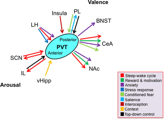
The paraventricular nucleus of the thalamus (PVT) has for decades been acknowledged to be an important node in the limbic system, but studies of emotional processing generally fail to incorporate it into their investigational framework. Here, we propose that the PVT should be considered as an integral part of the emotional processing network. Through its distinct subregions, cell populations, and connections with other limbic nuclei, the PVT participates in both major features of emotion: arousal and valence. The PVT, particularly the anterior PVT, can through its neuronal activity promote arousal, both as part of the sleep-wake cycle and in response to novel stimuli. It is also involved in reward, being both responsive to rewarding stimuli and itself affecting behavior reflecting reward, likely via specific populations of cells distributed throughout its subregions. Similarly, neuronal activity in the PVT contributes to depression-like behavior, through yet undefined subregions. The posterior PVT in particular demonstrates a role in anxiety-like behavior, generally promoting but also inhibiting this behavior. This subregion is also especially responsive to stressors, and it functions to suppress the stress response following chronic stress exposure. In addition to participating in unconditioned or primary emotional responses, the PVT also makes major contributions to conditioned emotional behavior. Neuronal activity in response to a reward-predictive cue can be detected throughout the PVT, and endogenous activity in the posterior PVT strongly predicts approach or seeking behavior. Similarly, neuronal activity during conditioned fear retrieval is detected in the posterior PVT and its activation facilitates the expression of conditioned fear. Much of this involvement of the PVT in arousal and valence has been shown to occur through the same general afferents and efferents, including connections with the hypothalamus, prelimbic and infralimbic cortices, nucleus accumbens, and amygdala, although a detailed functional map of the PVT circuits that control emotional responses remains to be delineated. Thus, while caveats exist and more work is required, the PVT, through its extensive connections with other prominent nuclei in the limbic system, appears to be an integral part of the emotional processing network.
Keywords: anterior; anxiety; arousal; depression; fear; posterior; reward; stress.
Copyright © 2020 Barson, Mack and Gao.
Figures
References
-
- Arluison M., Brochier G., Vankova M., Leviel V., Villalobos J., Tramu G. (1994). Demonstration of peptidergic afferents to the bed nucleus of the stria terminalis using local injections of colchicine. A combined immunohistochemical and retrograde tracing study. Brain Res. Bull. 34, 319–337. 10.1016/0361-9230(94)90026-4 - DOI - PubMed
Publication types
Grants and funding
LinkOut - more resources
Full Text Sources