. 2020 Oct 22:11:531933.
doi: 10.3389/fphys.2020.531933. eCollection 2020.

The Mediator Subunit, Med23 Is Required for Embryonic Survival and Regulation of Canonical WNT Signaling During Cranial Ganglia Development

Affiliations

The Mediator Subunit, Med23 Is Required for Embryonic Survival and Regulation of Canonical WNT Signaling During Cranial Ganglia Development

Soma Dash et al. Front Physiol. .

Abstract

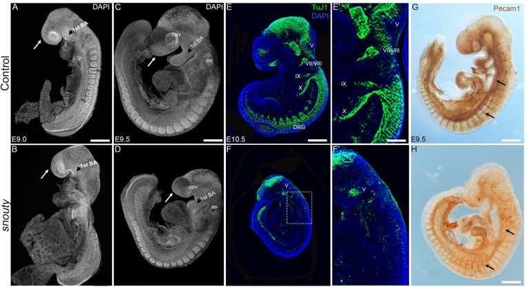
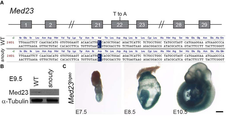
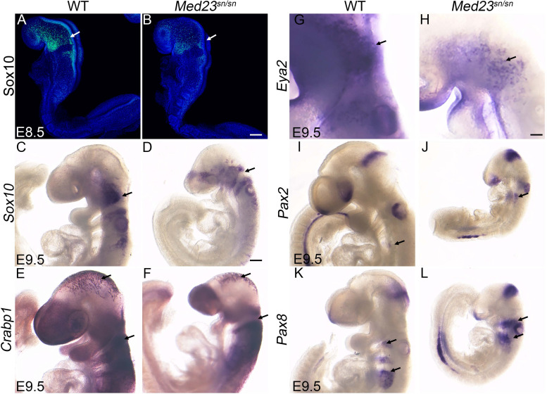
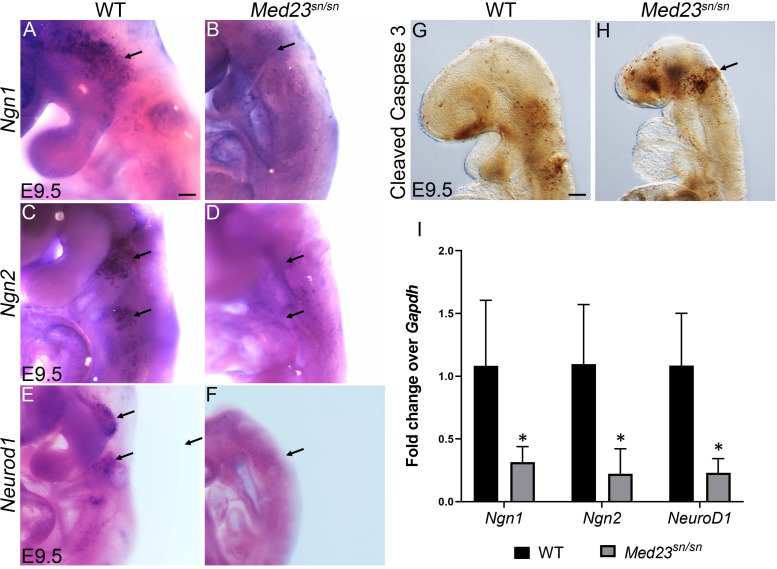
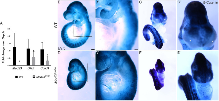
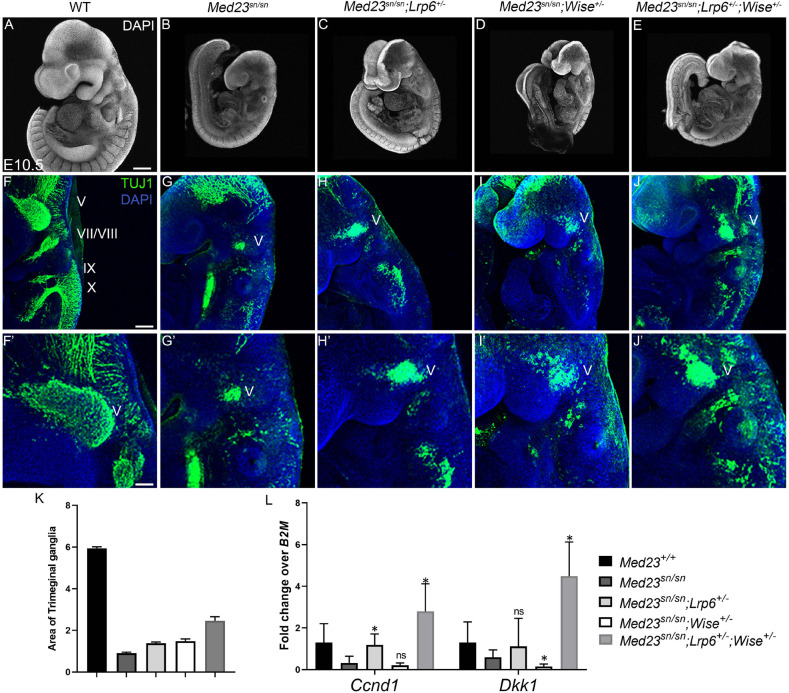
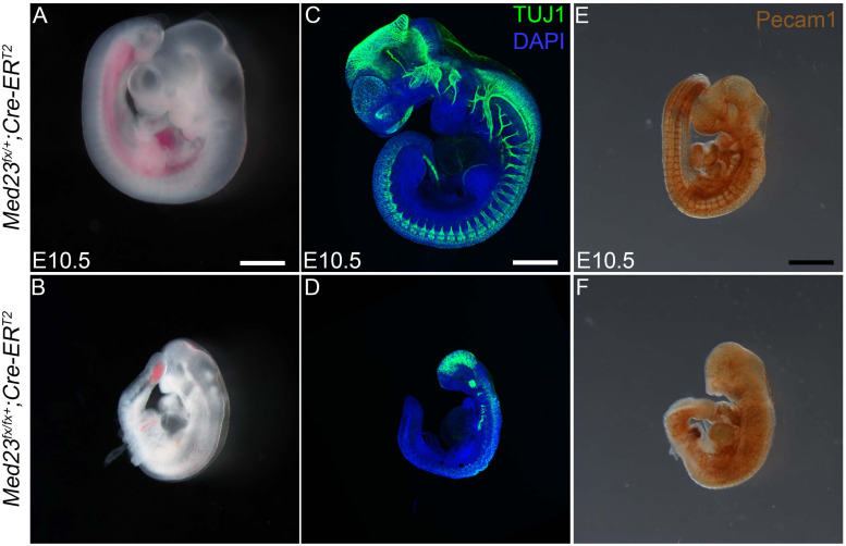
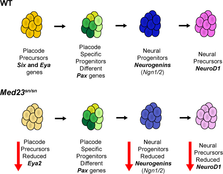
Development of the vertebrate head is a complex and dynamic process, which requires integration of all three germ layers and their derivatives. Of special importance are ectoderm-derived cells that form the cranial placodes, which then differentiate into the cranial ganglia and sensory organs. Critical to a fully functioning head, defects in cranial placode and sensory organ development can result in congenital craniofacial anomalies. In a forward genetic screen aimed at identifying novel regulators of craniofacial development, we discovered an embryonically lethal mouse mutant, snouty, which exhibits malformation of the facial prominences, cranial nerves and vasculature. The snouty mutation was mapped to a single nucleotide change in a ubiquitously expressed gene, Med23, which encodes a subunit of the global transcription co-factor complex, Mediator. Phenotypic analyses revealed that the craniofacial anomalies, particularly of the cranial ganglia, were caused by a failure in the proper specification of cranial placode neuronal precursors. Molecular analyses determined that defects in cranial placode neuronal differentiation in Med23 sn/sn mutants were associated with elevated WNT/β-catenin signaling, which can be partially rescued through combined Lrp6 and Wise loss-of-function. Our work therefore reveals a surprisingly tissue specific role for the ubiquitously expressed mediator complex protein Med23 in placode differentiation during cranial ganglia development. This highlights the importance of coupling general transcription to the regulation of WNT signaling during embryogenesis.

Keywords: MED23; Wnt signaling; cranial ganglia; cranial placodes; neural crest cells.


Figures

FIGURE 1
FIGURE 1
snouty embryos exhibit small body size and craniofacial defects at E9.5. (A–D) Control and snouty mouse embryo littermates at E9.0 and E9.5 were stained with DAPI and imaged with a confocal microscope demonstrating the smaller body size and shortened frontonasal prominences (arrows) in snouty embryos. (E–F′) Neuronal marker TuJ1 staining of control and snouty embryos at E9.5 suggests that snouty embryos exhibit disrupted cranial ganglia. (E′,F′) Are higher magnification images of (E,F). (G,H) PECAM1 immunostaining of E9.5 control and snouty embryos reveal abnormal development of the vascular tree like network in the head and epibranchial regions of snouty mutants. Intersomitic vessel formation (arrow, G,H) is not affected.opv, optic vesicle; ov, otic capsules; BA, branchial arch; DRG, dorsal root ganglia. Scale bars for (A–D) is 300 um, (E,F) is 350 um, (G) is 300 um, (H) is 200 um.
FIGURE 2
FIGURE 2
snouty mutant mice have a mutation in the Med23 gene. (A) The snouty mouse mutant phenotype was determined to be caused by a single base pair mutation in Exon 22 of the Med23 gene, which leads to a valine to aspartic acid amino acid change in the protein. Chromosome position of the mutation is noted based on mm10 mouse assembly. (B) Western blot against Med23 shows a drastic reduction of Med23 protein levels in snouty mutant embryos. (C) Ubiquitous expression of LacZ throughout E7.5 to E10.5 embryos is indicative of the Med23 expression. Scale bar is 250 um.
FIGURE 3
FIGURE 3
Med23sn/sn mutants display neural crest cell and placodal defects. (A–D) Sox10 expression in E8.5 and E9.5 control and Med23sn/sn embryos shows reduced Sox10 + cells entering the cranial ganglia region near the 1st branchial arch and around the otic vesicle in Med23sn/sn embryos. (E,F) Crabp1 ISH reveals proper neural crest cell migration in the cranial and trunk regions of E9.5 control and Med23sn/sn embryos. (G,H) Eya2 expression in control and Med23sn/sn embryos shows proper formation of anterior pre-placodal area. However, very few Eya2+ cells are observed in the trigeminal and epibranchial regions of Med23sn/sn embryos (arrow, H). (I,J) Pax2+ cells are present in the optic, otic, and isthmus of both control and Med23sn/sn embryos. (K,L) Pax8+ cells are present but loosely organized in the epibranchial region, otic placode and isthmus of Med23sn/sn embryos in comparison to controls. Scale bar for (A,B) is 120 um, (C–F,I–L) is 300 um, and (G–H) is 150 um.
FIGURE 4
FIGURE 4
Med23sn/sn mutants display defects in neuronal differentiation. (A,B) Ngn1 is expressed in trigeminal (arrow) and epibranchial cells in control embryos, however is downregulated or lost in Med23sn/sn embryos. (C,D) Ngn2+ cells are observed in the epibranchial ganglia of control embryos (arrows, C). Ngn2+ cell number is drastically reduced in the epibranchial ganglia of Med23sn/sn embryos (arrows, D). (E,F) Neurod1+ cells are considerably reduced in the developing trigeminal and epibranchial ganglia of Med23sn/sn embryos at E9.5 compared to controls. (G,H) A higher number of cleaved Caspase 3 positive cells are observed in the developing trigeminal and frontonasal prominence of E9.5 Med23sn/sn embryos compared to controls. (I) qPCR analyses of E9.5 wild-type and Med23sn/sn littermate embryos revealed downregulation of Ngn1, Ngn2, and NeuroD1 transcripts in the mutants. Statistical analysis was performed using ANOVA and denotes a p-value < 0.05. Scale bar for (A–F) is 150 um, (G,H) is 300 um. *p < 0.05.
FIGURE 5
FIGURE 5
WNT signaling is upregulated and ectopically expressed in the lateral nasal processes of Med23sn/sn embryos. (A) qPCR analysis of cDNA obtained from E9.5 wild-type and Med23sn/sn littermate embryos shows a reduction in the levels of Med23, Dkk1, and Ccnd1. (B–E′) Lateral and frontal images of control BATGal and Med23sn/sn;BATGal embryos reveal an increase in WNT signaling in the frontonasal processes at E9.5. The lateral nasal processes, which are usually devoid of active WNT signaling (*, asterisk, control) exhibit positive LacZ staining (WNT activity) in Med23sn/sn embryos (*, asterisk, Med23sn/sn). (B′–E′) are high magnification images of (B–E). Scale bar for (B–E) is 450 um, (B′–E′) is 100 um.
FIGURE 6
FIGURE 6
Rescue of the Med23sn/sn phenotype by modulating WNT signaling. DAPI stained E9.5 (A) wild-type, (B) Med23sn/sn, (C) Med23sn/sn; Lrp6+/–, (D) Med23sn/sn; Wise+/– and (E) Med23sn/sn; Lrp6+/–; Wise+/– embryos. TuJ1 immunostained (green) E9.5 (F) wild-type (G) Med23sn/sn (H) Med23sn/sn; Lrp6+/–, (I) Med23sn/sn; Wise+/– and (J) Med23sn/sn; Lrp6+/–; Wise+/–, embryos. Combinatorial loss of Lrp6 and Wise restores neuronal differentiation in the epibranchial region of Med23sn/sn embryos. (F′–J′) are higher magnification images of (F–J) focused on the trigeminal ganglia. (K) Quantification of the area of Tuj1 staining in the trigeminal ganglia is shown as a ratio of the length of the embryo for wild-type, Med23sn/sn, Med23sn/sn; Lrp6+/–, Med23sn/sn; Wise+/– and Med23sn/sn; Lrp6+/–; Wise+/– (L) qPCR analysis of cDNA obtained from E9.5 wild-type, Med23sn/sn, Med23sn/sn; Lrp6+/–, Med23sn/sn; Wise+/–, and Med23sn/sn; Lrp6+/–; Wise+/– embryos indicates a significant increase in Ccnd1 and Dkk1 transcripts in Med23sn/sn; Lrp6+/–; Wise+/– embryos compared to WT and Med23sn/sn. Statistical analysis was performed using Students t-test with Med23sn/sn as one of the nominal variables. Scale bars for (A–E) is 300 um, (F–J) is 200 um, (F′–J′) is 50 um. *p < 0.05.
FIGURE 7
FIGURE 7
Conditional deletion of Med23 in neural crest cells. (A,B) Brightfield images of P0 Med23fx/+;Wnt1-Cre and Med23fx/fx;Wnt1-Cre embryos indicate that the mutants exhibit micrognathia. (C,D) TuJ1 staining of E10.5 Med23fx/+;Wnt1-Cre and Med23fx/fx;Wnt1-Cre embryos however revealed proper formation of the cranial ganglia in mutant embryos. Scale bars for (A,B) is 450 um, (C,D) is 375 um.
FIGURE 8
FIGURE 8
Conditional deletion of Med23 in endothelial cells. (A,B) Brightfield images of E16.5 Med23fx/+;Tek-Cre and Med23fx/fx;Tek-Cre embryos indicate that the mutant embryos exhibit severe hemorrhaging and edema, probably resulting in lethality. (C,D) Immunostaining for endothelial cells using PECAM1 suggests development in Med23fx/fx;Tek-Cre embryos is normal. Scale bars for (A,B) is 450 um, (C,D) is 300 um.
FIGURE 9
FIGURE 9
Temporal systemic deletion of Med23 at E6.5 phenocopies Med23sn/sn embryos. (A,B) Med2fx/fx;Cre-ERT2 embryos treated with tamoxifen at E5.5 are small in size compared to Med2fx/+;Cre-ERT2 embryos. These embryos also display a shortened frontonasal prominence similar to Med23sn/sn embryos. (C,D) TuJ1 staining revealed severely hypoplastic cranial as well as vagal ganglia in Med2fx/fx;Cre-ERT2 embryos. (E,F) PECAM1 staining revealed that endothelial cells are disorganized in the Med2fx/fx;Cre-ERT2 embryos compared to control Med2fx/+;Cre-ERT2 embryos. Scale bars for (A,B) is 250 um, (C,D) is 375 um, (E,F) is 300 um.
FIGURE 10
FIGURE 10
Model for Med23-mediated regulation of cranial placode development. In wild-type embryos, placode precursors express Six and Eya genes. Their subsequent expression of different Pax genes defines them as placode specific progenitor cells. Some of these cells then express Ngn1 and Ngn2 thus becoming neural progenitor cells. Ngn1 and Ngn2 regulate the expression of NeuroD1 which then defines these cells as neural precursors. However, in Med23sn/sn embryos, placode precursors exhibit reduced Eya2 expression, and although Pax genes are similarly expressed in Med23sn/sn embryos as they are in wild-type embryos, Ngn1, Ngn2, and NeuroD1 are downregulated. Collectively, this reduces the number of neural precursors resulting in defects in cranial ganglia development in Med23sn/sn embryos.

Similar articles

Cited by

  • Complex Feline Disease Mapping Using a Dense Genotyping Array.
    Hernandez I, Hayward JJ, Brockman JA, White ME, Mouttham L, Wilcox EA, Garrison S, Castelhano MG, Loftus JP, Gomes FE, Balkman C, Brooks MB, Fiani N, Forman M, Kern T, Kornreich B, Ledbetter EC, Peralta S, Struble AM, Caligiuri L, Corey E, Lin L, Jordan J, Sack D, Boyko AR, Lyons LA, Todhunter RJ. Hernandez I, et al. Front Vet Sci. 2022 Jun 16;9:862414. doi: 10.3389/fvets.2022.862414. eCollection 2022. Front Vet Sci. 2022. PMID: 35782544 Free PMC article.
  • Central Med23 deficiency leads to malformation of dentate gyrus and ADHD-like behaviors in mice.
    Zhou BY, Li ZX, Li YW, Li JN, Liu WT, Liu XY, Hu ZB, Zhao L, Chen JY, Hu L, Song NN, Feng X, Wang G, Xu L, Ding YQ. Zhou BY, et al. Neuropsychopharmacology. 2025 Jul;50(8):1224-1236. doi: 10.1038/s41386-025-02088-1. Epub 2025 Mar 20. Neuropsychopharmacology. 2025. PMID: 40114018 Free PMC article.
  • Med23 Regulates Sox9 Expression during Craniofacial Development.
    Dash S, Bhatt S, Falcon KT, Sandell LL, Trainor PA. Dash S, et al. J Dent Res. 2021 Apr;100(4):406-414. doi: 10.1177/0022034520969109. Epub 2020 Nov 6. J Dent Res. 2021. PMID: 33155500 Free PMC article.
  • Bi-allelic MED16 variants cause a MEDopathy with intellectual disability, motor delay, and craniofacial, cardiac, and limb malformations.
    Guillouet C, Agostini V, Baujat G, Cocciadiferro D, Pippucci T, Lesieur-Sebellin M, Georget M, Schatz U, Fauth C, Louie RJ, Rogers C, Davis JM, Konstantopoulou V, Mayr JA, Bouman A, Wilke M, VanNoy GE, England EM, Park KL, Brown K, Saenz M, Novelli A, Digilio MC, Mastromoro G, Rongioletti MCA, Piacentini G, Kaiyrzhanov R, Guliyeva S, Hasanova L, Shears D, Bhatnagar I, Stals K, Klaas O, Horvath J; University of Washington Center for Mendelian Genomics; Bouvagnet P, Witmer PD, MacCarrick G, Cisarova K, Good JM, Gorokhova S, Boute O, Smol T, Bruel AL, Patat O, Broadbent JR, Tan TY, Tan NB, Lyonnet S, Busa T, Graziano C, Amiel J, Gordon CT. Guillouet C, et al. Am J Hum Genet. 2025 Apr 3;112(4):829-845. doi: 10.1016/j.ajhg.2025.02.016. Epub 2025 Mar 12. Am J Hum Genet. 2025. PMID: 40081376
  • Genetically Engineered Mice Unveil In Vivo Roles of the Mediator Complex.
    Ilchuk LA, Kubekina MV, Okulova YD, Silaeva YY, Tatarskiy VV, Filatov MA, Bruter AV. Ilchuk LA, et al. Int J Mol Sci. 2023 May 26;24(11):9330. doi: 10.3390/ijms24119330. Int J Mol Sci. 2023. PMID: 37298278 Free PMC article. Review.

References

    1. Ahn Y. (2015). Signaling in tooth, hair, and mammary placodes. Curr. Top. Dev. Biol. 111 421–459. 10.1016/bs.ctdb.2014.11.013 - DOI - PubMed
    1. Ahn Y., Sanderson B. W., Klein O. D., Krumlauf R. (2010). Inhibition of Wnt signaling by Wise (Sostdc1) and negative feedback from Shh controls tooth number and patterning. Development 137 3221–3231. 10.1242/dev.054668 - DOI - PMC - PubMed
    1. Ahn Y., Sims C., Logue J. M., Weatherbee S. D., Krumlauf R. (2013). Lrp4 and Wise interplay controls the formation and patterning of mammary and other skin appendage placodes by modulating Wnt signaling. Development 140 583–593. 10.1242/dev.085118 - DOI - PMC - PubMed
    1. Ahn Y., Sims C., Murray M. J., Kuhlmann P. K., Fuentes-Antrás J., Weatherbee S. D., et al. (2017). Multiple modes of Lrp4 function in modulation of Wnt/β-catenin signaling during tooth development. Development 144 2824–2836. 10.1242/dev.150680 - DOI - PMC - PubMed
    1. Allen B. L., Taatjes D. J. (2015). The Mediator complex: a central integrator of transcription. Nat. Rev. Mol. Cell Biol. 16 155–166. 10.1038/nrm3951 - DOI - PMC - PubMed

LinkOut - more resources