. 2020 Oct 22:11:587709.
doi: 10.3389/fphys.2020.587709. eCollection 2020.

Beta-Adrenergic Receptor Stimulation Modulates the Cellular Proarrhythmic Effects of Chloroquine and Azithromycin

Affiliations

Beta-Adrenergic Receptor Stimulation Modulates the Cellular Proarrhythmic Effects of Chloroquine and Azithromycin

Henry Sutanto et al. Front Physiol. .

Abstract

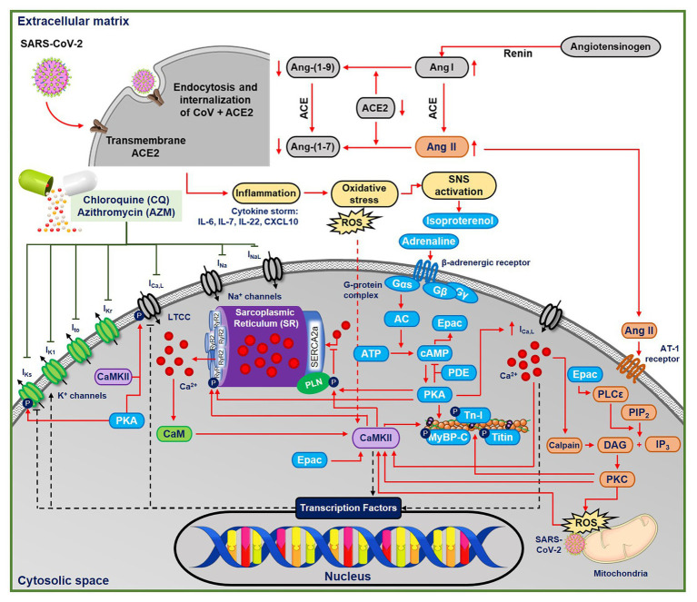
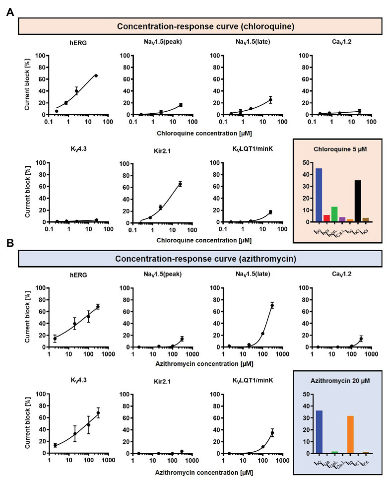
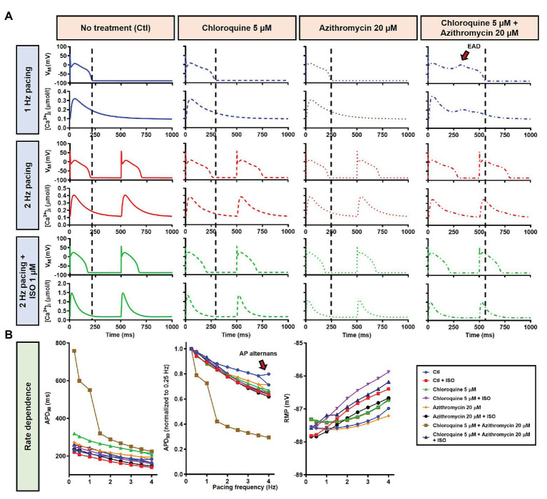
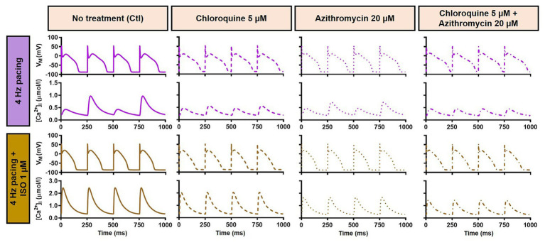
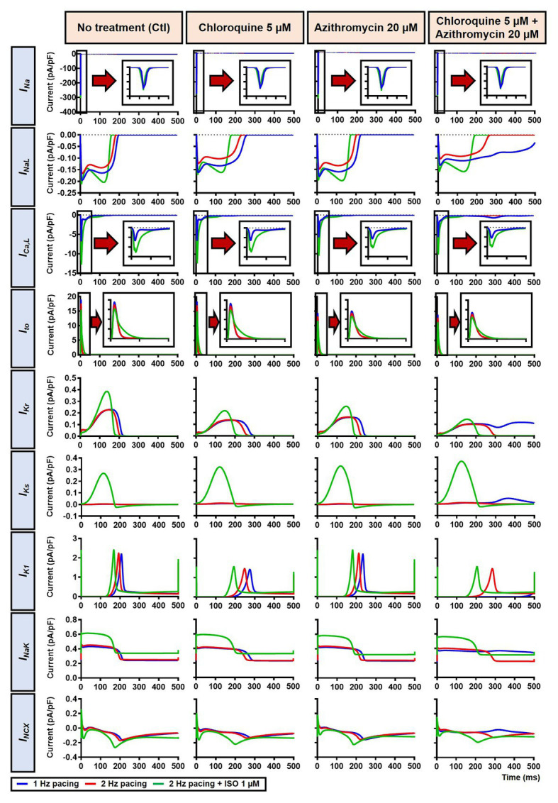
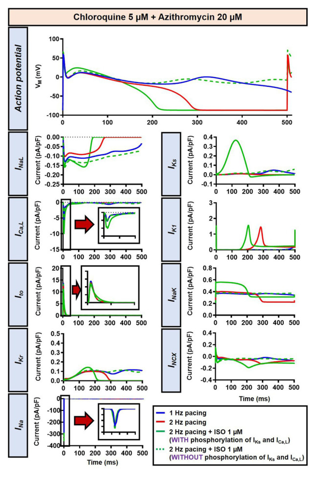
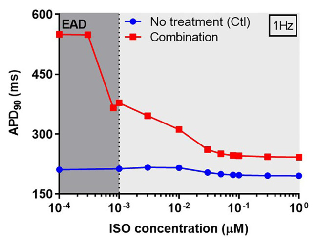
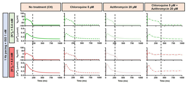
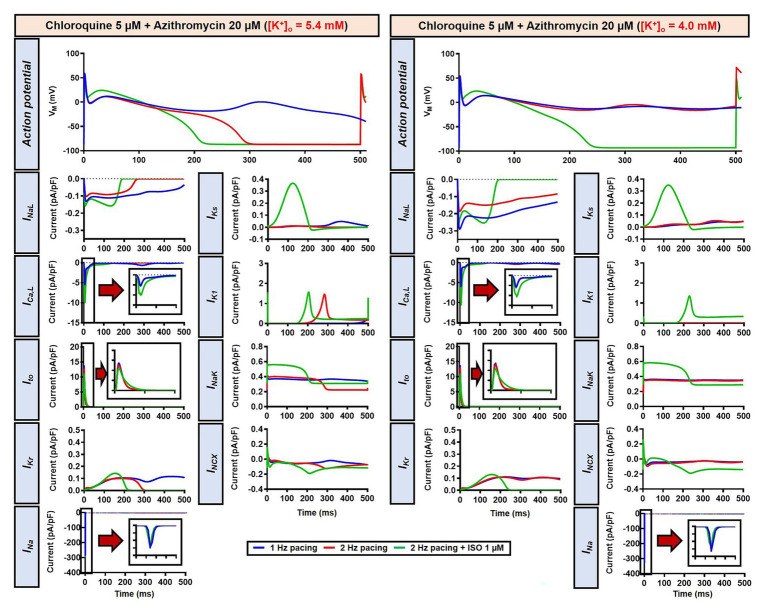
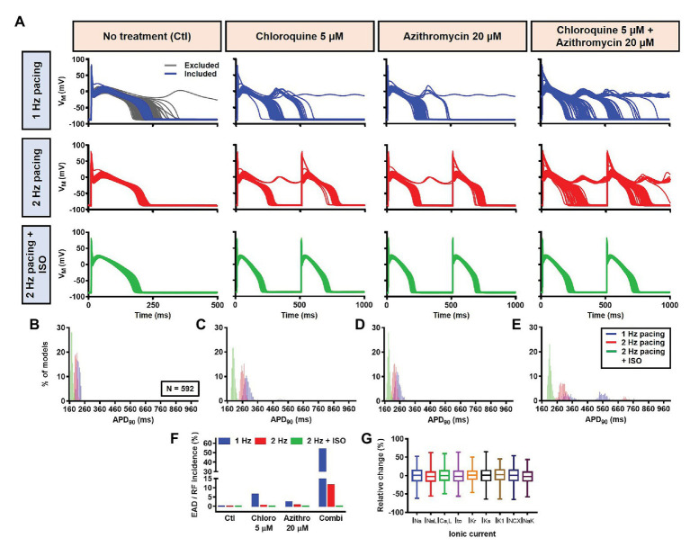
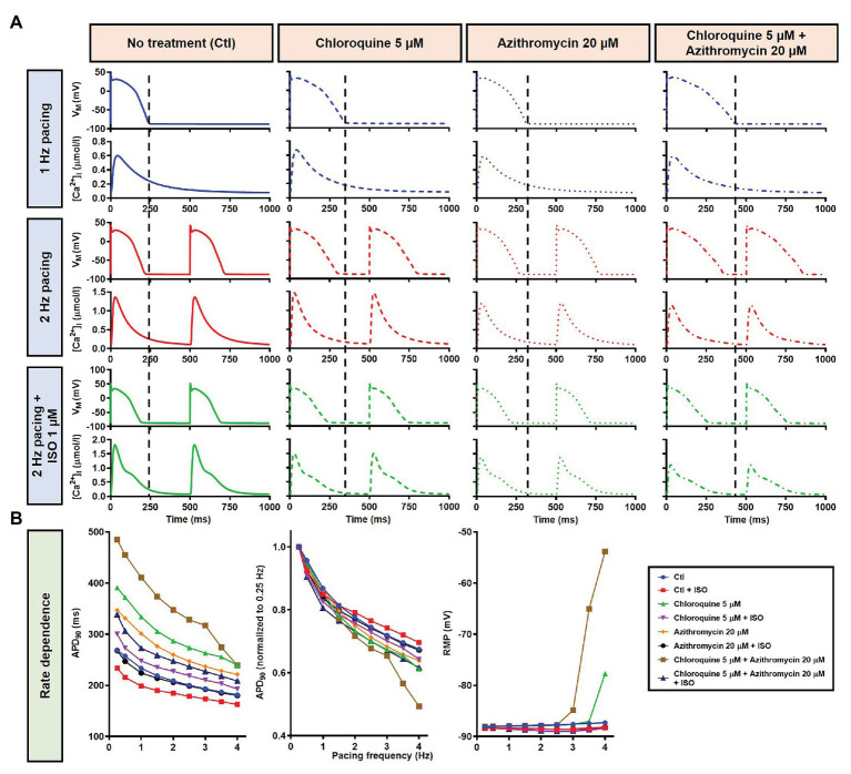
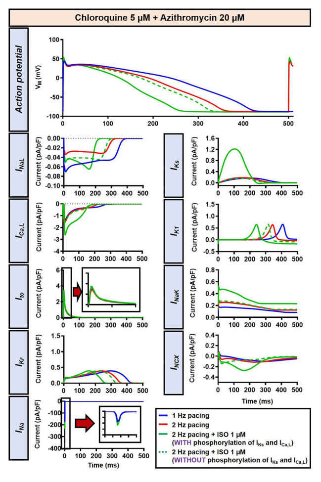
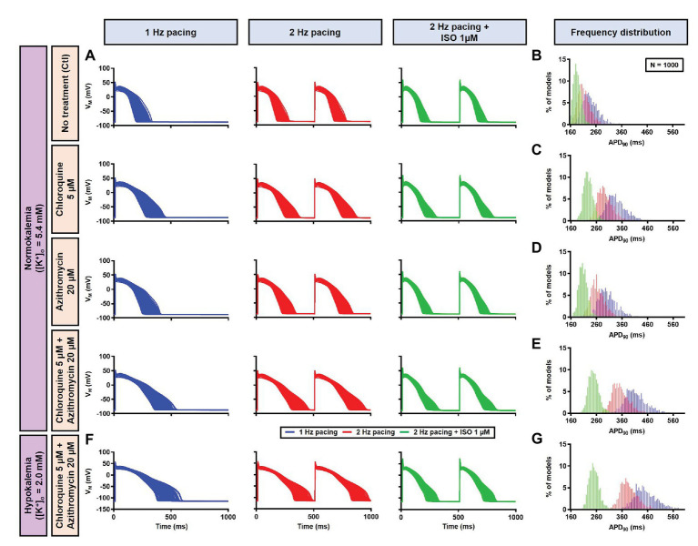
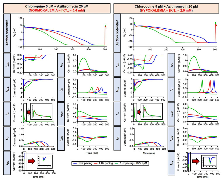
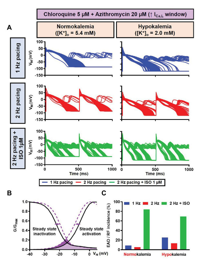
The antimalarial drug, chloroquine (CQ), and antimicrobial drug, azithromycin (AZM), have received significant attention during the COVID-19 pandemic. Both drugs can alter cardiac electrophysiology and have been associated with drug-induced arrhythmias. Meanwhile, sympathetic activation is commonly observed during systemic inflammation and oxidative stress (e.g., in SARS-CoV-2 infection) and may influence the electrophysiological effects of CQ and AZM. Here, we investigated the effect of beta-adrenergic stimulation on proarrhythmic properties of CQ and AZM using detailed in silico models of ventricular electrophysiology. Concentration-dependent alterations in ion-channel function were incorporated into the Heijman canine and O'Hara-Rudy human ventricular cardiomyocyte models. Single and combined drug effects on action-potential (AP) properties were analyzed using a population of 1,000 models accommodating inter-individual variability. Sympathetic stimulation was simulated by increasing pacing rate and experimentally validated isoproterenol (ISO)-induced changes in ion-channel function. In the canine ventricular model at 1 Hz pacing, therapeutic doses of CQ and AZM (5 and 20 μM, respectively) individually prolonged AP duration (APD) by 33 and 13%. Their combination produced synergistic APD prolongation (+161%) with incidence of proarrhythmic early afterdepolarizations in 53.5% of models. Increasing the pacing frequency to 2 Hz shortened APD and together with 1 μM ISO counteracted the drug-induced APD prolongation. No afterdepolarizations occurred following increased rate and simulated application of ISO. Similarly, CQ and AZM individually prolonged APD by 43 and 29% in the human ventricular cardiomyocyte model, while their combination prolonged APD by 76% without causing early afterdepolarizations. Consistently, 1 μM ISO at 2 Hz pacing counteracted the drug-induced APD prolongation. Increasing the ICa,L window current produced afterdepolarizations, which were exacerbated by ISO. In both models, reduced extracellular K+ reduced the repolarization reserve and increased drug effects. In conclusion, CQ- and AZM-induced proarrhythmia is promoted by conditions with reduced repolarization reserve. Sympathetic stimulation limits drug-induced APD prolongation, suggesting the potential importance of heart rate and autonomic status monitoring in particular conditions (e.g., COVID-19).

Keywords: COVID-19; arrhythmia; azithromycin; beta-adrenergic; chloroquine; computational modeling; electrophysiology-basic.


Figures

Figure 1
Figure 1
The multifactorial effects of coronavirus disease 2019 (COVID-19) in the ventricular cardiomyocyte and the ionic targets of chloroquine (CQ) and azithromycin (AMZ). The severe acute respiratory syndrome-associated coronavirus type-2 (SARS-CoV-2) leads to the endocytosis and internalizations of the transmembrane angiotensin converting enzyme type 2 (ACE2) receptors, preventing the conversion of angiotensin I and II into their metabolites. Thus, angiotensin II binds to the AT-II receptor, initiating protein kinase-C (PKC)-dependent pathways, which may further activate Ca2+/calmodulin-dependent protein kinase II (CaMKII)-dependent signaling cascades. In COVID-19, the systemic inflammation and cytokine storm can also increase oxidative stress, leading to reactive oxygen species (ROS)-mediated CaMKII activation. CQ and AZM alter action potential properties through inhibition of multiple cardiac ion channels [fast Na+ current (INa), late Na+ current (INaL), rapid delayed-rectifier K+ current (IKr), slow delayed-rectifier K+ current (IKs), inward-rectifier K+ current (IK1), transient-outward K+ current (Ito), and L-type Ca2+ current (ICa,L)]. AC, adenylyl cyclase; ACE, angiotensin converting enzyme; Ang II, angiotensin II; ATP, adenosine triphosphate; CaM, calmodulin; CaMKII, Ca2+/calmodulin-dependent protein kinase II; cAMP, cyclic adenosine monophosphate; DAG, diacyl glycerol; IL, interleukin; IP3, inositol triphosphate; PDE, phosphodiesterase; PIP2, phosphatidylinositol biphosphate; PKA, protein kinase A; PKC, protein kinase C; PLC, phospholipase C; ROS, reactive oxygen species; Tn-I, troponin-I.
Figure 2
Figure 2
The concentration-dependent effects of CQ and AZM on cardiac ion channels. (A) CQ mainly blocks IKr and IK1, with minor effects on INa, INaL, Ito, ICa,L, and IKs. (B) AZM mainly blocks IKr and Ito, with minimal effects on INa, INaL, ICa,L, IK1, and IKs. The experimental data (black symbols) were obtained from previous experiments (Crumb et al., 2016) and were fitted using Hill equations in the model (black lines). Bar charts show percentage inhibition of different ion channels using the clinically relevant concentrations employed in subsequent simulations.
Figure 3
Figure 3
The effects of CQ and AZM on action potential (AP) properties of canine ventricular epicardium. (A) The AP and Ca2+ transient of non-treated, CQ 5 μM, AZM 20 μM, and combined groups. The dashed vertical lines indicate the end of the AP with 1 Hz pacing to provide a clearer depiction of the effects of increasing pacing rate and isoproterenol (ISO) on action potential duration (APD). The early afterdepolarization (EAD) is indicated with an arrow. (B) APD and resting membrane potential (RMP) for different pacing rates in the four groups with and without simulated beta-adrenergic stimulation. The occurrence of alternans at 4 Hz pacing is marked with an arrow. AP, action potential; APD, action potential duration; Ctl, control; ISO, isoproterenol; RMP, resting membrane potential.
Figure 4
Figure 4
Action potential and Ca2+-transient alternans at 4 Hz pacing. The upper panels exemplify the AP and Ca2+-transient alternans at 4 Hz pacing in non-treated, CQ alone, and AZM alone groups. The application of 1 μM ISO abolishes the alternans, as shown in the lower panels. AP, action potential; AZM, azithromycin; Ctl, control; CQ, chloroquine; ISO, isoproterenol.
Figure 5
Figure 5
The effects of beta-adrenergic stimulation on cardiac ion channels of canine ventricular epicardium. The effects were assessed in four groups: non-treated, CQ, AZM, and combined groups. The blue lines represent the ionic currents during 1 Hz pacing, the red lines represent the ionic currents during 2 Hz pacing, and the green lines represent the currents during 2 Hz pacing with ISO 1 μM. The fast Na+ current (INa), L-type Ca2+ current (ICa,L), and transient-outward K+ current (Ito) are shown at an expanded scale in the insets. Ctl, control; ISO, isoproterenol.
Figure 6
Figure 6
The impact of IKs and ICa,L phosphorylation on the action potential of canine ventricular epicardium in the presence of CQ and AZM. The effect of CQ 5 μM in combination with AZM 20 μM in the presence of ISO-induced IKs and ICa,L phosphorylation is shown in solid green lines. The green dashed lines represent the effect of CQ 5 μM in combination with AZM 20 μM in the absence of ISO-induced IKs and ICa,L phosphorylation.
Figure 7
Figure 7
The concentration-dependent effect of ISO on APD of canine ventricular epicardium. The APD in the presence of various concentrations of ISO from 0.1 nM to 1 μM in non-treated group (blue line) was compared to the combined group (CQ 5 μM + AZM 20 μM; red line). The simulations were performed with 1 Hz pacing. APD, action potential duration; Ctl, control; ISO, isoproterenol.
Figure 8
Figure 8
The effects of CQ and AZM on canine AP in the presence of an intermediate concentration of ISO. The AP and Ca2+ transient of non-treated, CQ 5 μM, AZM 20 μM and combined groups with 1 Hz pacing and 1 nM ISO in the presence of low extracellular K+ ([K+]o = 4.0 mM) were compared to [K+]o = 5.4 mM. The dashed vertical lines indicate the end of AP in models with [K+]o = 5.4 mM to provide a clearer depiction of the effects of reduced extracellular K+ on APD. AP, action potential; APD, action potential duration; Ctl, control; ISO, isoproterenol.
Figure 9
Figure 9
The impact of reduced extracellular K+ on the action potential of canine ventricular epicardium in the presence of CQ and AZM. The left panels show the effect of CQ 5 μM in combination with AZM 20 μM with 5.4 mM [K+]o. The right panels show the effect of CQ 5 μM in combination with AZM 20 μM in the presence of reduced [K+]o (4.0 mM).
Figure 10
Figure 10
The cellular effects of CQ and AZM in the population of 1,000 canine ventricular epicardial myocyte models. (A) The APs of 592 models included in the study, with the 408 excluded non-physiological APs shown as gray lines. (B–E) The frequency distribution of APD in non-treated, CQ, AZM and combined groups. (F) The incidence of EAD/repolarization failure (RF) observed in the population-based study (as percentage of models). (G) Boxplot showing the distribution of relative changes in ionic currents to accommodate the interindividual variability. AP, action potential; APD, action potential duration; Ctl, control; EAD, early afterdepolarization; ISO, isoproterenol; RF, repolarization failure.
Figure 11
Figure 11
The effects of CQ and AZM on AP properties of human ventricular epicardium. (A) The AP and Ca2+ transient of non-treated, CQ 5 μM, AZM 20 μM, and combined groups with 1 Hz pacing (blue), 2 Hz pacing (red), or 2 Hz pacing with electrophysiological effects of maximal beta-adrenergic stimulation (green). The dashed vertical lines indicate the end of AP with 1 Hz pacing to provide a clearer depiction of the effects of increasing pacing rate and ISO on APD. (B) APD and RMP for different pacing rates in the four groups with and without simulated beta-adrenergic stimulation. AP, action potential; APD, action potential duration; Ctl, control; ISO, isoproterenol; RMP, resting membrane potential.
Figure 12
Figure 12
The impact of IKs and ICa,L phosphorylation on the action potential of human ventricular epicardium in the presence of CQ and AZM. The effect of CQ 5 μM in combination with AZM 20 μM in the presence of ISO-induced IKs and ICa,L phosphorylation is shown in solid green lines. The green dashed lines represent the effect of CQ 5 μM in combination with AZM 20 μM in the absence of ISO-induced IKs and ICa,L phosphorylation.
Figure 13
Figure 13
The cellular effects of CQ and AZM in the population of 1,000 human ventricular epicardial myocyte models in the absence and presence of hypokalemia. (A–E) The effects of CQ and AZM on the human ventricular myocyte AP in normokalemia ([K+]o = 5.4 mM). (A) The APs of 1,000 models included in the study under Ctl conditions, with 5 μM CQ, 20 μM AZM, or a combination (top to bottom) at 1 Hz pacing, 2 Hz pacing, or 2 Hz pacing with electrophysiological effects of maximal beta-adrenergic stimulation (left to right). (B–E) The frequency distribution of APD in non-treated, CQ, AZM, and combined groups. (F,G) The effects of CQ and AZM in the presence of severe hypokalemia ([K+]o = 2.0 mM) on human ventricular APs (F) and the frequency distributions of APD (G). AP, action potential; APD, action potential duration; Ctl, control; ISO, isoproterenol.
Figure 14
Figure 14
The impact of severe hypokalemia on the action potential of human ventricular epicardium in the presence of CQ and AZM. The left panels show the effect of CQ 5 μM in combination with AZM 20 μM in the absence of hypokalemia ([K+]o = 5.4 mM). The right panels show the effect of CQ 5 μM in combination with AZM 20 μM in the presence of severe hypokalemia ([K+]o = 2.0 mM).
Figure 15
Figure 15
The impact of severe hypokalemia on the population of 1,000 human ventricular epicardium APs with increased ICa,L window in the presence of CQ and AZM. (A) The effect of CQ 5 μM in combination with AZM 20 μM in the absence of hypokalemia ([K+]o = 5.4 mM; left panel) and in the presence of severe hypokalemia ([K+]o = 2.0 mM; right panel). (B) A 3 mV shift in the steady-state activation and inactivation of ICa,L was introduced in the human ventricular epicardium model to increase the ICa,L window (shift from black to purple lines). (C) The incidence of EAD/RF observed in the population-based study (as percentage of models). EAD, early afterdepolarization; ICa,L, L-type Ca2+ current; ISO, isoproterenol; RF, repolarization failure; VM, membrane potential.

References

    1. Adameova A., Shah A. K., Dhalla N. S. (2020). Role of oxidative stress in the genesis of ventricular arrhythmias. Int. J. Mol. Sci. 21:4200. 10.3390/ijms21124200, PMID: - DOI - PMC - PubMed
    1. Anyukhovsky E. P., Sosunov E. A., Rosen M. R. (1996). Regional differences in electrophysiological properties of epicardium, midmyocardium, and endocardium: in vitro and in vivo correlations. Circulation 94, 1981–1988. 10.1161/01.cir.94.8.1981, PMID: - DOI - PubMed
    1. Arshad S., Kilgore P., Chaudhry Z. S., Jacobsen G., Wang D. D., Huitsing K., et al. (2020). Treatment with hydroxychloroquine, azithromycin, and combination in patients hospitalized with COVID-19. Int. J. Infect. Dis. 97, 396–403. 10.1016/j.ijid.2020.06.099, PMID: - DOI - PMC - PubMed
    1. Atkinson J. G. (2020). Problems with the analysis in “treatment with hydroxychloroquine, azithromycin, and combination in patients hospitalized with COVID-19”. Int. J. Infect. Dis. 99:37. 10.1016/j.ijid.2020.07.057, PMID: - DOI - PMC - PubMed
    1. Bartos D. C., Giudicessi J. R., Tester D. J., Ackerman M. J., Ohno S., Horie M., et al. (2014). A KCNQ1 mutation contributes to the concealed type 1 long QT phenotype by limiting the Kv7.1 channel conformational changes associated with protein kinase A phosphorylation. Heart Rhythm 11, 459–468. 10.1016/j.hrthm.2013.11.021, PMID: - DOI - PMC - PubMed