Review
. 2020 Oct 21:11:596914.
doi: 10.3389/fmicb.2020.596914. eCollection 2020.

Naturally Occurring tRNAs With Non-canonical Structures

Affiliations
Review

Naturally Occurring tRNAs With Non-canonical Structures

Natalie Krahn et al. Front Microbiol. .

Abstract

Transfer RNA (tRNA) is the central molecule in genetically encoded protein synthesis. Most tRNA species were found to be very similar in structure: the well-known cloverleaf secondary structure and L-shaped tertiary structure. Furthermore, the length of the acceptor arm, T-arm, and anticodon arm were found to be closely conserved. Later research discovered naturally occurring, active tRNAs that did not fit the established 'canonical' tRNA structure. This review discusses the non-canonical structures of some well-characterized natural tRNA species and describes how these structures relate to their role in translation. Additionally, we highlight some newly discovered tRNAs in which the structure-function relationship is not yet fully understood.

Keywords: genetic code expansion; identity elements; mitochondria; non-canonical; pyrrolysine; selenocysteine; tRNA; translation.


Figures

FIGURE 1
FIGURE 1
(A) Secondary and (B) tertiary representation of a tRNA molecule colored based on specific regions: acceptor arm (green), T-arm (dark blue), D-arm (light blue), variable arm (yellow), anticodon arm (pink). The cloverleaf (secondary structure) model includes the standard tRNA numbering. Dashed lines correspond to the tertiary interactions which form the L-shape observed with tRNAs.
FIGURE 2
FIGURE 2
(A) Aminoacylation pathway for tRNASec is different in bacteria compared to archaea and eukaryotes. An additional step is required in the latter, resulting in an intermediate tRNASec containing a phosphoserine moiety. (B) Bacterial and (C) archaeal and eukaryotic elongation pathways for tRNASec. A selenocysteine insertion sequence (SECIS) element is required in the mRNA sequence which forms a hairpin in the 3′ translated region for bacteria or 3′ untranslated region for archaea and eukaryotes. A unique elongation factor [SelB (sometimes referred to as EFSec)] is required in all systems.
FIGURE 3
FIGURE 3
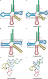
Comparing cloverleaf structures of tRNASer with tRNASec from bacterial and eukaryotic species with specific focus on the tertiary structure of tRNASec. Cloverleaf structures of (A) E. coli tRNASer and (B) E. coli tRNASec highlight differences in the acceptor domain and D-arm as well as different tertiary interactions. (C) Bacterial A. aeolicus tRNASec (PDB ID: 3W3S; Itoh et al., 2013) forms the canonical L-shaped tertiary structure. A similar comparision is shown of (D) H. sapiens tRNASer and (E) H. sapiens tRNASec. (F) The tertiary structure of H. sapiens tRNASec (PDB ID: 3A3A; Itoh et al., 2009) also shows a similar L-shape. The tRNA structure elements are colored accordingly: acceptor arm (green), T-arm (dark blue), D-arm (light blue), variable arm (yellow), and anticodon arm (pink). Tertiary interactions are represented by dashed lines with black lines being conserved interactions between the two tRNAs while magenta lines are unique to that tRNA. Magenta boxes highlight important regions of tRNASec for interaction with aminoacylation and elongation machinery.
FIGURE 4
FIGURE 4
(A) Cloverleaf structure of SelC tRNACys highlights its unique structure compared with canonical tRNAs. Magenta boxes emphasize these specific regions. R and Y denote A/G and U/C, respectively and empty circles represent no conservation in sequence. (B) Genomic structure of selC reveals an additional elongation factor (selB) in Syntrophobacterales and Desulfobacterales while an additional aaRS (cysS) is present in only Desulfobacterales. (C) cysS (which codes for CysRS) is an aaRS that has a mutated connective polypeptide (CP) domain and the anticodon binding domain (ABD) is absent. The Rossman fold (RF) and stem-contact (SC) fold are consistent between CysRS and CysRS
FIGURE 5
FIGURE 5
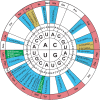
Genetic code wheel highlights the codons and subsequent amino acids which can be incorporated by allo-tRNAs of 9/3 structure (yellow), 8/4 structure (blue) or both (green) (adapted from Mukai et al., 2017). The number of allo-tRNAs found with the indicated anticodon (subscript) is shown as a bold number in front of the allo-tRNA name. Some allo-tRNAs with a specific anticodon are thought to be able to read through multiple codons.
FIGURE 6
FIGURE 6
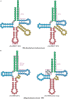
Allo-tRNA secondary structures for (A) Silvibacterium bohemicum and (B) Edaphobacter strain C40 have unique features from canonical tRNAs. (A) Both 8/4 and 9/3 structures are observed for allo-tRNASer with anticodons (boxed in magenta) that do not correlate with decoding of the Ser amino acid. (B) Two completely different tRNA secondary structures are proposed with either a G at the –1 position to form an additional base pair (boxed in magenta) providing either a 8/4 or 9/4 structure and a tRNA structure with an additional fifth loop between the acceptor arm and D-arm (gray boxed in magenta).
FIGURE 7
FIGURE 7
Domain organization and binding mode of PylRS. (A) PylRS is composed of two domains, an N-terminal domain (PylSn) and a catalytic domain (PylSc). PylRS is either composed of a fusion of these two domains, two standalone proteins, or as a lone PylSc. (B) PylSc interacts with the acceptor stem and catalyzes the aminoacylation of tRNAPyl. PylSn forms a tight interaction with the variable arm.
FIGURE 8
FIGURE 8
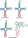
Cloverleaf structures of tRNAPyl from (A) M. barkeri, (B) D. hafniense, and (C) M. alvus. Identity elements for each tRNAPyl are highlighted by magenta boxes. The crystal structure of D. hafniense tRNAPyl (PDB ID: 2ZNI; Nozawa et al., 2009) is also shown in (B).
FIGURE 9
FIGURE 9
Mammalian mt-tRNA can be classified into three types. (A) Type I mt-tRNA, represented by mt-tRNASerUCN, shares similarities with canonical tRNAs, featuring the same conserved D- and T-loop interactions. (B) Type II mt-tRNA, represented by mt-tRNAAsp, is the most commonly found mt-tRNA. (C) Type III mt-tRNA, represented by mt-tRNASerAGY, do not have a D-arm. Instead, the connecting region between the acceptor and anticodon stem interacts with the variable and T-loop to promote folding.
FIGURE 10
FIGURE 10
Nematode mt-tRNAs have diverse and highly unusual secondary structures. Examples of these abnormal mt-tRNA structures are shown here. (A) R. culicivorax mt-tRNAIle has no D- or T-arm. (B) C. elegans mt-tRNATyr has a D-arm, but no T-arm. (C) A. suum mt-tRNASerUCU has a short T-arm and a variable loop, but no D-arm.

References

    1. Alfonzo J. D., Söll D. (2009). Mitochondrial tRNA import–the challenge to understand has just begun. Biol. Chem. 390 717–722. - PMC - PubMed
    1. Amberg R., Mizutani T., Wu X. Q., Gross H. J. (1996). Selenocysteine synthesis in mammalia: an identity switch from tRNASer to tRNASec. J. Mol. Biol. 263 8–19. 10.1006/jmbi.1996.0552 - DOI - PubMed
    1. Ambrogelly A., Gundllapalli S., Herring S., Polycarpo C., Frauer C., Söll D. (2007). Pyrrolysine is not hardwired for cotranslational insertion at UAG codons. Proc. Natl. Acad. Sci. U.S.A. 104 3141–3146. 10.1073/pnas.0611634104 - DOI - PMC - PubMed
    1. Baron C., Böck A. (1991). The length of the aminoacyl-acceptor stem of the selenocysteine-specific tRNASec of Escherichia coli is the determinant for binding to elongation factors SELB or Tu. J. Biol. Chem. 266 20375–20379. - PubMed
    1. Baron C., Westhof E., Bock A., Giegé R. (1993). Solution structure of selenocysteine-inserting tRNASec from Escherichia coli. Comparison with canonical tRNASer. J. Mol. Biol. 231 274–292. 10.1006/jmbi.1993.1282 - DOI - PubMed

LinkOut - more resources