Review
. 2020 Oct 22:11:592010.
doi: 10.3389/fimmu.2020.592010. eCollection 2020.

Roles of HLA-G in the Maternal-Fetal Immune Microenvironment

Affiliations
Review

Roles of HLA-G in the Maternal-Fetal Immune Microenvironment

Xiuxiu Xu et al. Front Immunol. .

Abstract

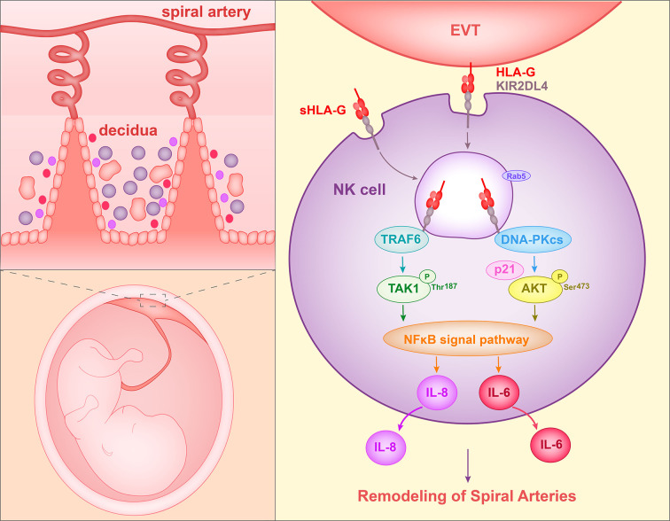
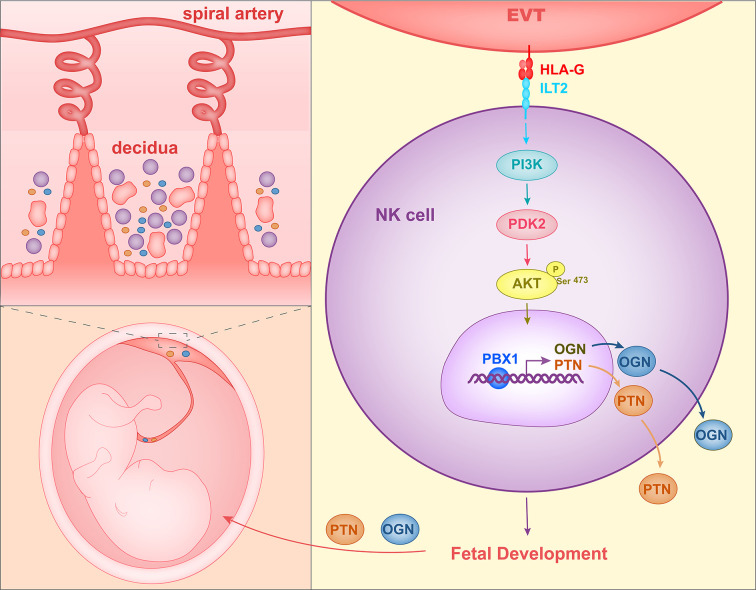
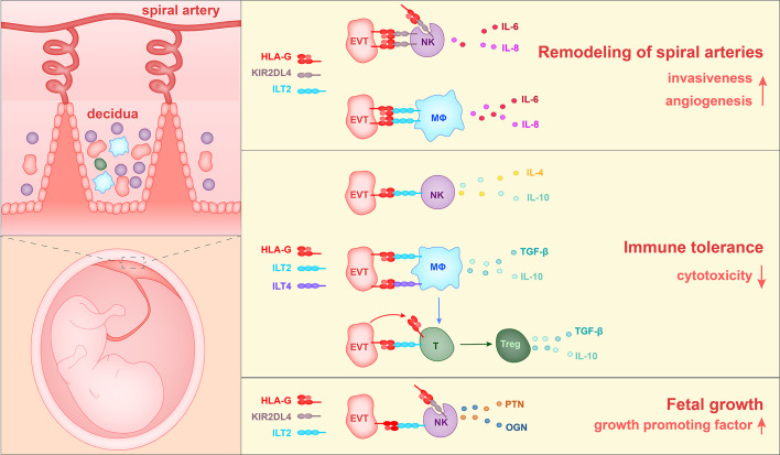
During pregnancy, the maternal uterus and fetus form a special microenvironment at the maternal-fetal interface to support fetal development. Extravillous trophoblasts (EVTs), differentiated from the fetus, invade into the decidua and interact with maternal cells. Human leukocyte antigen (HLA)-G is a non-classical MHC-I molecule that is expressed abundantly and specifically on EVTs in physiological conditions. Soluble HLA-G (sHLA-G) is also found in maternal blood, amniotic fluid, and cord blood. The abnormal expression and polymorphisms of HLA-G are related to adverse pregnancy outcomes such as preeclampsia (PE) and recurrent spontaneous abortion (RSA). Here we summarize current findings about three main roles of HLA-G during pregnancy, namely its promotion of spiral artery remodeling, immune tolerance, and fetal growth, all resulting from its interaction with immune cells. These findings are not only of great significance for the treatment of pregnancy-related diseases but also provide clues to tumor immunology research since HLA-G functions as a checkpoint in tumors.

Keywords: extravillous trophoblasts; fetal development; human leukocyte antigen G; immunology; natural killer cells; pregnancy; spiral artery remodeling.


Figures

Figure 1
Figure 1
Proposed interaction of HLA-G with decidual NK cells to promote spiral artery remodeling. HLA-G on EVTs or sHLA-G secreted by EVTs binds to KIR2DL4 on NK cells, and HLA-G and KIR2DL4 are then endocytosed into Rab5+ endosomes of NK cells. The endocytosed KIR2DL4 binds to TRAF6, induces phosphorylation of TAK1 at Thr187 and activates the NF-κB pathway. In addition, KIR2DL4 interacts with DNA-PKcs, triggering phosphorylation of Akt at Ser473 and upregulating p21. Phosphorylated Akt then activates the NF-κB pathway and results in the expression of the senescence-associated secretory phenotype (SASP) and production of, for example, IL-6 and IL-8. The SASP promotes vascular permeability, angiogenesis and invasion of EVT.
Figure 2
Figure 2
Mechanism of HLA-G interacting with decidual NK cells to promote fetal growth. HLA-G on EVTs binds to ILT2 on NK cells, activates the PI3K-AKT signal pathway and induces expression of transcription factor PBX1. PBX1 upregulates the secretion of growth-promoting factor PTN and OGN, facilitating early fetal growth.
Figure 3
Figure 3
The currently identified roles of HLA-G in the pregnancy microenvironment. 1) HLA-G on extravillous trophoblasts (EVTs) binds to KIR2DL4 on NK cells or ILT2 on macrophages, and in this way stimulates the production of IL-6, IL-8, and VEGFα. Soluble HLA-G binds to KIR2DL4 on NK cells and induces the production of IL-6 and IL-8. Thus HLA-G promotes vascular permeability, angiogenesis and EVT invasiveness, and thereby participates in the remodeling of spiral arteries. 2) HLA-G on EVTs binds to ILT2 on NK cells, ILT2 and ILT4 on macrophages, and ILT2 on Treg cells, and reduces levels of cytotoxicity towards fetal tissues. 3) HLA-G on EVTs binds to ILT2 and KIR2DL4 on NK cells and promotes the secretion of growth-promoting factors PTN and OGN, thereby facilitating fetal growth.

References

    1. Orr HT, Bach FH, Ploegh HL, Strominger JL, Kavathas P, DeMars R. Use of HLA loss mutants to analyse the structure of the human major histocompatibility complex. Nature (1982) 296(5856):454–6. 10.1038/296454a0 - DOI - PubMed
    1. Bodmer JG, Marsh SG, Albert E. Nomenclature for factors of the HLA system, 1989. Immunol Today (1990) 11(1):3–10. 10.1016/0167-5699(90)90003-R - DOI - PubMed
    1. Fujii T, Ishitani A, Geraghty DE. A Soluble Form of the Hla-G Antigen Is Encoded by a Messenger-Ribonucleic-Acid Containing Intron 4. J Immunol (1994) 153(12):5516–24. - PubMed
    1. Paul P, Cabestre FA, Ibrahim E, Lefebvre S, Khalil-Daher I, Vazeux G, et al. Identification of HLA-G7 as a new splice variant of the HLA-G mRNA and expression of soluble HLA-G5, -G6, and -G7 transcripts in human transfected cells. Hum Immunol (2000) 61(11):1138–49. 10.1016/S0198-8859(00)00197-X - DOI - PubMed
    1. Carosella ED, Moreau P, Le Maoult J, Le Discorde M, Dausset J, Rouas-Freiss N. HLA-G molecules: from maternal-fetal tolerance to tissue acceptance. Adv Immunol (2003) 81:199–252. 10.1016/S0065-2776(03)81006-4 - DOI - PubMed

Publication types

MeSH terms