Review
. 2020 Sep;15(5):529-557.
doi: 10.1016/j.ajps.2019.11.003. Epub 2019 Dec 17.

Development of 3D bioprinting: From printing methods to biomedical applications

Affiliations
Review

Development of 3D bioprinting: From printing methods to biomedical applications

Zeming Gu et al. Asian J Pharm Sci. 2020 Sep.

Abstract

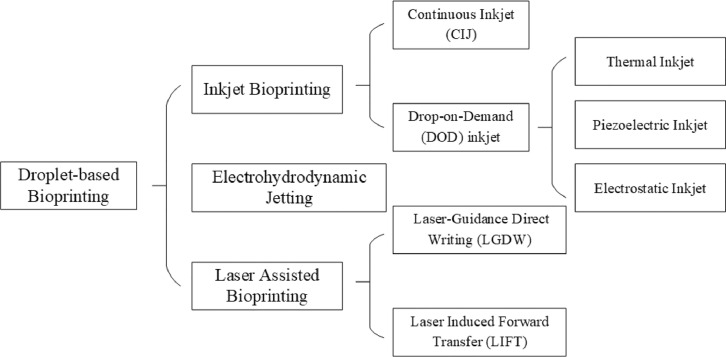
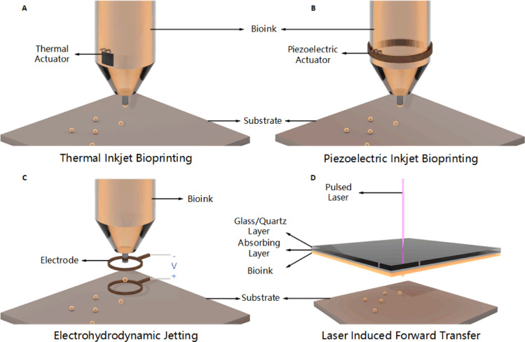
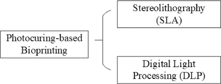
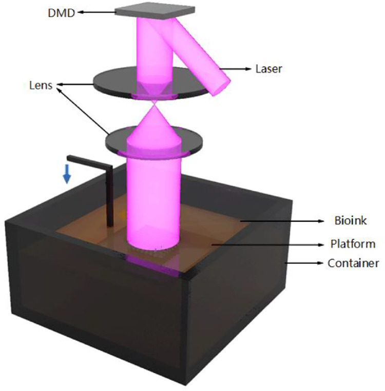
Biomanufacturing of tissues/organs in vitro is our big dream, driven by two needs: organ transplantation and accurate tissue models. Over the last decades, 3D bioprinting has been widely applied in the construction of many tissues/organs such as skins, vessels, hearts, etc., which can not only lay a foundation for the grand goal of organ replacement, but also be served as in vitro models committed to pharmacokinetics, drug screening and so on. As organs are so complicated, many bioprinting methods are exploited to figure out the challenges of different applications. So the question is how to choose the suitable bioprinting method? Herein, we systematically review the evolution, process and classification of 3D bioprinting with an emphasis on the fundamental printing principles and commercialized bioprinters. We summarize and classify extrusion-based, droplet-based, and photocuring-based bioprinting methods and give some advices for applications. Among them, coaxial and multi-material bioprinting are highlighted and basic principles of designing bioinks are also discussed.

Keywords: 3D bioprinting; Bioink; Droplet-based bioprinting; Extrusion-based bioprinting; Photocuring-based bioprinting.


Conflict of interest statement

The authors report no conflicts of interest. The authors alone are responsible for the content and writing of this article.

Figures

Fig. 1
Fig. 1
Four types of cutting potato corresponding to four typical 3D printing processes.
Fig. 2
Fig. 2
A brief history of bioprinting.
Fig. 3
Fig. 3
3D bioprinting process (A) X-ray machine, (B) CT machine, (C) MRI machine, (D) Alginate, (E) Scanning electron microscope (SEM) image of GelMA (reproduced with permission from , Copyright 2018 WILEY‐VCH Verlag GmbH & Co. KGaA, Weinheim), (F) Image of human umbilical vein endothelial cells (HUVECs) (reproduced with permission from , Copyright 2018 WILEY‐VCH Verlag GmbH & Co. KGaA, Weinheim), (G) Principle of extrusion-based bioprinting, (H) Principle of piezoelectric inkjet bioprinting, (I) Principle of digital light processing (DLP), (J) Culture of endothelial progenitor cell-laden blood vessel (reproduced with permission from , Copyright 2017 Gao et al.), (K) In vivo implantation of cardiac patches fabricated by laser induced forward transfer (LIFT) in rats (reproduced with permission from , Copyright 2011 Elsevier), (L) Biochip used for in vitro testing, adapted from unpublished work of our research team.
Fig. 4
Fig. 4
Classification of extrusion-based bioprinting.
Fig. 5
Fig. 5
Principles of extrusion-based bioprinting.
Fig. 6
Fig. 6
(A) Extrusion-based bioprinter EFL-BP6601; (B) High-precision printer EFL-BP5800 (3 µm resolution).
Fig. 7
Fig. 7
(A) Alternately bioprinted fibroblasts and keratinocytes mimicking natural skin structures (reproduced with permission from , Copyright 2008 Elsevier); (B) Handheld 3D skin bioprinting device combining microfluidic technique (reproduced with permission from , Copyright 2018 the Royal Society of Chemistry); (C) Multi-nozzle bioprinting bone tissue (reproduced with permission from , Copyright 2016 WILEY‐VCH Verlag GmbH & Co. KGaA, Weinheim); (D) Scaffold-free construction of neural grafts (reproduced with permission from , Copyright 2013 IOP Publishing Ltd).
Fig. 8
Fig. 8
(A) Vascular structures fabrication using agarose as sacrificial material (reproduced with permission from , Copyright 2009 Elsevier); (B) Vascular network printed in suspended hydrogel (reproduced with permission from , Copyright 2015 Hinton et al.); (C) 3D bioprinting whole heart containing major blood vessels (reproduced with permission from , Copyright 2019 Noor et al.); (D) Nanoclay and GelMA hybrid bioprinting complex structures (reproduced with permission from , Copyright 2019 IOP Publishing Ltd.).
Fig. 9
Fig. 9
(A) Typical coaxial nozzle configuration (reproduced with permission from , Copyright 2016 IOP Publishing Ltd.); (B) Perfusable hollow vascular structure (reproduced with permission from , Copyright 2015 Elsevier); (C) 2D patterns built by perfusable tubes (reproduced with permission from , Copyright 2016 Elsevier); (D) Vessel-like structures with multilevel fluidic channels (reproduced with permission from , Copyright 2017 American Chemical Society).
Fig. 10
Fig. 10
(A) Three-channel coaxial nozzles constructing tubular structure with switchable single/double layers (reproduced with permission from , Copyright 2018 WILEY‐VCH Verlag GmbH & Co. KGaA, Weinheim); (B) Mice experiment of bioprinted vascular structures using Pluronic as sacrificial material (reproduced with permission from , Copyright 2017 Gao et al.); (C) Coaxial printing of double-layered, multi-layered, helical GelMA microfibers (reproduced with permission from , Copyright 2018 WILEY-VCH Verlag GmbH & Co. KGaA, Weinheim); (D) Coaxial printing to build microfluidic bioreactor (reproduced with permission from , Copyright 2016 Elsevier).
Fig. 11
Fig. 11
Classification of droplet-based bioprinting.
Fig. 12
Fig. 12
Principles of droplet-based bioprinting.
Fig. 13
Fig. 13
(A) Simulation cell proliferation state of skin by LIFT (reproduced with permission from , Copyright 2013 Michael et al.); (B) Inkjet printing zigzag tubular structure (reproduced with permission from , Copyright 2012 Wiley Periodicals, Inc.); (C) Inkjet printing horizontally and vertically branched vascular structures (reproduced with permission from , Copyright 2014 Wiley Periodicals, Inc.); (D) Cell-laden Y-shaped tubular structures using MAPLE-DW technique (reproduced with permission from , Copyright 2015 IOP Publishing Ltd.).
Fig. 14
Fig. 14
(A) Implantation of cardiac patches fabricated by LIFT in rats (reproduced with permission from , Copyright 2011 Elsevier); (B) EHDJ constructing microspheres with complex structures (reproduced with permission from , Copyright 2018 WILEY‐VCH Verlag GmbH & Co. KGaA, Weinheim); (C) Self-folding droplet networks simulating tissue structures (reproduced with permission from , Copyright 2013 Springer Nature Limited); (D) Inkjet printing vascularized tissue structure (reproduced with permission from , Copyright 2012 Elsevier); (E) EHDJ manufacturing cell-laden microspheres and patterns (reproduced with permission from , Copyright 2018 WILEY‐VCH Verlag GmbH & Co. KGaA, Weinheim).
Fig. 15
Fig. 15
Classification of photocuring-based bioprinting.
Fig. 16
Fig. 16
Principle of DLP bioprinting.
Fig. 17
Fig. 17
DLP bioprinter EFL-BP8600.
Fig. 18
Fig. 18
(A) NGCs built by µSLA and rats experiment (reproduced with permission from , Copyright 2015 Pateman et al.); (B) NGCs fabricated by DLP (reproduced with permission from , Copyright 2018 Elsevier); (C) Hydrogel-based liver structure (reproduced with permission from , Copyright 2016 National Academy of Sciences).
Fig. 19
Fig. 19
Evaluation for bioinks performance (adapted with permission from , Copyright 2019 IOP Publishing Ltd.).

References

    1. He Y., Gao Q., Liu A., Sun M., Fu J. 3D bioprinting: from structure to function. J Zhejiang Univ (Eng Sci) 2019;53(3):1–12.
    1. Dong H., Fang Y., Wang D., Zhang H., Lei L., Luo Y. Current situation and thinking of organ donation at home and abroad. J Nurs. 2017;24(11):23–26.
    1. Mironov V., Boland T., Trusk T., Forgacs G., Markwald R.R. Organ printing: computer-aided jet-based 3D tissue engineering. Trends Biotechnol. 2003;21(4):157–161. - PubMed
    1. Boland T., Mironov V., Gutowska A., Roth E.A., Markwald R.R. Cell and organ printing 2: fusion of cell aggregates in three‐dimensional gels. Anat Rec Part A. 2003;272(2):497–502. - PubMed
    1. Mironov V. Printing technology to produce living tissue. Exp Opin Biol Ther. 2003;3(5):701–704. - PubMed