CLOCK-BMAL1 regulates circadian oscillation of ventricular arrhythmias in failing hearts through β1 adrenergic receptor
- PMID: 33194018
- PMCID: PMC7653582
CLOCK-BMAL1 regulates circadian oscillation of ventricular arrhythmias in failing hearts through β1 adrenergic receptor
Abstract
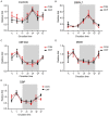
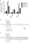
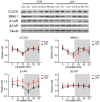
The incidence of ventricular arrhythmias (VAs) in chronic heart failure (CHF) exhibits a notable circadian rhythm, for which the underlying mechanism has not yet been well defined. Thus, we aimed to investigate the role of cardiac core circadian genes on circadian VAs in CHF. First, a guinea pig CHF model was created by transaortic constriction. Circadian oscillation of core clock genes was evaluated by RT-PCR and was found to be unaltered in CHF (P > 0.05). Using programmed electrical stimulation in Langendorff-perfused failing hearts, we discovered that the CHF group exhibited increased VAs with greater incidence at CT3 compared to CT15 upon isoproterenol (ISO) stimulation. Circadian VAs was blunted by a β1-AR-selective blocker rather than a β2-AR-selective blocker. Circadian oscillation of β1-AR was retained in CHF (P > 0.05) and a 4-h phase delay between β1-AR and CLOCK-BMAL1 was recorded. Therefore, when CLOCK-BMAL1 was overexpressed using adenovirus infection, an induced overexpression of β1-AR also ensued, which resulted in prolonged action potential duration (APD) and enhanced arrhythmic response to ISO stimulation in cardiomyocytes (P < 0.05). Finally, chromatin immunoprecipitation and luciferase assays confirmed that CLOCK-BMAL1 binds to the enhancer of β1-AR gene and upregulates β1-AR expression. Therefore, in this study, we discovered that CLOCK-BMAL1 regulates the expression of β1-AR on a transcriptional level and subsequently modulates circadian VAs in CHF.
Keywords: Arrhythmia; chronic heart failure; circadian clock; β1 adrenergic receptor.
AJTR Copyright © 2020.
Conflict of interest statement
None.
Figures


Similar articles
-
Molecular basis of Period 1 regulation by adrenergic signaling in the heart.FASEB J. 2021 Oct;35(10):e21886. doi: 10.1096/fj.202100441R. FASEB J. 2021. PMID: 34473369
-
Activation of β3-adrenergic receptor inhibits ventricular arrhythmia in heart failure through calcium handling.Tohoku J Exp Med. 2010 Nov;222(3):167-74. doi: 10.1620/tjem.222.167. Tohoku J Exp Med. 2010. PMID: 20975248
-
β1 -Adrenoceptor autoantibodies increase the susceptibility to ventricular arrhythmias involving abnormal repolarization in guinea-pigs.Exp Physiol. 2017 Jan 1;102(1):25-33. doi: 10.1113/EP085778. Epub 2016 Dec 15. Exp Physiol. 2017. PMID: 27862484
-
Regulation of cyclic adenosine monophosphate release by selective β2-adrenergic receptor stimulation in human terminal failing myocardium before and after ventricular assist device support.J Heart Lung Transplant. 2012 Oct;31(10):1127-35. doi: 10.1016/j.healun.2012.07.005. J Heart Lung Transplant. 2012. PMID: 22975104
-
The cardiomyocyte molecular clock regulates the circadian expression of Kcnh2 and contributes to ventricular repolarization.Heart Rhythm. 2015 Jun;12(6):1306-14. doi: 10.1016/j.hrthm.2015.02.019. Epub 2015 Feb 19. Heart Rhythm. 2015. PMID: 25701773 Free PMC article.
Cited by
-
Harm of circadian misalignment to the hearts of the adolescent wistar rats.J Transl Med. 2022 Aug 6;20(1):352. doi: 10.1186/s12967-022-03546-w. J Transl Med. 2022. PMID: 35933342 Free PMC article.
-
Research progress of circadian rhythm in cardiovascular disease: A bibliometric study from 2002 to 2022.Heliyon. 2024 Mar 23;10(7):e28738. doi: 10.1016/j.heliyon.2024.e28738. eCollection 2024 Apr 15. Heliyon. 2024. PMID: 38560247 Free PMC article. Review.
References
-
- Saour B, Smith B, Yancy CW. Heart failure and sudden cardiac death. Card Electrophysiol Clin. 2017;9:709–723. - PubMed
-
- Mozaffarian D, Benjamin EJ, Go AS, Arnett DK, Blaha MJ, Cushman M, Das SR, de Ferranti S, Despres JP, Fullerton HJ, Howard VJ, Huffman MD, Isasi CR, Jimenez MC, Judd SE, Kissela BM, Lichtman JH, Lisabeth LD, Liu S, Mackey RH, Magid DJ, McGuire DK, Mohler ER 3rd, Moy CS, Muntner P, Mussolino ME, Nasir K, Neumar RW, Nichol G, Palaniappan L, Pandey DK, Reeves MJ, Rodriguez CJ, Rosamond W, Sorlie PD, Stein J, Towfighi A, Turan TN, Virani SS, Woo D, Yeh RW, Turner MB. Heart disease and stroke statistics-2016 update: a report from the American Heart Association. Circulation. 2016;133:e38–360. - PubMed
-
- Takeda N, Maemura K. Circadian clock and the onset of cardiovascular events. Hypertens Res. 2016;39:383–390. - PubMed
LinkOut - more resources
Full Text Sources
Research Materials
