. 2020 Oct 22:10:554883.
doi: 10.3389/fonc.2020.554883. eCollection 2020.

Host CD39 Deficiency Affects Radiation-Induced Tumor Growth Delay and Aggravates Radiation-Induced Normal Tissue Toxicity

Affiliations

Host CD39 Deficiency Affects Radiation-Induced Tumor Growth Delay and Aggravates Radiation-Induced Normal Tissue Toxicity

Alina V Meyer et al. Front Oncol. .

Abstract

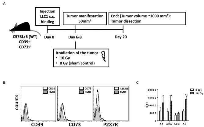
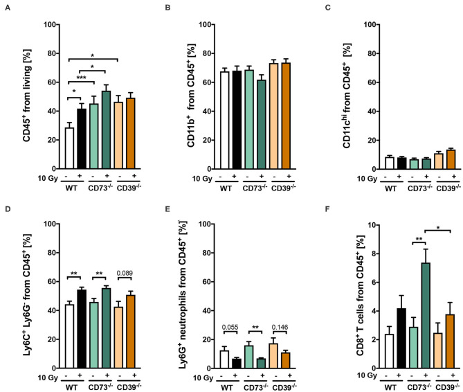
The ectonucleoside triphosphate diphosphohydrolase (CD39)/5' ectonuclotidase (CD73)-dependent purinergic pathway emerges as promising cancer target. Yet, except for own previous work revealing a pathogenic role of CD73 and adenosine in radiation-induced lung fibrosis, the role of purinergic signaling for radiotherapy outcome remained elusive. Here we used C57BL/6 wild-type (WT), CD39 knockout (CD39-/-), and CD73 knockout (CD73-/-) mice and hind-leg tumors of syngeneic murine Lewis lung carcinoma cells (LLC1) to elucidate how host purinergic signaling shapes the growth of LLC1 tumors to a single high-dose irradiation with 10 Gy in vivo. In complementary in vitro experiments, we examined the radiation response of LLC1 cells in combination with exogenously added ATP or adenosine, the proinflammatory and anti-inflammatory arms of purinergic signaling. Finally, we analyzed the impact of genetic loss of CD39 on pathophysiologic lung changes associated with lung fibrosis induced by a single-dose whole-thorax irradiation (WTI) with 15 Gy. Loss of CD73 in the tumor host did neither significantly affect tumor growth nor the radiation response of the CD39/CD73-negative LLC1 tumors. In contrast, LLC1 tumors exhibited a tendency to grow faster in CD39-/- mice compared to WT mice. Even more important, tumors grown in the CD39-deficient background displayed a significantly reduced tumor growth delay upon irradiation when compared to irradiated tumors grown on WT mice. CD39 deficiency caused only subtle differences in the immune compartment of irradiated LLC1 tumors compared to WT mice. Instead, we could associate the tumor growth and radioresistance-promoting effects of host CD39 deficiency to alterations in the tumor endothelial compartment. Importantly, genetic deficiency of CD39 also augmented the expression level of fibrosis-associated osteopontin in irradiated normal lungs and exacerbated radiation-induced lung fibrosis at 25 weeks after irradiation. We conclude that genetic loss of host CD39 alters the tumor microenvironment, particularly the tumor microvasculature, and thereby promotes growth and radioresistance of murine LLC1 tumors. In the normal tissue loss of host, CD39 exacerbates radiation-induced adverse late effects. The suggested beneficial roles of host CD39 on the therapeutic ratio of radiotherapy suggest that therapeutic strategies targeting CD39 in combination with radiotherapy have to be considered with caution.

Keywords: ATP; CD73; cancer; ionizing radiation; purinergic signaling.


Figures

Figure 1
Figure 1
LLC1 murine lung cancer cells are deficient for CD39, CD73, and P2X7 but express the four Ado receptors. For in vivo experiments mouse syngeneic tumors were generated by subcutaneous injection of 0.5 × 10⁶ LLC1 cells into the hindlimb of C57BL/6 (WT), CD73−/−, and CD39−/− mice. On days 6–8 after tumor manifestation, tumors were irradiated with 0 Gy (sham) or 10 Gy. Tumors were dissected at day 20 or when the tumor volume reached 1,000 mm3. (A) Schematic depiction of the experimental setup. (B,C) LLC1 cells were stained against (B) CD39, CD73, P2X7R; FMO = fluorescence minus one) or (C) AdoR- A1, A2A, A2B, and A3 and were further analyzed by flow cytometry (data show means ± SD; *P ≤ 0.05, **P ≤ 0.01 by one-way ANOVA followed by post hoc Newman–Keuls test).
Figure 2
Figure 2
Host CD39 deficiency accelerates LLC1 tumor growth and reduces radiation-induced tumor growth delay. LLC1 tumor cells (0.5 × 10⁶ cells each) were subcutaneously transplanted onto the hindlimb of C57BL/6 wild-type (circles) CD73-knockout [knockout (−/−), triangles] and CD39−/− (diamonds) mice. Hind-leg single-dose irradiation with 0 or 10 Gy was conducted at the timepoint of tumor manifestation (~100 mm3). LLC1 tumor volume in mm3 in respective experimental groups was determined at indicated time points using a sliding caliper (A/C). Growth delay was calculated as time (days) until the 6-fold tumor volume was reached (B/D). Shown are means ± SEM, *P ≤ 0.05, **P ≤ 0.01, #,****P ≤ 0.0001 analyzed by two-way ANOVA followed by a post hoc Tukey test, n (A) = 15/17/17/15; n (B) = 17/15/13/16; n (C) = 16/14/13/16, n (D) = 13/14/15/15. In (A,C), significant results between the animal of the same groups are marked with asterisks (0 and 10 Gy) and between the strains (C57BL/6 10 Gy and CD73−/− or CD39−/− 10 Gy) with hashtags.
Figure 3
Figure 3
Effects of extracellular ATP and Ado on LLC1 tumor cells in vitro. LLC1 cells were incubated with ATP (2 mg/mL) or Ado (1 mg/mL) 1 h prior to 0-, 5-, or 10-Gy irradiation. Forty-eight hours later, cells were stained with TMRE or PI and further analyzed with flow cytometry to investigate the (A) mitochondrial potential (ΔΨm), (B) the sub-G1 fraction (apoptosis), and (C) PI-positive cells (cell death). Data show means ± SEM; *P ≤ 0.05, **P ≤ 0.01, ***P ≤ 0.001 by two-way ANOVA followed by post hoc Tukey test.
Figure 4
Figure 4
(A-F) Host CD39 deficiency causes only subtle changes in radiation-induced tumor immune cell infiltration. LLC1 tumor cells (0.5 × 10⁶ cells each) were subcutaneously transplanted onto the hindlimb of C57BL/6 wild-type (white/black) CD73-knockout (CD73−/−; green, triangles) and CD39-knockout mice (CD39−/−; orange circles) mice. Hind-leg single-dose irradiation with 0 or 10 Gy was conducted at the timepoint of tumor manifestation. Three days after irradiation, tumors were dissected, and single-cell suspensions were generated. Tumor-infiltrating leukocytes were analyzed via flow cytometry. Shown in bar diagrams are the percentages of diverse immune cell populations. Shown are means ± SEM, *P ≤ 0.05, **P ≤ 0.01, ***P ≤ 0.001, analyzed by one-way ANOVA followed by a post hoc Tukey test, n = (13/14) (C57BL/6), (9/9) (CD73−/−), (10/9) (CD39−/−).
Figure 5
Figure 5
Host CD39 deficiency causes a reduced regressive phenotype of irradiated tumors. LC1 tumor cells (0.5 × 10⁶ cells each) were subcutaneously transplanted onto the hindlimb of C57BL/6 wild-type and CD39- and CD73-deficient [knockout (−/−)] mice. Hind-leg single-dose irradiation with 0 or 10 Gy was conducted at the timepoint of tumor manifestation. Mice were sacrificed at the timepoint of maximal tumor volume (1,000 mm3). (A) Hematoxylin-eosin (H&E) or (B) Masson Goldner trichrome (MT) staining of paraffin-embedded LLC1 tumor tissue, scale bar 100 μm. Magnified pictures (400-fold) highlight special structures. n = 15/17 (C57BL/6), 17/13 (CD39−/−), 7/13 (CD73−/−). Asterisks and open arrows indicate areas of a higher degree of epithelial structures (orange colored).
Figure 6
Figure 6
LLC1 tumor grown on CD39−/− mice are not impaired in their proliferative activity after irradiation. LLC1 tumor cells (0.5 × 10⁶ cells each) were subcutaneously transplanted onto the hindlimb of C57BL/6 wild-type and CD39- and CD73-deficient [knockout (−/−)] mice. Hind-leg single-dose irradiation with 0 or 10 Gy was conducted at the timepoint of tumor manifestation. Mice were sacrificed at the timepoint of maximal tumor volume (1,000 mm3). IHC staining of proliferation cell nuclear antigen (PCNA) of paraffin-embedded LLC1 tumor tissue, magnification 200-fold, scale bar 100 μm (A). Shown in bar diagrams are the absolute numbers of DAB+ cell counts per tissue section (B). Magnified pictures (400-fold) highlight special structures. Shown are means ± SEM, **P ≤ 0.01, ****P ≤ 0.0001, analyzed by one-way ANOVA followed by a post hoc Tukey test n = 15/17 (C57BL/6), n = 17/13 (CD39−/−), n = 7/13 (CD73−/−).
Figure 7
Figure 7
LLC1 tumor grown on CD39−/− mice show an altered microvasculature. LLC1 tumor cells (0.5 × 10⁶ cells each) were subcutaneously transplanted onto the hindlimb of C57BL/6 wild-type and CD39- and CD73-deficient [knockout (−/−)] mice. Hind-leg single-dose irradiation with 0 or 10 Gy was conducted at the timepoint of tumor manifestation. Mice were sacrificed at the timepoint of maximal tumor volume (1,000 mm3). (A) Immune fluorescent staining of CD34 (green) and DAPI (blue) of paraffin-embedded LLC1 tumor tissue, magnification 200-fold, scale bar 100 μm. 15/17 (C57BL/6), n = 17/13 (CD39−/−), n = 7/13 (CD73−/−). (B) RT qPCR analysis to reveal the relative expression of VE Cad, VegfR2, and VCAM1 to actin. The relative expression is shown in bar diagrams. Shown are means ± SEM, *P ≤ 0.05, **P ≤ 0.01, ***P ≤ 0.001, ****P ≤ 0.0001, analyzed by one-way ANOVA followed by a post hoc Tukey test n = 12/12 (C57BL/6), n = 12/12 (CD39−/−), n = 12/12 (CD73−/−).
Figure 8
Figure 8
CD39−/− mice develop enhanced pulmonary fibrosis 25 weeks after irradiation. C57BL/6 (WT) and CD39−/− mice received 0- or 15-Gy whole-thorax irradiation (WTI) and were sacrificed at 25 weeks after irradiation. (A) Hematoxylin-eosin (H&E) or (B) Masson Goldner trichrome (MT) stained lung sections at 25 weeks after irradiation. Lung sections were evaluated for fibrosis by using 10 non-overlapping randomized pictures (scale bar = 100 μm) per slide. Ashcroft scoring was done by three independent observers on slides blinded to the genotype and treatment condition. Magnified pictures (400-fold) highlight special structures. Asterisks emphasize thickening of alveolar wall structures and arrowheads fibrotic regions. (C) Quantification of fibrosis in WT (n ≥ 7) and CD73−/− (n = 7) mice, horizontal lines represent mean values. *P ≤ 0.05, ***P ≤ 0.001, **** P ≤ 0.0001 by one-way ANOVA followed by post hoc Newman–Keuls test, n = 9/9/15/15.
Figure 9
Figure 9
A CD39 deficiency leads to an increased osteopontin expression and TGF-β accumulation after irradiation. C57BL/6 (WT) and CD39−/− mice received 0- or 15-Gy whole-thorax irradiation (WTI) and were sacrificed at 25 weeks after irradiation. Immunohistochemical staining of paraffin-embedded lung sections with a primary antibody for (A,B) osteopontin (OPN) and (C,D) transforming growth factor β (TGF-β). Shown in (A,C right panels) are representative overview pictures of each indicated group (scale bar = 100 μm, magnified pictures 400-fold). (B,D) Using the Software Orbit Image Analysis version 2.65, positive areas for OPN and TGF-β were analyzed and are shown in percentage of the whole lung section. *P ≤ 0.05, **P ≤ 0.01, ****P ≤ 0.0001 by one-way ANOVA followed by post hoc Newman–Keuls test, n = 8/7/8/8.

Similar articles

Cited by

References

    1. Curran WJ, Jr, Paulus R, Langer CJ, Komaki R, Lee JS, Hauser S, et al. . Sequential vs. concurrent chemoradiation for stage III non-small cell lung cancer: randomized phase III trial RTOG 9410. J Natl Cancer Inst. (2011) 103:1452–60. 10.1093/jnci/djr325 - DOI - PMC - PubMed
    1. Bradley JD, Paulus R, Komaki R, Masters G, Blumenschein G, Schild S, et al. Standard-dose versus high-dose conformal radiotherapy with concurrent and consolidation carboplatin plus paclitaxel with or without cetuximab for patients with stage IIIA or IIIB non-small-cell lung cancer (RTOG 0617): a randomised, two-by-two factorial phase 3 study. Lancet Oncol. (2015) 16:187–99. 10.1016/S1470-2045(14)71207-0 - DOI - PMC - PubMed
    1. Ohri N. Radiotherapy dosing for locally advanced non-small cell lung carcinoma: “MTD” or “ALARA” ? Front Oncol. (2017) 7:205 10.3389/fonc.2017.00205 - DOI - PMC - PubMed
    1. Pfeifer GP. How the environment shapes cancer genomes. Curr Opin Oncol. (2015) 27:71–7. 10.1097/CCO.0000000000000152 - DOI - PMC - PubMed
    1. Morgan MA, Parsels LA, Maybaum J, Lawrence TS. Improving the efficacy of chemoradiation with targeted agents. Cancer Discov. (2014) 4:280–91. 10.1158/2159-8290.CD-13-0337 - DOI - PMC - PubMed