. 2020 Oct 30:8:581331.
doi: 10.3389/fcell.2020.581331. eCollection 2020.

Initial WNT/β-Catenin Activation Enhanced Mesoderm Commitment, Extracellular Matrix Expression, Cell Aggregation and Cartilage Tissue Yield From Induced Pluripotent Stem Cells

Affiliations

Initial WNT/β-Catenin Activation Enhanced Mesoderm Commitment, Extracellular Matrix Expression, Cell Aggregation and Cartilage Tissue Yield From Induced Pluripotent Stem Cells

Ursula Kreuser et al. Front Cell Dev Biol. .

Abstract

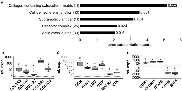
Mesodermal differentiation of induced pluripotent stem cells (iPSCs) in vitro and subsequent specification into mesodermal derivatives like chondrocytes is currently afflicted with a substantial cell loss that severely limits tissue yield. More knowledge on the key players regulating mesodermal differentiation of iPSCs is currently needed to drive all cells into the desired lineage and to overcome the current need for intermediate cell selection steps to remove misdifferentiated cells. Using two independent human iPSC lines, we here report that a short initial WNT/β-catenin pulse induced by the small molecule CHIR99021 (24 h) enhanced expression of mesodermal markers (PDGFRα, HAND1, KDR, and GATA4), supported the exit from pluripotency (decreased OCT4, SOX2, and LIN28A) and inhibited ectodermal misdifferentiation (reduced PAX6, TUBB3, and NES). Importantly, the initial CHIR pulse increased cell proliferation until day 14 (five-fold), adjusted expression of adhesion-related genes (CDH3 up, CDH6 down) and increased extracellular matrix (ECM)-related gene expression (COL6, COL1, COL3, COL5, DCN, NPNT, LUM, MGP, MATN2, and VTN), thus yielding more matrix-interacting progenitors with a high aggregation capability. Enhanced contribution to chondrogenic pellet formation increased the cell yield after eight weeks 200-fold compared to controls. The collagen type II and proteoglycan-positive area was enlarged in the CHIR group, indicating an increased number of cartilage-forming cells. Conclusively, short initial WNT activation improved mesoderm commitment and our data demonstrated for the first time to our knowledge that, acting via stimulation of cell proliferation, ECM expression and cell aggregation, WNT pulsing is a key step to make cell selection steps before chondrogenesis obsolete. This advanced understanding of the WNT/β-catenin function is a major step toward robust and efficient generation of high-quality mesodermal progenitors from human iPSCs and toward rescuing low tissue yield during subsequent in vitro chondrogenesis, which is highly desired for clinical cartilage regeneration, disease modeling and drug screening.

Keywords: WNT/β-catenin; aggregation; cartilage; cell survival; extracellular matrix; induced pluripotent stem cells.


Figures

FIGURE 1
FIGURE 1
Effects of short initial CHIR treatment on β-catenin regulation and proliferation during IMR-iPSC mesodermal predifferentiation. (A) Schematic representation of the iPSC differentiation protocol. Subconfluent iPSC cultures were induced into mesoderm differentiation by switching to serum-containing medium. During the first 24 h cells were additionally treated with 5 μM CHIR99021 (CHIR). Controls received either 0.025% DMSO or remained untreated (Ctrl). After CHIR treatment, the CHIR-IWP2 group received additionally 5 μM IWP2 for 48 h from day 3 to day 5. (B) Western blot analysis for active and total β-catenin during the first 7 days of mesoderm differentiation using β-actin as loading control. One representative blot of three is shown. (C) Densitometric analysis of Western blots in (A) (n = 3–5, mean ± SEM, *p < 0.05 vs. Ctrl, paired t-test + Bonferroni; #p < 0.05 vs. day 0, ANOVA + Bonferroni). (D) Western blot analysis of the CHIR and the CHIR-IWP2 group. One representative blot of three is shown. (E) Densitometric analysis of Western blots described in (D) (n = 3, mean ± SEM). (F) DNA quantification during mesoderm differentiation from day 7 to day 14, day 7 set to 1 (n = 5, mean ± SEM, *p < 0.05 vs. Ctrl, paired t-test + Bonferroni; #p < 0.05 vs. day 7, ANOVA + Bonferroni). (G) Cell count on day 14 of differentiation relative to day 7 determined using the hemocytometer (n = 8). Medians are depicted as horizontal lines, boxes represent first and third quartiles and whiskers are maximal and minimal values. White circle indicates an outlier with a value between 1.5 and 3 times the interquartile range (IQR) (*p < 0.05 vs. Ctrl, Wilcoxon test + Bonferroni).
FIGURE 2
FIGURE 2
Regulation of the pluripotency-associated genes after CHIR treatment. Gene expression of SOX2, OCT4, and LIN28A at day 14 of IMR-iPSC mesodermal predifferentiation in CHIR-treated cells compared to control (Ctrl) was determined by qPCR and RPL13 was used as reference. Relative gene expression compared to expression in iPSCs at d0 of differentiation (dashed line) is depicted. White circle indicates an outlier with a value between 1.5 and 3 times the IQR, black square indicates an extreme outlier above 3 times IQR (n = 5, *p < 0.05, Wilcoxon test).
FIGURE 3
FIGURE 3
Effects of CHIR treatment on differentiation toward the three germ layers during IMR-iPSC mesoderm differentiation. Mesoderm differentiation was assessed by (A) gene expression on day 14 of the mesoderm markers KDR, GATA4, and HAND1, using qPCR and RPL13 serving as reference (n = 5); and by (B) flow cytometric analysis of PDGFRα expression at day 7 of differentiation. Representative histograms depict immuno-labeled cells (black lines) compared to unstained control cells (light gray lines), which were used to adjust the gates. (C) Quantification of percentage of PDGFRα-positive cells at day 7 and day 14 in Ctrl and CHIR groups (n = 5). Gene expression of specified (D) ectoderm markers and (E) endoderm markers was assessed by qPCR at day 14 and RPL13 served as reference gene. Circles in all boxplots depict outliers with values between 1.5 and 3 times the IQR, black square indicates an extreme outlier above 3 times IQR (n = 5, *p < 0.05, Wilcoxon test), Ctrl = control.
FIGURE 4
FIGURE 4
Effects of short initial CHIR treatment on aggregation and condensation of mesodermal progenitors. Condensation assay for control (Ctrl) and CHIR-treated IMR-iPSCs after (A) 24 h and (B) after cells completed aggregation. Representative experiments are shown, and numbers of typical experiments are indicated (scale bar = 200 μm). (C) Time until cells finished assembling into 3D structures (considered as completed aggregation) was determined microscopically (n = 7, *p < 0.05, Wilcoxon test).
FIGURE 5
FIGURE 5
Chondrogenic differentiation of control (Ctrl) and CHIR-treated IMR-iPSCs. (A) DNA quantification at day 15, 21, and 56 of differentiation relative to day 14 (mean ± SEM, n = 3–10, p < 0.05 vs. Ctrl, paired t-test). (B) Histological analysis after 42 days of chondrogenesis. Collagen type II deposition was examined by immunostaining and proteoglycan deposition by safranin O staining (insert). Two representative experiments (i and ii) shown and the numbers of typical experiments are indicated (n = 8, scale bar = 200 μM). (C) Tissue volume at day 56 calculated from histomorphometric data of cartilage pellets, white square indicates an extreme outlier above 3 times the IQR (n = 11, p < 0.05, Wilcoxon test).
FIGURE 6
FIGURE 6
Global gene expression analysis of control (Ctrl) and CHIR-treated IMR-iPSC mesodermal progenitors (day 14). (A) PANTHER statistical overrepresentation analysis of all genes with a more than two-fold difference in mean expression levels between Ctrl and CHIR cells according to cDNA microarray data (n = 1). All significantly and more than two-fold overrepresented classes in the Gene Ontology-Slim Cellular Component category are depicted according to their overrepresentation score. Numbers in brackets indicate the number of genes included in each class (see also Tables 3–5) numbers behind each bar indicate the p-value. Differential regulation of designated collagen (B), extracellular matrix (C) and adhesion-related (D) genes of interest was confirmed by qPCR analysis and RPL13 served as reference gene. Relative gene expression (rel. expr.) compared to controls (dotted line) is depicted. Circles indicate outliers with a value between 1.5 and 3 times the IQR, the square indicates an extreme outlier above 3 times IQR (n = 5, *p < 0.05 vs. Ctrl, Wilcoxon test).

References

    1. Adkar S. S., Wu C. L., Willard V. P., Dicks A., Ettyreddy A., Steward N., et al. (2019). Step-wise chondrogenesis of human induced pluripotent stem cells and purification via a reporter allele generated by CRISPR-Cas9 genome editing. Stem Cells 37 65–76. 10.1002/stem.2931 - DOI - PMC - PubMed
    1. Baek S., Lee Y. W., Yoon S., Baek S. Y., Kim B. S., Oh S. O. (2010). CDH3/P-Cadherin regulates migration of HuCCT1 cholangiocarcinoma cells. Anat. Cell Biol. 43 110–117. 10.5115/acb.2010.43.2.110 - DOI - PMC - PubMed
    1. Biechele S., Cox B. J., Rossant J. (2011). Porcupine homolog is required for canonical Wnt signaling and gastrulation in mouse embryos. Dev. Biol. 355 275–285. 10.1016/j.ydbio.2011.04.029 - DOI - PubMed
    1. Birk D. E. (2001). Type V collagen: heterotypic type I/V collagen interactions in the regulation of fibril assembly. Micron 32 223–237. 10.1016/s0968-4328(00)00043-3 - DOI - PubMed
    1. Brandenberger R., Schmidt A., Linton J., Wang D., Backus C., Denda S., et al. (2001). Identification and characterization of a novel extracellular matrix protein nephronectin that is associated with integrin alpha8beta1 in the embryonic kidney. J. Cell Biol. 154 447–458. 10.1083/jcb.200103069 - DOI - PMC - PubMed