Review
. 2020 Oct 22:7:588338.
doi: 10.3389/fvets.2020.588338. eCollection 2020.

Diagnostic Imaging in Intervertebral Disc Disease

Collaborators, Affiliations
Review

Diagnostic Imaging in Intervertebral Disc Disease

Ronaldo C da Costa et al. Front Vet Sci. .

Abstract

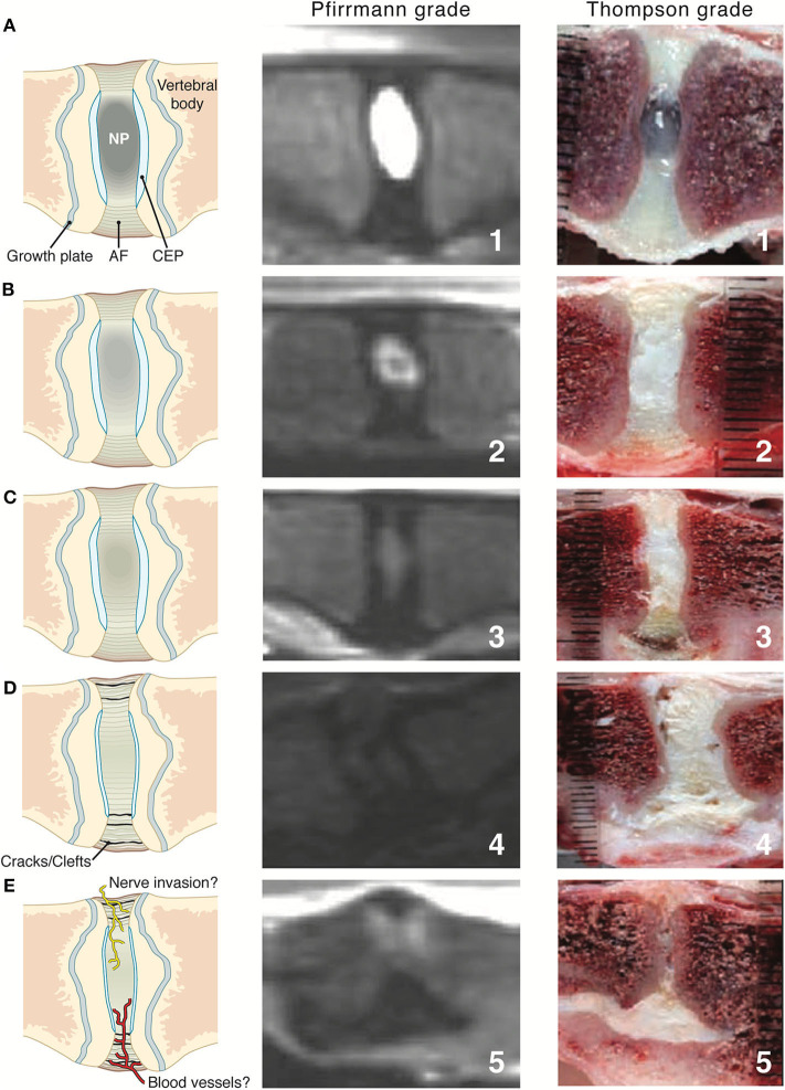
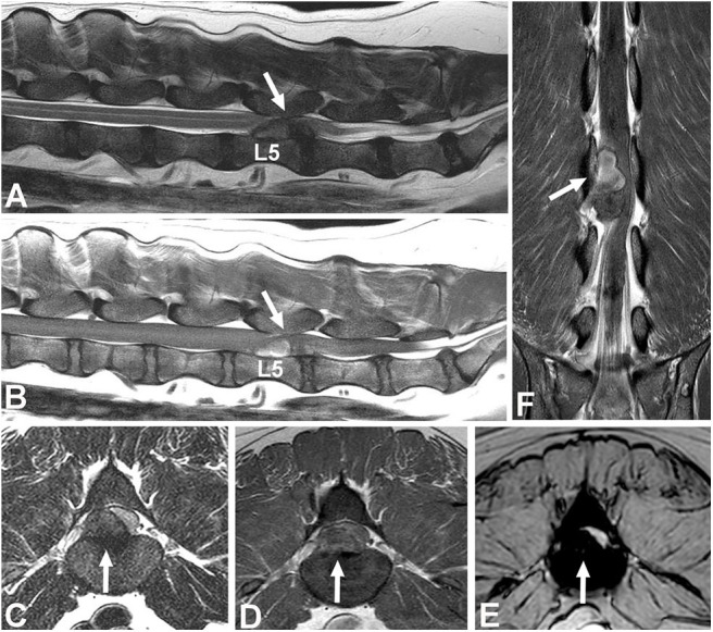
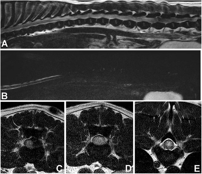
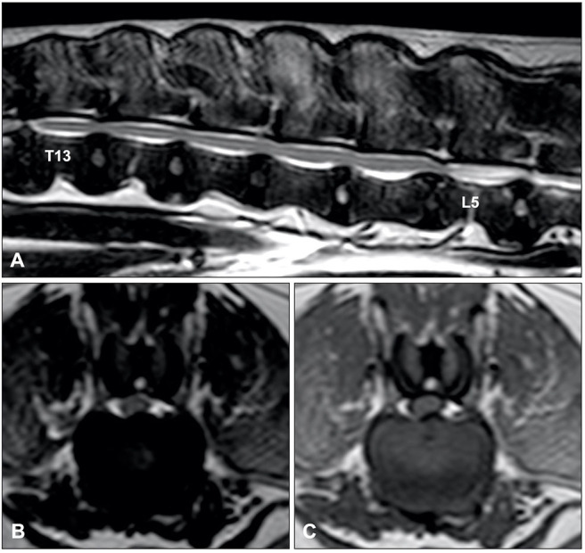
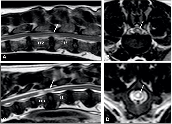
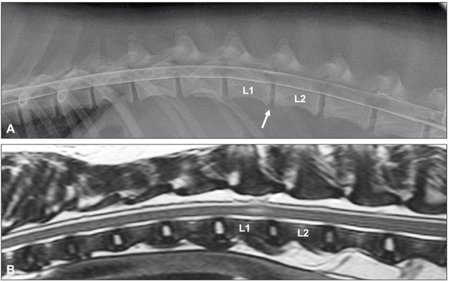
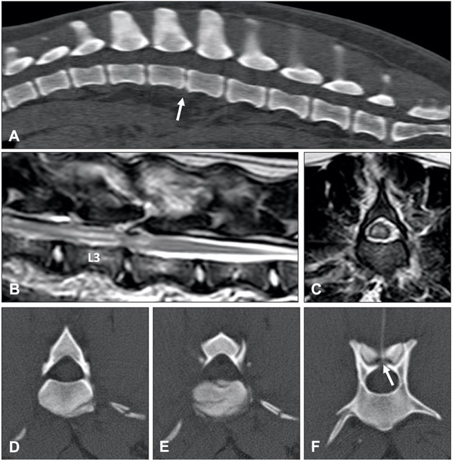
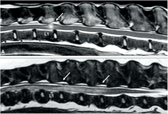
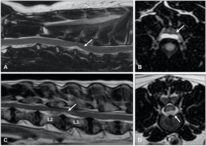
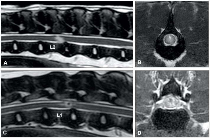
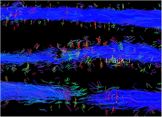
Imaging is integral in the diagnosis of canine intervertebral disc disease (IVDD) and in differentiating subtypes of intervertebral disc herniation (IVDH). These include intervertebral disc extrusion (IVDE), intervertebral disc protrusion (IVDP) and more recently recognized forms such as acute non-compressive nucleus pulposus extrusion (ANNPE), hydrated nucleus pulposus extrusion (HNPE), and intradural/intramedullary intervertebral disc extrusion (IIVDE). Many imaging techniques have been described in dogs with roles for survey radiographs, myelography, computed tomography (CT), and magnetic resonance imaging (MRI). Given how common IVDH is in dogs, a thorough understanding of the indications and limitations for each imaging modality to aid in diagnosis, treatment planning and prognosis is essential to successful case management. While radiographs can provide useful information, especially for identifying intervertebral disc degeneration or calcification, there are notable limitations. Myelography addresses some of the constraints of survey radiographs but has largely been supplanted by cross-sectional imaging. Computed tomography with or without myelography and MRI is currently utilized most widely and have become the focus of most contemporary studies on this subject. Novel advanced imaging applications are being explored in dogs but are not yet routinely performed in clinical patients. The following review will provide a comprehensive overview on common imaging modalities reported to aid in the diagnosis of IVDH including IVDE, IVDP, ANNPE, HNPE, and IIVDE. The review focuses primarily on canine IVDH due to its frequency and vast literature as opposed to feline IVDH.

Keywords: computed tomography; extrusion; herniation; intervertebral disc; magnetic resonance imaging; protrusion.


Figures

Figure 1
Figure 1
(A) Lateral myelogram of a 5-year old Basset Hound with an L1-L2 IVDE. Thinning of the contrast columns is severe and the ventral contrast column is almost discontinued at L1-L2. Thinning of the contrast column is beyond the boundaries of the affected disc and distributed asymmetrical with a longer dorsal deviation of the ventral contrast column caudal to the affected disc. (B) Lateral myelogram of a 9-year old Basset Hound with IVDPs between L1-L2 and L2-L3. Thinning of the contrast columns is only mild, dorsal deviation of the ventral contrast column is centered over the affected disc spaces and distribution of contrast thinning is cranio-caudally symmetrical over the disc spaces.
Figure 2
Figure 2
Computed tomographic (CT) images showing the appearance of mineralized intervertebral disc extrusion in the vertebral canal. (A) Sagittal reconstructed non-contrast CT image showing a hyperattenuating mass suggestive of extruded disc material into the vertebral canal between C2-3 (arrow). The intervertebral disc space also mineralization at C2-3. (B) Transverse non-contrast CT image showing a large hyperattenuating mass into the vertebral canal at the intervertebral disc level C2-3. (C) Sagittal reconstructed non-contrast CT image showing hyperattenuating material into the vertebral canal between T1-12 (arrow). (D) Transverse non-contrast CT image showing a large hyperattenuating mass disc extrusion occupying most of the vertebral canal at the intervertebral disc level T11-12.
Figure 3
Figure 3
Images of a 10-year old female spayed Beagle presented with an acute onset of non-ambulatory paraparesis and spinal pain. (A) Sagittal non-contrast CT image revealing areas of spondylosis and no obvious compressive lesion. (B) CT-myelography shows areas of ventral subarachnoid space compression at T13-L1, L1-2, and L2-3. Thinning of dorsal and ventral contrast columns is noted between L1-2. (C–E) images show transverse non-contrast CT images representing T13-L1, L1-2, and L2-3 intervertebral levels, respectively. Peridural fat is lost at L1-2 level. (F–H) CT-myelographic images at levels T13-L1, L1-2, and L2-3. Ventral, lateral and dorsal spinal cord compressions are noted at L1-2. Surgical decompression and biopsy confirmed that the compressive material was extruded disc material.
Figure 4
Figure 4
Images of a dog with cervical intervertebral disc extrusion. (A) Sagittal T2W image. (B) Sagittal HASTE image. (C) Sagittal STIR image. (D) Sagittal T1W image. All sagittal images show ventral extradural compression in the ventral aspect of the vertebral canal at the intervertebral level C2-3. Note how the compressive material is hypointense in all images, though image resolution varies with different sequences. The signal of intervertebral discs in situ also varies in all sequences. (E) Transverse T2W image showing a large hypointense mass causing spinal cord compression at the C2-3 level. (F) Transverse T1W image showing the hypointense compressive material at C2-3. (G) Transverse T2W image showing the C3-4 level with no spinal cord compression.
Figure 5
Figure 5
Intervertebral disc (IVD) maturation from young to early and late stage IVDD where the first column shows illustrative representations throughout the stages (A–E), middle column shows Pfirrmann grading via T2W MR images and last column showing Thompson grading of canine IVD. AF, annulus fibrosus; CEP, cartilaginous end plate; NP, nucleus pulposus. Pfirrmann grade and Thompson grade images adapted with permission from Bergknut et al. (90) and Thompson et al. (91).
Figure 6
Figure 6
Images of a female spayed, 6-year-old, mixed breed dog with an acute onset of paraparesis, spinal pain, and fecal and urinary incontinence. (A) Sagittal T2W image showing a large mass with mixed signal intensity between L5-6 (arrow). (B) Sagittal T1W image showing that the cranial aspect of the mass is hyperintense (arrow). (C) Transverse T2 image showing severe spinal cord compression caused by a large hypointense mass between L5-6 (arrow). (D) Transverse T1W contrast-enhanced image showing mild heterogeneous contrast enhancement of the hypointense lesion. (E) Gradient echo image showing marked hypointensity. (F) Dorsal T1W contrast-enhanced image showing contrast enhancement of the cranial aspect of the compressive lesion. Note the length of the compressive lesion. Surgical decompression and biopsy confirmed that the compressive material was extruded intervertebral disc with hemorrhage.
Figure 7
Figure 7
Images of female spayed, 13-year-old Cocker Spaniel with an acute onset of paraplegia. (A) Sagittal T2W image showing a large area of diffuse spinal cord hyperintensity. (B) Sagittal HASTE image showing diffuse spinal cord swelling. (C) Transverse T2W image at the L5-6 level showing lateralized spinal cord compression and spinal cord hyperintensity. (D) Transverse T2W image at the L3-4 level showing diffuse spinal cord hyperintensity and apparent swelling. (E) Transverse T2W image at T9-10 level showing centrally located spinal cord hyperintensity. Myelomalacia and intervertebral disc extrusion at L5-6 were confirmed at post-mortem examination.
Figure 8
Figure 8
Sagittal T2-weighted (A), and transverse T2-weighted (B) and T1-weighted MR (C) images of a 10-year old Dalmatian with multiple IVDPs between T13 and L6. The nucleus pulposus of most intervertebral discs has maintained some T2-weighted hyperintense signal, multiple discs are affected, intervertebral disk herniations are contained within the boundaries of the intervertebral disc space, and have resulted in midline spinal cord compression.
Figure 9
Figure 9
Sagittal (A,C) and transverse (B,D) T2-weighted MR images of a 9-year old Labrador retriever (A,B) and an 11-year old Poodle (C,D) with thoracolumbar IVDP. A poorly demarcated, diffuse intraparenchymal hyperintensity is seen overlying the affected intervertebral disc (arrow) in the Labrador retriever, while a well-demarcated and bright intraparenchymal hyperintensity is seen in the Poodle (arrow). It is currently unclear if this difference in intraparenchymal intensity characteristics is associated with differences in clinical presentation or outcome.
Figure 10
Figure 10
Lateral myelogram (A) and sagittal T2-weighted image (B) of a 4-year old Soft-Coated Wheaten terrier with an L1-L2 ANNPE. (A) Although the L1-L2 intervertebral disc space is narrowed (arrow), the myelographic pattern appears to be normal. (B) A focal intramedullary hyperintense lesion just cranial from the L1-L2 disc space can be observed. The homogenously hyperintense nucleus pulposus has a reduced volume and the intervertebral disc space is narrowed.
Figure 11
Figure 11
ANNPE can occur after external trauma. CT can be used to exclude or characterize concurrent vertebral fractures and luxations. (A) Sagittal reconstructed CT study of a 6-year old Boxer who became paraplegic immediately after a road traffic accident. Although a narrowed intervertebral disc space (arrow) is in itself not sufficient to confirm a diagnosis of ANNPE, the CT study was able to exclude the presence of a vertebral fracture and luxation. Sagittal (B) and transverse (C) T2-weighted MR images and transverse CT images at the levels of caudal L3 (D), the caudal vertebral endplate of L3 (E) and cranial L4 (F) of a 4-year old Border collie who ran into a tree. Although the MRI (A,B) reveals abnormalities suggestive for ANNPE, there are also abnormalities suggestive for external trauma, such as diffuse and bilateral hyperintensities in the paravertebral muscles. (C–E) The CT study revealed multiple fractures and (D) a mildly increased distance between the cranial and caudal articular processes.
Figure 12
Figure 12
T2-weighted sagittal MR images of (A) an 8-year old cross breed with ANNPE and (B) a 4-year old Staffordshire Bull terrier with ischemic myelopathy. (A) Focal hyperintensity can be seen overlying the L1-L2 intervertebral disc. The L1-L2 intervertebral disc space is narrowed and has a reduced volume of T2W hyperintense nucleus pulposus. (B) A relatively well-demarcated longitudinal hyperintensity can be seen between L1 and L4.
Figure 13
Figure 13
T2-weighted (A) sagittal and (B) transverse MR images of a 6-year old Border collie with a C4-C5 HNPE. The intervertebral disc space is narrowed and contains a reduced volume of hyperintense nucleus pulposus. There is a ventral extradural homogenously hyperintense compressive lesion. (B) The compressive material is midline and has a bilobed, “seagull” appearance. T2-weighted (C) sagittal and (D) transverse images of 7-year old Beagle with acute onset paraplegia. Although HNPE occurs most often in the cervical region, this dog was diagnosed with an L2-L3 HNPE.
Figure 14
Figure 14
(A) T2-weighted sagittal and (B) BALT GRAD (T2-weighted thin-slice gradient echo) transverse MR images of an 8-year old Greyhound with an L2-L3 IIVDE. (A) The intervertebral disc has a decreased volume of homogenously hyperintense nucleus pulposus. A ventrocaudal to dorsocranial intramedullary linear hyperintensity, starting from the L2-L3 intervertebral disc can be seen. (B) Hyperintense linear tract can be seen through the spinal cord. (C) T2-weighted sagittal and (D) BALT GRAD transverse MR images of a 6-year old Beagle with an L1-L2 IIVDE. The intramedullary lesion has a hypointense center, suggestive for intraparenchymal hemorrhage.
Figure 15
Figure 15
Tractography of the spinal cord from a normal dog (top) compared to dogs with SCI showing moderate fiber disruption (middle) and complete trans-lesional discontinuity (bottom). Blue depicts cranial to caudal oriented fibers.

References

    1. Bergknut N, Egenvall A, Hagman R, Gustas P, Hazewinkel HAW, Meij BP, et al. . Incidence of intervertebral disk degeneration-related diseases and associated mortality rates in dogs. J Am Vet Med Assoc. (2012) 240:1300–9. 10.2460/javma.240.11.1300 - DOI - PubMed
    1. Priester WA. Canine intervertebral disc disease – occurrence by age, breed, and sex among 8,117 cases. Theriogenology. (1976) 6:293–303. 10.1016/0093-691X(76)90021-2 - DOI
    1. Olsson SE. On disc protrusion in dog (enchondrosis intervertebralis); a study with special reference to roentgen diagnosis and to the value of disc fenestration. Acta Orthop Scand Suppl. (1951) 8:1–95 10.3109/ort.1951.22.suppl-8.01 - DOI - PubMed
    1. Hansen H. A pathologic-anatomical study on disc degeneration in the dog. Acta Orthop Scand Suppl. (1952) 11:1–117. 10.3109/ort.1952.23.suppl-11.01 - DOI - PubMed
    1. Konar M, Lang J, Fluhmann G, Forterre F. Ventral intraspinal cysts associated with the intervertebral disc: magnetic resonance observations in seven dogs. Vet Surg. (2008) 37:94–101. 10.1111/j.1532-950X.2007.00353.x - DOI - PubMed