Review
. 2020 Sep 21;31(Suppl 2):259-267.
doi: 10.31138/mjr.31.3.259. eCollection 2020 Sep.

Anti-Rheumatic Drugs for the Fight Against the Novel Coronavirus Infection (SARSCoV-2): What is the Evidence?

Affiliations
Review

Anti-Rheumatic Drugs for the Fight Against the Novel Coronavirus Infection (SARSCoV-2): What is the Evidence?

Eleftherios Pelechas et al. Mediterr J Rheumatol. .

Abstract

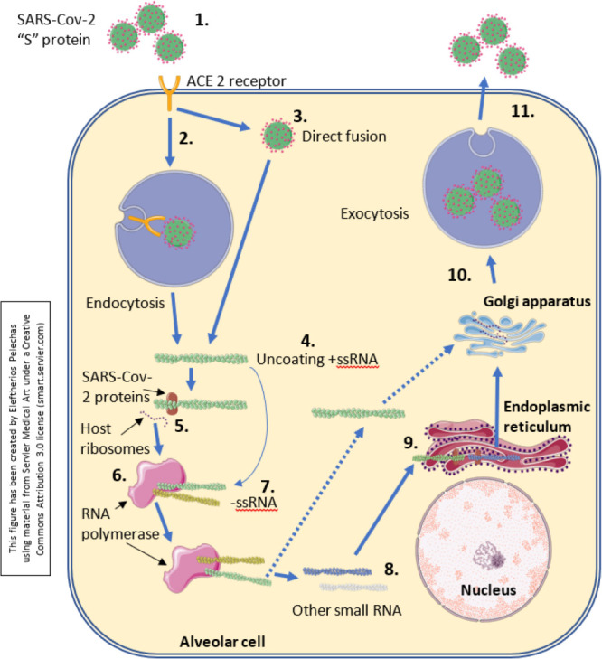
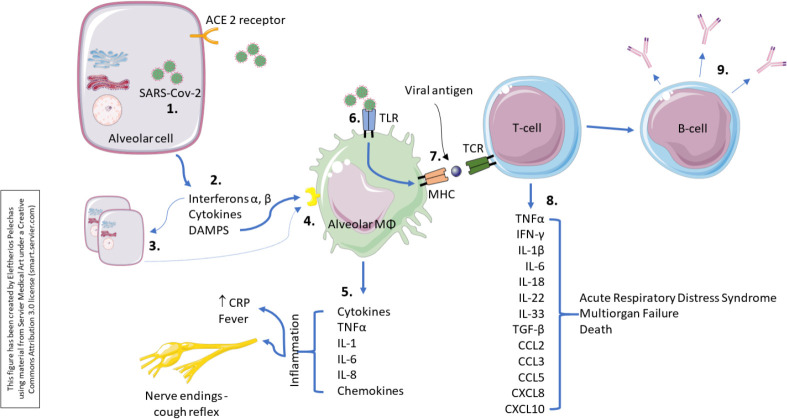
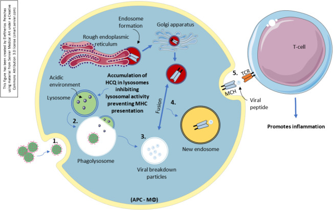
SARS-CoV-2 is a positive-sense single-stranded RNA virus that causes the COVID-19 infection. Spike proteins are the most important proteins found on its capsule using the host's ACE2 receptors to invade respiratory cells. The natural course of the COVID-19 infection is variable, from asymptomatic to severe and potentially fatal. A small percentage of the severely infected patients will end up in an intensive care unit for ventilatory support. Elderly male patients with pre-existing medical conditions and smokers are at a disproportionate high risk to develop severe complications. Studies have shown that deaths occur due to a dysregulated immune system that overreacts, producing a plethora of cytokines, leading to the so-called "cytokine storm" phenomenon. In this direction, many drugs that are used in the everyday practice of Rheumatologists have been used. Indeed, pro-inflammatory cytokines such as the IL-1 and IL-6 have been shown to be the pivotal cytokines expressed, and anti-cytokine treatment has been tried so far with various results. In addition, hydroxychloroquine, an antimalarial drug, has been shown to reduce COVID-19 symptoms. Other drugs have also been used, such as intravenous pulses of immunoglobulins, and colchicine. Robust clinical trials are needed in order to find the suitable treatment. Current data indicate that hydroxychloroquine and cytokine targeting therapies may prove helpful in the fight of SARS-CoV-2 in appropriately selected patients.

Keywords: ACE2 receptor; COVID-19; IL-6; SARS-CoV-2; colchicine; cytokine storm; hydroxychloroquine; spike protein; tocilizumab.


Figures

Figure 1.
Figure 1.
Invasion and replication of the SARS-CoV-2 within an alveolar cell. The virus targets and binds onto ACE 2 receptors on the surface of type 2 alveolar cells via its surface protein “spikes”. ACE 2 is needed for virus to gain entry inside the cell. This is a representation of the corona virus life cycle within the cell. Corona viruses will enter via endocytosis or direct fusion of the viral envelope with the host membrane. Once inside, the virus particle is uncoated, and its genome enters the cell cytoplasm. Since coronavirus has a single positive stranded RNA genome, it can directly produce its proteins and new genome in the cytoplasm by attaching to the host’s ribosomes. The host’s ribosomes will translate the viral RNA to make proteins that will make RNA polymerase. The RNA polymerase will read the positive strand of RNA of the virus again to make a negative RNA strand. The negative strand will be used by the RNA polymerase again to make a positive RNA strand as well other small positive RNA strands. These small RNA strands will be read by the host’s ribosomes again in the endoplasmic reticulum to help make the structural components of the virus. The endoplasmic reticulum will transfer these accessory and structural proteins into the Golgi apparatus where it will be packaged up with the positive RNA strand formed, to essentially form a new virus. These viruses are then released from the host cell by exocytosis through secretory vesicles. While the virus is self-replicating in the alveolar cells, it also damages it, and this will initiate the inflammatory response. Injured alveolar cells release interferons, cytokines as well as its intracellular components (not shown, continued in Figure 2).
Figure 2.
Figure 2.
Immunological response to SARS-CoV-2. While the virus is self-replicating in the alveolar cells, it also damages it, and this will initiate the inflammatory response. Injured alveolar cells release interferons, cytokines as well as its intracellular components. Interferons α, β act in a paracrine manner, and have numerous effects on the surrounding cells preparing them for the ongoing infection. The primary function is to induce protection against viruses in neighbouring non-infected cells. Alveolar macrophages detect cell injury via damage-associated molecular patterns from the alveolar cells. They also respond to the cytokines released by injured alveolar cells. This causes the alveolar macrophages themselves to secrete cytokines such as TNFα, IL-1, IL-6, IL-8, as well as other chemokines. The inflammatory process occurring within the lung parenchyma stimulates nerve endings responsible for initiating the cough reflex. Thus, people often present with a dry cough early on. TNFα and IL-1β are pro-inflammatory cytokines and cause increased vascular permeability and increase in expression of adhesion molecules. This allows recruitment of more immune cells including neutrophils and monocytes. The alveolar macrophage can also detect the virus using its special receptors, called Toll-like receptors (TLRs). It can engulf the virus particles to phagocytosis, process it, and then present it on its surface. Studies have shown that the viral proteins can be presented. By presenting the viral proteins, specific T-cells may recognise them and mount an adaptive immune response consisting of T-cell activation and production of a plethora of proinflammatory cytokines and chemokines that may lead to a cytokine storm. In addition, B-cells or the plasma cells will then be activated and produce antibodies against the viral proteins.
Figure 3.
Figure 3.
Antigen-processing onto macrophage/APC and the action of hydroxychloroquine. An antigen-presenting cell takes up the pathogen (SARS-CoV-2) end engulfs it with phagocytosis. Lysosomes that contain acids create an acidic environment and fuse with the phagocytosed “invader”, creating the phagolysosome which has an acidic environment as well. Because of the acidic environment, the content of the phagolysosome breaks down, leaving various particles that will be processed as antigens. In the meantime, within the endoplasmic reticulum the ribosomes are synthesizing MHC and allow the formation of an endosome which passes also through the Golgi apparatus to form a new endosome. The viral breakdown particles will then fuse with the new endosome and the particles will bind onto the groove of the MHC. Finally, the antigen presenting cell will express it on its self-surface. Hydroxychloroquine accumulates in lysosomes, raising the pH establishes a non-acidic environment, and inhibits the lysosomal activity. In this way, it prevents the MHC presentation to the surface of the antigen-presenting cell.

References

    1. Jin Z, Du X, Xu Y, Deng Y, Liu M, Zhao Y, et al. Structure of Mpro from COVID-19 virus and discovery of its inhibitors. Nature 2020; 10.1038/s41586-020-2223-y. - DOI - PubMed
    1. Dai H, Zhang X, Xia J, Zhang T, Shang Y, Huang R, et al. High-resolution Chest CT Features and Clinical Characteristics of Patients Infected with COVID-19 in Jiangsu, China. Int J Infect Dis 2020;S1201-9712(20)30218-6. - PMC - PubMed
    1. Cohen FS. How viruses invade cells. Biophys J 2016; 110 (5): 1028– 32. - PMC - PubMed
    1. The Novel Coronavirus Pneumonia Emergency Response Epidemiology Team The epidemiological characteristics of an outbreak of 2019 novel coronavirus diseases (COVID-19) in China. Chin J Epidemiol 2020; 41. - PMC - PubMed
    1. Centres for Disease Control and Prevention Morbidity and Mortality Weekly Report, Hospitalization Rates and Characteristics of Patients Hospitalized with Laboratory-confirmed Coronavirus Disease 2019 – COVID-NET, 14 States, March 1–30, 2020. https://www.cdc.gov/mmwr/volumes/69/wr/mm6915e3.htm . Accessed on 10.04.2020. - PMC - PubMed

LinkOut - more resources