. 2020 Nov 16;16(11):e1009038.
doi: 10.1371/journal.ppat.1009038. eCollection 2020 Nov.

Tick-transmitted thogotovirus gains high virulence by a single MxA escape mutation in the viral nucleoprotein

Affiliations

Tick-transmitted thogotovirus gains high virulence by a single MxA escape mutation in the viral nucleoprotein

Jonas Fuchs et al. PLoS Pathog. .

Abstract

Infections with emerging and re-emerging arboviruses are of increasing concern for global health. Tick-transmitted RNA viruses of the genus Thogotovirus in the Orthomyxoviridae family have considerable zoonotic potential, as indicated by the recent emergence of Bourbon virus in the USA. To successfully infect humans, arboviruses have to escape the restrictive power of the interferon defense system. This is exemplified by the high sensitivity of thogotoviruses to the antiviral action of the interferon-induced myxovirus resistance protein A (MxA) that inhibits the polymerase activity of incoming viral ribonucleoprotein complexes. Acquiring resistance to human MxA would be expected to enhance the zoonotic potential of these pathogens. Therefore, we screened a panel of 10 different thogotovirus isolates obtained from various parts of the world for their sensitivity to MxA. A single isolate from Nigeria, Jos virus, showed resistance to the antiviral action of MxA in cell culture and in MxA-transgenic mice, whereas the prototypic Sicilian isolate SiAr126 was fully MxA-sensitive. Further analysis identified two amino acid substitutions (G327R and R328V) in the viral nucleoprotein as determinants for MxA resistance. Importantly, when introduced into SiAr126, the R328V mutation resulted in complete MxA escape of the recombinant virus, without causing any viral fitness loss. The escape mutation abolished viral nucleoprotein recognition by MxA and allowed unhindered viral growth in MxA-expressing cells and in MxA-transgenic mice. These findings demonstrate that thogotoviruses can overcome the species barrier by escaping MxA restriction and reveal that these tick-transmitted viruses may have a greater zoonotic potential than previously suspected.


Conflict of interest statement

The authors have declared that no competing interests exist.

Figures

Fig 1
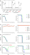
Fig 1. JOSV but not SiAr126 is resistant to the antiviral action of MxA.
(A) Vero cells stably expressing MxA (MxA) or control Vero cells (CTRL) were infected with 50 pfu of each virus. A plaque assay was performed to determine the plaque reduction in the presence of MxA. Counted plaques were normalized to the control for each virus (mean ± SD (n = 3), one-way ANOVA, Tukey’s multiple comparison test, ***p<0.001, ns–not significant) (B) Growth kinetics of JOSV and SiAr126 in the presence or absence of MxA. Vero-MxA or control Vero cells were infected with JOSV or SiAr126 (MOI 0.001). At the indicated time points the supernatant was harvested and the viral load determined by plaque assay (mean ± SD (n = 3), two-way ANOVA, Tukey’s multiple comparison test, ***p<0.001, ns–not significant). (C) Western blot analysis of MxA expression in Vero-MxA or control Vero cells in comparison to Huh7 or A549 cells treated with the indicated amounts of human IFNα2a or IFNαB/D for 24 h [42]. (D, E) The in vivo protective effect of mouse MX1 and human MxA against SiAr126 and JOSV was analyzed by i.p. infections of MX1-negative C57BL/6 mice (Mx1-/-), MX1-positive C57BL/6 mice congenic for murine A2G-Mx1 (Mx1+/+) or MxA-transgenic mice carrying the human MX1 locus (hMX1-tg+/+, red) with 100 pfu of SiAr126 (D) or 1000 pfu of JOSV (E) for 4 days (n = 5). Organs were harvested and the viral load determined by plaque assay. (F) Mx1-/- mice or hMX1-tg+/+ mice were treated i.p. with 20,000 IU IFNαB/D for 16 h prior to infection with 1,000 pfu of JOSV (n = 5) or were left untreated. The viral load in the organs was determined 4 days after infection by plaque assay. (D-F) Shown are the geometric means. Statistical analyses were performed with a one-way ANOVA on log-transformed values (Tukey’s multiple comparison test, ***p<0.001, ns–not significant) and displayed in comparison to the Mx1-/- C57BL/6 group (D, E) or untreated animals (F).
Fig 2
Fig 2. The viral nucleoprotein determines MxA sensitivity.
(A-E) 293T cells were co-transfected with the components of the polymerase reconstitution system for either SiAr126 or JOSV, consisting of 10 ng of the expression plasmids encoding the polymerase subunits PB1, PB2, PA, 50 ng for the respective NPs, 50 ng for a viral minigenome coding for firefly luciferase reporter under the control of the viral promotor (pPol-I FF-Luc) and 10 ng for Renilla luciferase under a constitutive promotor (RLuc). 24 h after transfection the cells were lysed and the firefly and Renilla luciferase activities determined. The firefly luciferase was normalized to the Renilla luciferase activity and the expression of NP, actin and MxA was controlled by Western blot. Significance was calculated with a one-way ANOVA (Tukey’s multiple comparison test, ***p<0.001, ns–not significant). (A) Activity of the SiAr126 and JOSV polymerase reconstitution system in the absence of MxA. 293T cells were co-transfected with the components of the SiAr126 or JOSV polymerase reconstitution system. The control without NP (–NP) was set to 1 (mean ± SD, n = 3). (B) Inhibition of the SiAr126 or JOSV system by MxA. The SiAr126 or JOSV systems were co-transfected with 50 ng expression plasmids for human MxA or the antivirally inactive mutant MxA(T103A). The empty vector controls were each set to 100% (mean ± SD, n = 3). (C) Schematic representation of the chimeric viral NPs. Grey represents SiAr126 NP and red JOSV NP. Fusion sites were selected in conserved regions of the proteins and are indicated as dotted lines with the corresponding amino acid positions. (D) Relative luciferase activity of the chimeric NPs in the SiAr126 or JOSV systems in the absence of MxA. The polymerase reconstitution systems were co-transfected with 50 ng of the chimeric NPs. The (–NP) control was set to 1 (mean ± SD, n = 3). (E) MxA sensitivity of the functional chimeric NPs. 293T cells were co-transfected with the components of the SiAr126 or JOSV system including 50 ng of the compatible chimeric NP expression plasmids and 50 ng MxA or MxA(T103A). The empty vector controls for the individual chimeric NPs were set to 100% (mean ± SD, n = 3).
Fig 3
Fig 3. Positions G327 and R328 are responsible for MxA sensitivity.
(A) Schematic representation of an amino acid alignment of JOSV and SiAr126 NP (green–identical, yellow–similar, white–non synonymous). Enlarged is the MxA sensitivity region corresponding to amino acids 289 to 370 of SiAr126 NP. Asterisks mark non-synonymous amino acid differences that could influence MxA sensitivity. (B-D) 293T cells were co-transfected with SiAr126 expression plasmids including 10 ng of PB1, PB2 and, PA, 50 ng of the individual NP mutants, 50 ng of pPol-I FF-Luc and 10 ng of RLuc. At 24 h after transfection the cells were lysed and firefly and Renilla luciferase activity determined. The firefly luciferase was normalized to the Renilla luciferase activity and the expression of NP, actin and MxA were controlled by Western blot. Significance was calculated with a one-way ANOVA (Tukey’s multiple comparison test, *p<0,05, ***p<0.001, ns–not significant). (B) The amino acid substitutions marked in (A) were introduced in SiAr126 NP and tested with the SiAr126 polymerase for activity in the absence of MxA. NP(WT) was set to 100% (mean ± SD, n = 3). (C) MxA sensitivity of the SiAr126 NP single mutants. 293T cells were transfected with the components of the SiAr126 system including the NP single mutants and 50 ng of MxA or MxA(T103A). Displayed is the ratio of the luciferase activity in the presence of MxA compared to MxA(T103A) (mean ± SD, n = 3). (D) Increased amounts of 300 ng MxA or MxA(T103A) were co-transfected with the SiAr126 system in the presence of 50 ng of NP expression plasmids coding for G327R, R328V or G327R/R328V. The empty vector control for the respective NP mutants was set to 100% (mean ± SD, n = 3). (E) Crystal structure of IAV (H5N1) NP (2q06.2.A) showing adaptive mutations responsible for MxA escape of pandemic IAVs [44] and a predicted SiAr126 NP structure (SWISS-MODEL; template H5N1 NP (2q06.2.A)) highlighting the positions 328V and 327R. Red–strong, yellow–weak influence on MxA sensitivity, for IAV according to Mänz et al. [44]. wt–wildtype.
Fig 4
Fig 4. Recombinant SiAr126 NP(R328V) is MxA resistant.
(A, B) Growth kinetics of rSiAr126(wt) or rSiAr126-NP(R328V) in the presence or absence of MxA. Vero-MxA or control Vero cells (A) or Huh7 cells stably expressing a control vector, MxA or MxA(T103A) (B) were infected with rSiAr126(wt) or rSiAr126-NP(R328V) (MOI 0.001). At the indicated time points the supernatants were harvested and the viral load determined by plaque assay (log-transformed values, mean ± SD, n = 3). Statistical analyses were performed with a two-way ANOVA (Tukey’s multiple comparison test, **p<0.01, ***p<0.001, ns–not significant). (C) As in (B) Huh7 cells were infected with rSiAr126(wt) or rSiAr126-NP(R328V) (MOI 0.01). At the indicated time points the cells were lysed and the expression of MxA, actin and viral NP controlled by Western blot.
Fig 5
Fig 5. R328V leads to MxA escape in vivo.
(A) C57BL/6 (Mx1-/-), congenic A2G-Mx1 (Mx1+/+) and transgenic human MX1 (hMX1-tg+/+) mice were infected with 100 pfu of rSiAr126(wt) or rSiAr126-NP(R328V) for 4 days. Organs were harvested and the viral load determined by plaque assay. Shown are the geometric means (n = 5). Statistical analyses were performed with a one-way ANOVA on log-transformed values (Tukey’s multiple comparison test, **p<0.01, ***p<0.001, ns–not significant) and displayed for each virus in comparison to the corresponding Mx1-/- group. (B-E) Weight loss and survival of Mx1-/- and hMX1-tg+/+ mice infected with rSiAr126(wt) (B, C) or rSiAr126-NP(R328V) (D, E). Mice were infected i.p. with the indicated inoculums (4 mice/group). Each day weight (mean ± SEM) and survival of the animals were monitored.
Fig 6
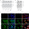
Fig 6. Loss of NP recognition by MxA is responsible for MxA escape.
(A) Co-immunoprecipitation (CoIP) of NP with MxA from transfected cells. 293T cells were co-transfected with the components of the SiAr126 polymerase system, including 100 ng of PB1, PB2, PA, 500 ng of the individual NP mutants, 500 ng of pPol-I FF-Luc and 700 ng of FLAG-MxA or FLAG-MxA(ΔL4). (B) CoIP of NP with MxA from rSiAr126(wt) and rSiAr126-NP(R328V) infected cells. 293T cells were transfected with 1,000 ng of FLAG-MxA or FLAG-MxA(ΔL4) and after 24 h infected with the recombinant viruses (MOI 10) for 24 h. (A and B) Cells were harvested and Flag-MxA was precipitated via anti-FLAG coupled beads from the lysates. MxA, viral NP and actin were detected in whole cell lysates (WCL) and the MxA precipitates (IP: FLAG) via Western blot. Representative Western blots of 3 individual experiments are shown. (C) Immunofluorescence pictures of Huh7 ctrl and MxA(wt)-expressing cells infected with rSiAr126(wt) and rSiAr126-NP(R328V). Huh7 ctrl and MxA cells were infected for 6 h (MOI 50), fixed on coverslips and stained for viral NP (green), MxA (red) and for the nucleus (DAPI, blue). Pictures were taken with an Apotome. wt–wildtype.
Fig 7
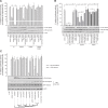
Fig 7. SiAr126 NP mutants escape restriction by different MX1 variants.
(A-C) 293T cells were co-transfected with the components of the polymerase reconstitution system, including 10 ng of PB1, PB2, PA, 50 ng of NP, 50 ng of pPol-I FF-Luc and 10 ng of RLuc. At 24 h after transfection the cells were lysed and the firefly and Renilla luciferase activity determined. Firefly luciferase activity was normalized to the Renilla luciferase activity and the expression of NP, actin and MxA was controlled by Western blot. Significance was calculated with a one-way ANOVA (Tukey’s multiple comparison test, ***p<0.001, ns–not significant). (A) 293T cells were transfected with the components of the SiAr126 system including 50 ng of wildtype NP, NP(G327R), NP(R328V) or NP(G327R/R328V). To analyze the influence of nuclear MxA, 300 ng of MxA, MxA(T103A), NLS-MxA or NLS-MxA(T103A) were co-transfected. The empty vector control for the respective NP mutants was set to 100% (mean ± SD, n = 3). (B) 293T cells were transfected with the components of the SiA126 system including 50 ng of wildtype NP, NP(G327R) and NP(R328V). To analyze the influence of MxA super-restrictors, 600 ng MxA, MxA(T103A) or MxA constructs with mutations in loop 4 (L4) were co-transfected. The empty vector control for the respective NP mutants was set to 100% (mean ± SD, n = 3). (C) 293T cells were transfected with the components of the SiAr126 or JOSV system. Different mammalian MX1 proteins were tested by transfecting 50 ng of bat MX1 (Eidolon helvum), ferret MX1, canine MX1, swine MX1, bovine MX1 with a repaired ORF, equine MX1, African green monkey MX1, orangutan MX1, human MxA, and MxA(T103A). The empty vector control for the respective polymerase systems was set to 100% (mean ± SD, n = 3). wt–wildtype.
Fig 8
Fig 8. Model for the MxA escape by thogotoviruses.
(A) Cytoplasmic MxA mono- and oligomers interact (direct or indirect) via their unstructured loop 4 (L4) with incoming vRNPs by recognizing surface exposed residues in NP leading to their retention in the cytoplasm. (B) Positions G327 and R328 are crucial for the MxA-NP interaction. Mutations at these positions lead to the loss of this interaction. Therefore incoming vRNPs can escape MxA recognition, translocate into the nucleus and initiate viral transcription and replication.

Similar articles

Cited by

References

    1. McCauley JW, Hongo S, Kaverin N V, Kochs G, Lamb RA, Matrosovich MN, et al. Family orthomyxoviridae. Virus Taxon ninth Rep Int Comm Taxon viruses. 2012;749–761.
    1. King AMQ, Lefkowitz EJ, Mushegian AR, Adams MJ, Dutilh BE, Gorbalenya AE, et al. Changes to taxonomy and the International Code of Virus Classification and Nomenclature ratified by the International Committee on Taxonomy of Viruses (2018). Arch Virol. 2018. 10.1007/s00705-018-3847-1 - DOI - PubMed
    1. Iuliano AD, Roguski KM, Chang HH, Muscatello DJ, Palekar R, Tempia S, et al. Estimates of global seasonal influenza-associated respiratory mortality: a modelling study. Lancet. 2018. 10.1016/S0140-6736(17)33293-2 - DOI - PMC - PubMed
    1. Anderson CR, Casals J. Dhori virus, a new agent isolated from Hyalomma dromedarii in India. Indian J Med Res. 1973. - PubMed
    1. Jackson KC, Gidlewski T, Root JJ, Bosco-Lauth AM, Lash RR, Harmon JR, et al. Bourbon Virus in Wild and Domestic Animals, Missouri, USA, 2012–2013. Emerg Infect Dis. 2019. 10.3201/eid2509.181902 - DOI - PMC - PubMed

Publication types

MeSH terms