. 2020 Oct 27;11(43):3849-3862.
doi: 10.18632/oncotarget.27766.

Predictive biomarkers for sacituzumab govitecan efficacy in Trop-2-expressing triple-negative breast cancer

Affiliations

Predictive biomarkers for sacituzumab govitecan efficacy in Trop-2-expressing triple-negative breast cancer

Thomas M Cardillo et al. Oncotarget. .

Abstract

Sacituzumab govitecan (SG) is an antibody-drug conjugate composed of a humanized anti-Trop-2 IgG antibody conjugated via a hydrolysable linker to SN-38, the topoisomerase I-inhibitory active component of irinotecan. We investigated whether Trop-2-expression and homologous recombination repair (HRR) of SN-38-mediated double-strand DNA (dsDNA) breaks play a role in the sensitivity of triple-negative breast cancer (TNBC) to SG. Activation of HRR pathways, as evidenced by Rad51 expression, was assessed in SG-sensitive cell lines with low and moderate Trop-2-expression (SK-MES-1 squamous cell lung carcinoma and HCC1806 TNBC, respectively), compared to a low Trop-2-expressing, less SG-sensitive TNBC cell line (MDA-MB-231). Further, two Trop-2-transfectants of MDA-MB-231, C13 and C39 (4- and 25-fold higher Trop-2, respectively), were treated in mice with SG to determine whether increasing Trop-2 expression improves SG efficacy. SG mediated >2-fold increase in Rad51 in MDA-MB-231 but had no effect in SK-MES-1 or HCC1806, resulting in lower levels of dsDNA breaks in MDA-MB-231. SG and saline produced similar effects in parental MDA-MB-231 tumor-bearing mice (median survival time (MST) = 21d and 19.5d, respectively). However, in mice bearing higher Trop-2-expressing C13 and C39 tumors after Trop-2 transfection, SG provided a significant survival benefit, even compared to irinotecan (MST = 97d vs. 35d for C13, and 81d vs. 28d for C39, respectively; P < 0.0007). These results suggest that SG could provide better clinical benefit than irinotecan in patients with HRR-proficient tumors expressing high levels of Trop-2, as well as to patients with HRR-deficient tumors expressing low/moderate levels of Trop-2.

Keywords: RAD51; Trop-2; biomarker; sacituzumab govitecan; triple-negative breast cancer.


Conflict of interest statement

CONFLICTS OF INTEREST All authors are employed or were employed by Immunomedics, Inc., and hold stock or stock options in Immunomedics, Inc. David M. Goldenberg holds patents with potential royalties. David M. Goldenberg is an inventor of numerous relevant patents on sacituzumab govitecan.

Figures

Figure 1
Figure 1. Changes in Rad51 expression and function correlate with SG-mediated resistance in MDA-MB-231.
(A) Three different cell lines were incubated with SG for 24 h at 0, 25, 50, and 100 nM SN-38 equivalents. Assessment of changes in Rad51 expression (Δ Rad51) and dsDNA breaks (Δp-H2A.X) for each cell line was calculated as ratios relative to untreated control normalized to β-actin protein loading control. (B) MDA-MB-231 cells were co-incubated with SG (0, 10, and 25 nM SN-38 equivalents) plus one of two different Rad51 inhibitors (B02 and RI-1) at 10 μM for 24 h. Western blot analysis of cell lysates for both (A) and (B) was performed as described in Materials and Methods. Each experiment was performed twice.
Figure 2
Figure 2. FACS analysis of various MDA-MB-231 Trop-2-transfectants and assessment of SG-mediated changes in Rad51 expression.
(A) MDA-MB-231 was transfected with human Trop-2 as described in Materials and Methods. After G418 selection, seven clones were isolated and analyzed for surface expression of Trop-2 via FACS. Two cell lines with high Trop-2 expression (MDA-MB-468 and HCC1954) were used as positive controls. Parental MDA-MB-231 histogram shaded in yellow. MFI = mean fluorescence intensity. (B) Parental MDA-MB-231 and clones C13 and C39 were incubated with SG at the indicated concentrations for 24 h. Cell lysates were analyzed by western blot as described in Materials and Methods. Assessment of changes in Rad51 expression (Δ Rad51) for each cell line was calculated as ratios relative to untreated control normalized to β-actin protein loading control.
Figure 3
Figure 3. Expression of Trop-2 and changes in Rad51 expression mediated by irinotecan and SG in tumor xenografts of parental MDA-MB-231 and clones 13 and 39.
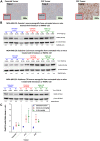
MDA-MB-231 tumors as well as those grown from C13 and C39 cells were established in NCr nu/nu mice as described in Materials and Methods. (A) Tumors were removed from mice and formalin-fixed prior to IHC staining with a goat anti-human Trop-2 polyclonal antibody as described in Materials and Methods. Negative control staining was with normal goat antibody. (B) Mice bearing parental MDA-MB-231, C13, or C39 tumors were injected i.v. with either irinotecan or SG (doses shown in SN-38 equivalents). Mice that received no therapy served as untreated control. After 24 h, mice were euthanized and tumors removed and flash-frozen. Frozen tumors were analyzed for Rad51 via Western blot as described in Materials and Methods. Relative ratio of Rad51 to β-actin loading control was based on untreated Animal 1 for each tumor type. The ratio for this mouse was set at 1.0 with all other ratios relative to this animal for each tumor type. (C) Mean Rad51:β-actin ratios for all the mice within a treatment group for each tumor-type (* P ≤ 0.05; one-tailed t-Test).
Figure 4
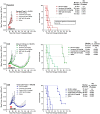
Figure 4. Increased Trop-2 expression in MDA-MB-231 tumors overcomes resistance to SG but not irinotecan.
NCr athymic nu/nu mice were injected s.c. with either MDA-MB-231 parental cells (parental), MDA-MB-231 clone 13 (C13) cells, or MDA-MB-231 clone 39 (C39) cells as described in Materials and Methods. Once tumors reached ~0.3 cm3 in size, mice were randomized into the various treatment groups. SG, control ADC, and parental hRS7 IgG antibody, were administered i.p., twice weekly for 4 weeks (black arrows). Irinotecan was administered i.v. at its MTD (q2dx5; red arrows). For all animal studies, the doses of SN-38 immunoconjugates and irinotecan are shown in SN-38 equivalents. The dose of hRS7 is shown at its protein dose equivalent to SG protein dose. Graphs to the left show mean tumor growth curves for each treatment group while those on the left indicate survival curves for these same groups of animals. **One mouse in SG group deemed an outlier via Grubbs’ test and removed from final analysis. Grey dotted line in survival curves indicates 50% survival line.

References

    1. Flashner-Abramson E, Vasudevan S, Adejumobi IA, Sonnenblick A, Kravchenko-Balasha N. Decoding cancer heterogeneity: studying patient-specific signaling signatures towards personalized cancer therapy. Theranostics. 2019; 9:5149–5165. 10.7150/thno.31657. - DOI - PMC - PubMed
    1. Li Y, Kang K, Krahn JM, Croutwater N, Lee K, Umbach DM, Li L. A comprehensive genomic pan-cancer classification using The Cancer Genome Atlas gene expression data. BMC Genomics. 2017; 18:508. 10.1186/s12864-017-3906-0. - DOI - PMC - PubMed
    1. Ramaker RC, Lasseigne BN, Hardigan AA, Palacio L, Gunther DS, Myers RM, Cooper SJ. RNA sequencing-based cell proliferation analysis across 19 cancers identifies a subset of proliferation-informative cancers with a common survival signature. Oncotarget. 2017; 8:38668–38681. 10.18632/oncotarget.16961. - DOI - PMC - PubMed
    1. Schink JC, Trosman JR, Weldon CB, Siziopikou KP, Tsongalis GJ, Rademaker AW, Patel JD, Benson AB 3rd, Perez EA, Gradishar WJ. Biomarker testing for breast, lung, and gastroesophageal cancers at NCI designated cancer centers. J Natl Cancer Inst. 2014; 106:dju256. 10.1093/jnci/dju256. - DOI - PMC - PubMed
    1. Lehmann BD, Pietenpol JA. Identification and use of biomarkers in treatment strategies for triple-negative breast cancer subtypes. J Pathol. 2014; 232:142–150. 10.1002/path.4280. - DOI - PMC - PubMed