Review
. 2022 May;42(4):917-930.
doi: 10.1007/s10571-020-01008-9. Epub 2020 Nov 16.

Mitochondria-ER Tethering in Neurodegenerative Diseases

Affiliations
Review

Mitochondria-ER Tethering in Neurodegenerative Diseases

Reza Raeisossadati et al. Cell Mol Neurobiol. 2022 May.

Abstract

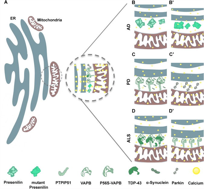
Organelles juxtaposition has been detected for decades, although only recently gained importance due to a pivotal role in the regulation of cellular processes dependent on membrane contact sites. Endoplasmic reticulum (ER) and mitochondria interaction is a prime example of organelles contact sites. Mitochondria-associated membranes (MAM) are proposed to harbor ER-mitochondria tether complexes, mainly when these organelles are less than 30 nm apart. Dysfunctions of proteins located at the MAM are associated with neurodegenerative diseases such as Parkinson's, Alzheimer's and amyotrophic lateral sclerosis, as well as neurodevelopmental disorders; hence any malfunction in MAM can potentially trigger cell death. This review will focus on the role of ER-mitochondria contact sites, regarding calcium homeostasis, lipid metabolism, autophagy, morphology and dynamics of mitochondria, mainly in the context of neurodegenerative diseases. Approaches that have been employed so far to study organelles contact sites, as well as methods that were not used in neurosciences yet, but are promising and accurate ways to unveil the functions of MAM during neurodegeneration, is also discussed in the present review.

Keywords: Autophagy; Calcium; Contact sites methodologies; Lipid metabolism; Mitochondria-associated membranes (MAM); Neurodegeneration.


Conflict of interest statement

The authors declare that they have no competing interests.

Figures

Fig. 1
Fig. 1
a ER-Mitochondria contact sites illustration and mitochondria-associated membranes (MAM) influence on Ca2+ homeostasis in neurodegenerative disease. b–b’ In Alzheimer’s disease (AD) mutation in Presenilin (PSEN1) located at the MAMs tightens contact sites and increases Ca2+ flux into the mitochondria. c–c’ In Parkinson’s disease, α-syn interacts with VAPB-PTPIP51, disturbing this tethering complex and decreasing MAM and Ca2+ shuttling into the mitochondria. Parkin overexpression increases the contact sites and elevates the Ca2+ flux into the mitochondria. d–d’ In ALS, TDP-43 reduces the binding of VAPB-PTPIP51; therefore, reducing Ca2+ shuttling into mitochondria. Mutation in VAPB (P56S) leads to a higher affinity to other tethering counterpart of PTPIP5 that triggers Ca2+ transfer to mitochondria
Fig. 2
Fig. 2
a Representation of endoplasmic reticulum (ER)-Mitochondria contact sites. Magnified area (dashed circle) illustrates tethering complexes and the methods using fluorescent approaches employed to study interaction between mitochondria and ER are exemplified in B to F. b Proximity ligation assay (PLA) is a broadly used method to detect the endogenous protein–protein interactions in fixed samples. It is based on the labeling of the target proteins with primary antibodies, followed by secondary antibodies conjugated to specific oligonucleotides; in close proximity targeted proteins can hybridize with connector oligonucleotides that serve as templates for rolling circular amplification by using fluorophore-labeled nucleotides (Soderberg et al. 2006). This technique has potential to detect MAM via tethering proteins in the presence of specific antibodies. c Fluorescence resonance energy transfer (FRET) is based on the transfer of energy between two fluorophores (donor/acceptor) fused to membrane proteins that has been used to visualize organelles' contact site, FRET provides high-resolution detection (approximately 10 nm) and is compatible to live cells analysis (Csordas et al. ; Scorrano et al. 2019). d Bioluminescence resonance energy transfer (BRET) is a new methodology that has been recently developed to quantify the contact site between mitochondria and ER using the so-called MERLIN (Mitochondria–ER Length Indicator Nanosensor). The Renilla Luciferase 8 (RLuc) acts as a donor linked to a truncated non-functional variant of calnexin (sCal) and mVenus as an acceptor fused to alpha-helical C-terminal domain of Bcl-xL (Hertlein et al. 2020). BRET provides higher resolution than fluorescence microscopy, it is applicable to live cell imaging and has limited phototoxicity as compared with FRET. e Biomolecular fluorescence complementation (BiFC) is based on split-fluorescent proteins such as split Venus or GFP composes of two complementary non-fluorescent residues fused to N or C terminal fragment of membranes bounded proteins, while targeted membranes locate in proximity the split fragments bind to each other and allowing the fluorescence to bright (Cieri et al. ; Kakimoto et al. ; Miller et al. 2015). The complementation is stable in this approach; therefore, it is not suitable to study the dynamic of the contact sites (Scorrano et al. 2019). The split probes irreversibly bind, which may alter the function of contact sites; on the other hand, it is applicable to live imaging. f Dimerization-dependent fluorescent protein (ddFP) works similar to BiFC, this approach is based on a reversible binding of weak or non-fluorescent protein monomers (Alford et al. ; Scorrano et al. 2019). Due to reversibility, the dynamic of contact sites can be monitored when using this approach

References

    1. AlSaif A, AlMohanna F, Bohlega S (2011) A mutation in sigma-1 receptor causes juvenile amyotrophic lateral sclerosis. Ann Neurol 70:913–919. 10.1002/ana.22534 - PubMed
    1. Alford SC, Ding Y, Simmen T, Campbell RE (2012) Dimerization-dependent green and yellow fluorescent proteins. ACS Synth Biol 1:569–575. 10.1021/sb300050j - PMC - PubMed
    1. Alza NP, Iglesias Gonzalez PA, Conde MA, Uranga RM, Salvador GA (2019) Lipids at the crossroad of alpha-synuclein function and dysfunction: biological and pathological implications. Front Cell Neurosci 13:175. 10.3389/fncel.2019.00175 - PMC - PubMed
    1. Arakhamia T et al (2020) Posttranslational modifications mediate the structural diversity of tauopathy strains. Cell 180:633–644. 10.1016/j.cell.2020.01.027 - PMC - PubMed
    1. Area-Gomez E et al (2018) A key role for MAM in mediating mitochondrial dysfunction in alzheimer disease. Cell Death Dis 9:335. 10.1038/s41419-017-0215-0 - PMC - PubMed

LinkOut - more resources