. 2021 Jan 1;53(1):1-11.
doi: 10.1152/physiolgenomics.00104.2020. Epub 2020 Nov 16.

A multimodal and integrated approach to interrogate human kidney biopsies with rigor and reproducibility: guidelines from the Kidney Precision Medicine Project

Affiliations

A multimodal and integrated approach to interrogate human kidney biopsies with rigor and reproducibility: guidelines from the Kidney Precision Medicine Project

Tarek M El-Achkar et al. Physiol Genomics. .

Abstract

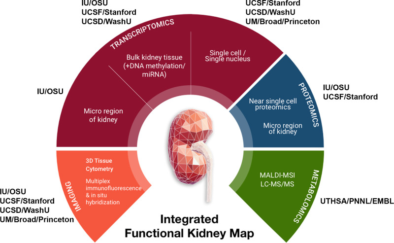
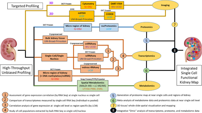
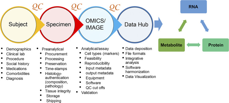
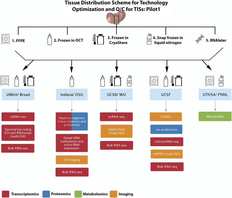
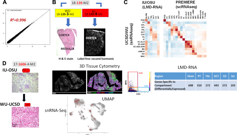
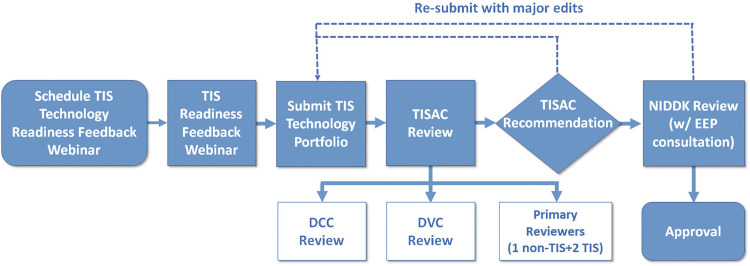
Comprehensive and spatially mapped molecular atlases of organs at a cellular level are a critical resource to gain insights into pathogenic mechanisms and personalized therapies for diseases. The Kidney Precision Medicine Project (KPMP) is an endeavor to generate three-dimensional (3-D) molecular atlases of healthy and diseased kidney biopsies by using multiple state-of-the-art omics and imaging technologies across several institutions. Obtaining rigorous and reproducible results from disparate methods and at different sites to interrogate biomolecules at a single-cell level or in 3-D space is a significant challenge that can be a futile exercise if not well controlled. We describe a "follow the tissue" pipeline for generating a reliable and authentic single-cell/region 3-D molecular atlas of human adult kidney. Our approach emphasizes quality assurance, quality control, validation, and harmonization across different omics and imaging technologies from sample procurement, processing, storage, shipping to data generation, analysis, and sharing. We established benchmarks for quality control, rigor, reproducibility, and feasibility across multiple technologies through a pilot experiment using common source tissue that was processed and analyzed at different institutions and different technologies. A peer review system was established to critically review quality control measures and the reproducibility of data generated by each technology before their being approved to interrogate clinical biopsy specimens. The process established economizes the use of valuable biopsy tissue for multiomics and imaging analysis with stringent quality control to ensure rigor and reproducibility of results and serves as a model for precision medicine projects across laboratories, institutions and consortia.

Keywords: imaging; kidney disease; metabolomics; proteomics; transcriptomics.


Conflict of interest statement

No conflicts of interest, financial or otherwise, are declared by the authors.

Figures

Figure. 1.
Figure. 1.
Overview of tissue interrogation sites and technologies in the Kidney Precision Medicine Project (KPMP). IU, Indiana University; OSU, Ohio State University; UCSD, University of California, San Diego; WU, Washington University in St. Louis; UM, University of Michigan; UCSF, University of California, San Francisco; UTHSA, University of Texas Health Sciences, San Antonio; PNNL, Pacific Northwest National Laboratory; EMBL, European Molecular Biology Laboratory.
Figure 2.
Figure 2.
Blueprint for interconnectivity for multimodal molecular assessment of kidney tissue at the initiation of the Kidney Precision Medicine Project (KPMP). IU, Indiana University; OSU, Ohio State University; UCSD, University of California, San Diego; WU, Washington University in St. Louis; UCSF, University of California, San Francisco.
Figure 3.
Figure 3.
Overview of “follow the tissue” pipeline with essential quality assurance/quality control (QA/QC) parameters.
Figure 4.
Figure 4.
Pilot 1 tissue processing and distribution. A single-source kidney tissue from a tumor nephrectomy was sectioned into equal blocks (schema in Supplemental Fig. S1) and processed into 5 tissue processing methods according to the downstream applications listed. Blocks of the tissue were distributed to the sites according to the technology, as indicated by the drawings. Formalin-fixed paraffin embedded (FFPE); in situ hybridization (ISH); single-cell (sc) and single nuclear (sn)RNA sequencing (RNA-seq). Near-single-cell (nsc)-proteomics; matrix-assisted laser desorption-ionization mass spectrometry imaging (MALDI-MSI). OSU, Ohio State University; UCSD, University of California, San Diego; UCSF, University of California, San Francisco; UTHSA, University of Texas Health Sciences, San Antonio; PNNL, Pacific Northwest National Laboratory
Figure 5.
Figure 5.
Testing the “follow the tissue” pipeline with data generated from pilot samples and demonstrating feasibility of multiple technologies on limited tissue sample across sites. A: high correlation of bulk RNA-seq from adjacent samples shipped and processed at different sites. B: adjacent samples from same patient shipped to different tissue investigative sites (TISs) show different types of information from different technologies in Kidney Precision Medicine Project (KPMP) with histological tissue composition verification for snRNAseq at WU-UCSD and label-free second harmonic generation highlighting extent of fibrosis at IU-OSU. C: correlation between cell types and regions among snRNA-seq, laser microdissection (LMD)transcriptomics of the indicated regions by IU/OSU, and between snRNA-seq and scRNA-seq. D: economizing tissue usage by rotating fresh optimal cutting temperature (OCT) frozen kidney tissue between IU and WU/UCSD. Here. frozen sections were processed and analyzed for 3-D tissue cytometry and LMD-RNA analysis, and then the block was shipped to WU/UCSD for histological registration and subsequently generating snRNA-seq. Genes specific to each LMD compartment were determined by their upregulation in that compartment compared with other compartments as well as differential expression at a nominal P < 0.001. The UMAP plot shows the sample (∼900 nuclei) contributing to many of the cortical cell types in the kidney snRNAseq atlas (∼18,000 nuclei) (11). IU, Indiana University; OSU, Ohio State University; UCSD, University of California, San Diego; WU, Washington University in St. Louis; UM, University of Michigan; UCSF.
Figure 6.
Figure 6.
Tissue interrogation site approval committee (TISAC) workflow. This workflow includes completion of a recorded webinar and submission of a portfolio that details the quality control metrics and technology outcomes. The TISAC reviews this material, providing recommendations to the tissue interrogation site for improvement before a site is approved to receive biopsies. The TISAC ultimately forwards their recommendations to the The National Institute of Diabetes and Digestive and Kidney Diseases (NIDDK)and external expert panel (EEP) for final approval. DVC, data visualization center; DCC, data coordination center.

References

    1. All of Us Research Program I, Denny JC, Rutter JL, Goldstein DB, Philippakis A, Smoller JW, Jenkins G, The DE. “All of Us”” Research Program. N Engl J Med 381: 668–676, 2019. - PMC - PubMed
    1. Brunskill EW, Potter SS. Gene expression programs of mouse endothelial cells in kidney development and disease. PLoS One 5: e12034, 2010. doi:10.1371/journal.pone.0012034. - DOI - PMC - PubMed
    1. Chen L, Clark JZ, Nelson JW, Kaissling B, Ellison DH, Knepper MA. Renal-tubule epithelial cell nomenclature for single-cell RNA-sequencing studies. JASN 30: 1358–1364, 2019. doi:10.1681/ASN.2019040415. - DOI - PMC - PubMed
    1. Chen L, Lee JW, Chou C-L, Nair AV, Battistone MA, Păunescu TG, Merkulova M, Breton S, Verlander JW, Wall SM, Brown D, Burg MB, Knepper MA. Transcriptomes of major renal collecting duct cell types in mouse identified by single-cell RNA-seq. Proc Natl Acad Sci USA 114: E9989–E9998, 2017. doi:10.1073/pnas.1710964114. - DOI - PMC - PubMed
    1. Fetting JL, Guay JA, Karolak MJ, Iozzo RV, Adams DC, Maridas DE, Brown AC, Oxburgh L. FOXD1 promotes nephron progenitor differentiation by repressing decorin in the embryonic kidney. Development 141: 17–27, 2014. doi:10.1242/dev.089078. - DOI - PMC - PubMed

Publication types

LinkOut - more resources