

Imaging algorithm for COVID-19: A practical approach
- PMID: 33197713
- PMCID: PMC7655027
- DOI: 10.1016/j.clinimag.2020.11.022
Imaging algorithm for COVID-19: A practical approach
Abstract
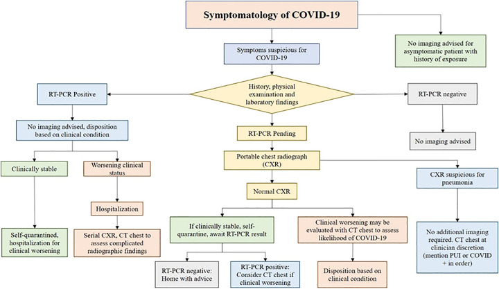
The global pandemic of COVID-19 pneumonia caused by the novel coronavirus (SARS-CoV-2) has strained healthcare resources across the world with emerging challenges of mass testing, resource allocation and management. While reverse transcriptase-polymerase chain reaction (RT-PCR) test is the most commonly utilized test and considered the current gold standard for diagnosis, the role of chest imaging has been highlighted by several studies demonstrating high sensitivity of computed tomography (CT). Many have suggested using CT chest as a first-line screening tool for the diagnosis of COVID-19. However, with advancement of laboratory testing and challenges in obtaining a CT scan without significant risk to healthcare providers, the role of imaging in diagnosis has been questioned. Several imaging societies have released consensus statements and guidelines on utilizing imaging resources and optimal reporting. In this review, we highlight the current evidence on various modalities in thoracic imaging for the diagnosis of COVID-19 and describe an algorithm on how to use these resources in an optimal fashion in accordance with the guidelines and statements released by major imaging societies.
Keywords: COVID-19; Chest radiograph; Computed tomography; SARS-CoV-2; Thoracic imaging in COVID-19.
Copyright © 2020 Elsevier Inc. All rights reserved.
Figures
Similar articles
-
Thoracic imaging tests for the diagnosis of COVID-19.Cochrane Database Syst Rev. 2020 Sep 30;9:CD013639. doi: 10.1002/14651858.CD013639.pub2. Cochrane Database Syst Rev. 2020. Update in: Cochrane Database Syst Rev. 2020 Nov 26;11:CD013639. doi: 10.1002/14651858.CD013639.pub3. PMID: 32997361 Updated.
-
Coronavirus Disease 2019 (COVID-19) diagnostic technologies: A country-based retrospective analysis of screening and containment procedures during the first wave of the pandemic.Clin Imaging. 2020 Nov;67:219-225. doi: 10.1016/j.clinimag.2020.08.014. Epub 2020 Aug 26. Clin Imaging. 2020. PMID: 32871426 Free PMC article. Review.
-
Thoracic imaging tests for the diagnosis of COVID-19.Cochrane Database Syst Rev. 2020 Nov 26;11:CD013639. doi: 10.1002/14651858.CD013639.pub3. Cochrane Database Syst Rev. 2020. Update in: Cochrane Database Syst Rev. 2021 Mar 16;3:CD013639. doi: 10.1002/14651858.CD013639.pub4. PMID: 33242342 Updated.
-
Evaluation of Diagnostic Performance of Chest CT with RT-PCR in Suspected Coronavirus Patients in Bangladesh.Mymensingh Med J. 2020 Oct;29(4):964-968. Mymensingh Med J. 2020. PMID: 33116103
-
Correlation between Chest Computed Tomography and Lung Ultrasonography in Patients with Coronavirus Disease 2019 (COVID-19).Ultrasound Med Biol. 2020 Nov;46(11):2918-2926. doi: 10.1016/j.ultrasmedbio.2020.07.003. Epub 2020 Jul 13. Ultrasound Med Biol. 2020. PMID: 32771222 Free PMC article.
Cited by
-
Fungal infection mimicking COVID-19 infection - A case report.Open Med (Wars). 2022 Apr 28;17(1):841-846. doi: 10.1515/med-2022-0443. eCollection 2022. Open Med (Wars). 2022. PMID: 35582198 Free PMC article.
-
Inter-Observer Agreement between Low-Dose and Standard-Dose CT with Soft and Sharp Convolution Kernels in COVID-19 Pneumonia.J Clin Med. 2022 Jan 27;11(3):669. doi: 10.3390/jcm11030669. J Clin Med. 2022. PMID: 35160121 Free PMC article.
-
Understanding COVID-19 Situation in Nepal and Implications for SARS-CoV-2 Transmission and Management.Environ Health Insights. 2022 Jun 7;16:11786302221104348. doi: 10.1177/11786302221104348. eCollection 2022. Environ Health Insights. 2022. PMID: 35694428 Free PMC article.
-
COVID-19 imaging: Diagnostic approaches, challenges, and evolving advances.World J Radiol. 2021 Jun 28;13(6):171-191. doi: 10.4329/wjr.v13.i6.171. World J Radiol. 2021. PMID: 34249238 Free PMC article. Review.
-
M-BLUE protocol for coronavirus disease-19 (COVID-19) patients: interobserver variability and correlation with disease severity.Clin Radiol. 2021 May;76(5):379-383. doi: 10.1016/j.crad.2021.02.003. Epub 2021 Feb 17. Clin Radiol. 2021. PMID: 33663912 Free PMC article.
References
-
- WHO . World Health Organization; 2020. World Health Organization coronavirus disease (COVID-19) situation Report-158.
-
- Beigel J.H., Tomashek K.M., Dodd L.E. Remdesivir for the treatment of Covid-19 — preliminary report. New England Journal of Medicine. 2020;383:1813–1826. - PubMed
Publication types
MeSH terms
LinkOut - more resources
Full Text Sources
Other Literature Sources
Medical
Miscellaneous
