. 2021 Jan:38:101773.
doi: 10.1016/j.redox.2020.101773. Epub 2020 Nov 4.

Engineering of a functional γ-tocopherol transfer protein

Affiliations

Engineering of a functional γ-tocopherol transfer protein

Walter Aeschimann et al. Redox Biol. 2021 Jan.

Abstract

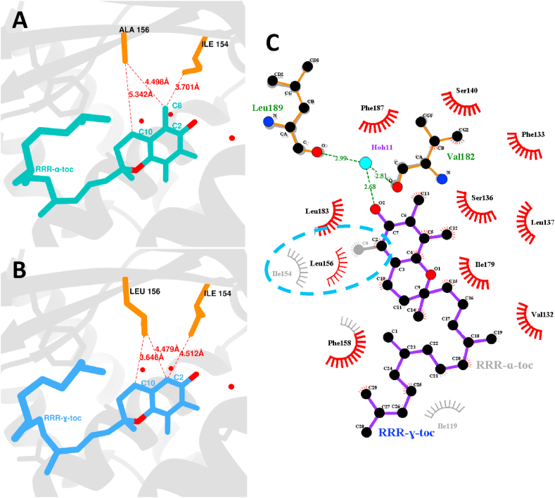
α-tocopherol transfer protein (TTP) was previously reported to self-aggregate into 24-meric spheres (α-TTPS) and to possess transcytotic potency across mono-layers of human umbilical vein endothelial cells (HUVECs). In this work, we describe the characterisation of a functional TTP variant with its vitamer selectivity shifted towards γ-tocopherol. The shift was obtained by introducing an alanine to leucine substitution into the substrate-binding pocket at position 156 through site directed mutagenesis. We report here the X-ray crystal structure of the γ-tocopherol specific particle (γ-TTPS) at 2.24 Å resolution. γ-TTPS features full functionality compared to its α-tocopherol specific parent including self-aggregation potency and transcytotic activity in trans-well experiments using primary HUVEC cells. The impact of the A156L mutation on TTP function is quantified in vitro by measuring the affinity towards γ-tocopherol through micro-differential scanning calorimetry and by determining its ligand-transfer activity. Finally, cell culture experiments using adherently grown HUVEC cells indicate that the protomers of γ-TTP, in contrast to α-TTP, do not counteract cytokine-mediated inflammation at a transcriptional level. Our results suggest that the A156L substitution in TTP is fully functional and has the potential to pave the way for further experiments towards the understanding of α-tocopherol homeostasis in humans.

Keywords: Antioxidant; Cytokine; Nanoparticle; Transcytosis; Vitamin E.


Figures

Fig. 1
Fig. 1
General view of γ-TTP (α-TTP A156L) in complex with γ-tocopherol. (A) View of the tetracosameric nano-sphere of γ-TTPS. (B) Zoom of the binding pocket with all its residues visualized; in orange are the residues highlighted that form the niche around C8 of α-tocopherol in the wild type α-TTP. The surface of the binding pocket is colored depending its hydrophobicity. (For interpretation of the references to color in this figure legend, the reader is referred to the Web version of this article.)
Fig. 2
Fig. 2
(A&B) Real space comparison of the binding pockets of wild type α-TTP complexed to RRR-α-tocopherol and of γ-TTP complexed to RRR-γ-tocopherol, respectively. (A) In the binding pocket of α-TTP in complex with α-tocopherol I154 is retracted, in order to create space for the niche surrounding the methyl group attached to C2 of α-tocopherol. (B) In contrast, in the binding pocket of γ-TTP bound to RRR-γ-tocopherol the void caused by the missing C8 is filled by L156 maintaining a similar distance (4.479 Å) to C2 as has A156 in the atomic model of wild type α-TTP (4.498 Å). (C) Schematic analysis (ligplot) of the binding pocket between α-TTP and γ-TTP; besides the newly introduced L156 very little discrepancies exist between both the atomic models.
Fig. 3
Fig. 3
Scheme of the thermodynamic cycle that emphasizes the relation between ligand binding and protein unfolding. (A) Classical thermodynamic cycle that describes a model in which the unfolding of the protein in complex does not influence the binding. Hence, in this model the ligand stays bound to the protein, despite of its structural integrity. (B) In this scheme unfolding of the protein causes the ligand to disassociate from the binding protein and the binding to the unfolded state is virtually inexistent (ΔGbU=0). (C) Schematic illustration of coupling between the unfolding equilibrium KU of the protein and the binding equilibrium Kb to the ligand. Increments in thermal stability reflect the addition of binding free energy ΔGbN to the free energy of the protein fold ΔGU; the resulting free energy is the unfolding free energy ΔGUT of the protein-ligand complex.
Fig. 4
Fig. 4
(A) Gibbs-Helmholtz plots of ΔGU and ΔGUT as functions of temperature. It can be observed that the general stability of α-TTP (apoprotein and holoprotein) is quite similar to γ-TTP. However, ΔGUT of γ-TTP features a strikingly sharper decline than that of the wild type. The difference between both curves at any temperature corresponds to ΔGb(T). The fact that ΔGb of γ-TTP decreases much faster with increasing temperature reflects the lower overall affinity of γ-TTP for γ-tocopherol than α-TTP for α-tocopherol. (B) Arrhenius plots of the dissociation equilibrium (Kd) for α-TTP + α-tocopherol and γ-TTP + γ-tocopherol, respectively. Values for the Kd at 24.85 C (298 K) and at 36.85 C (310 K) are highlighted within the plots.
Fig. 5
Fig. 5
(A) Thermodynamic signature of the binding of α-tocopherol to α-TTP and of γ-tocopherol to γ-TTP, respectively. In both case a strong favorable ethalpic contribution (ΔHb) exceeds the unfavourable entropic contribution (TΔSb), resulting in both cases in a negative binding free energy ΔGb; and thus in the binding of the ligand with a dissociation constant (Kd) at 36.85 C (310 K) of 14.25 nM for α-TTP/α-tocopherol and of 53.79 nM for γ-TTP/γ-tocopherol, respectively. (B) Illustration of the hydrogen bond network within the binding of α-TTP. The four trapped water molecules within the binding pocket together with residues Y100, Y117, S136, S140 V182, L189 and the ligand itself make up the large hydrogen bond network responsible for the strong ethalpic contribution (ΔHb) to the total binding (ΔGb).
Fig. 6
Fig. 6
The transcytotic efficacy of tetracosameric α-TTPs and γ-TTPs (α-TTP A156L) compared to its monomer form were monitored in a transwell model system comprising confluent and maturely developed monolayers of HUVECs. Human transferrin served as the positive control. Simultaneous application of rhodamine isothiocyanate - (RITC) labeled dextran confirmed the integrity of the HUVEC cell monolayers and served to determine the paracellular flux. Measurements report a 10-fold–18-fold increase in the flux through the endothelial cell layer of both α- and γ-TTPs compared to that of RITC-dextran. Our data also show that α- and γ-TTPs cross the endothelium at a flux rate 2–6 times faster than human transferrin with increasing speed the more complex the protein assemblies.
Fig. 7
Fig. 7
Primary human umbilical vein endothelial cells (HUVECs) were allowed to form a tight monolayer within 7 days of pre-culturing. At day 7 tocopherol transfer proteins (TTPs) carrying either α-tocopherol (α-TTP) or γ-tocopherol (α-TTP A156L, referred as γ-TTP) were added for 4 h followed by cytokine (TNF: 50 ng/ml, IL1F2: 1 ng/ml, IFNG: 50 ng/ml) stimulation for another 4 h mRNA levels of IL6 (A), CCL2 (B), TNF(C), COX2 (D) and ICAM1 (E) were significantly increased by cytokine stimulation (controls, left boxes) while α-TTP significantly reduced cytokine-induced mRNA expression of both IL6, CCL2, TNF and ICAM1 (central boxes). γ-TTP was not able to attenuate cytokine-mediated transcription of any pro-inflammatory cytokine or protein measured (right boxes). Boxplots display outliers, if applicable. N = 1 in triplicate. Statistical evaluation with R with assumed significance at p < 0.05. Relevant significant differences are designated with asterisks as follows: *p < 0.05, **p < 0.01, ***p < 0.001.

References

    1. Ingold K., Webb A., Witter D., Burton G., Metcalfe T., Muller D. Vitamin e remains the major lipid-soluble, chain-breaking antioxidant in human plasma even in individuals suffering severe vitamin e deficiency. Arch. Biochem. Biophys. 1987;259(1):224–225. - PubMed
    1. Howard A.C., McNeil A.K., McNeil P.L. Promotion of plasma membrane repair by vitamin e. Nat. Commun. 2011;2:597. - PMC - PubMed
    1. Traber M.G. Mechanisms for the prevention of vitamin e excess. J. Lipid Res. 2013;54(9):2295–2306. - PMC - PubMed
    1. Sattler S.E., Cahoon E.B., Coughlan S.J., DellaPenna D. Characterization of tocopherol cyclases from higher plants and cyanobacteria. evolutionary implications for tocopherol synthesis and function. Plant Physiol. 2003;132(4):2184–2195. - PMC - PubMed
    1. Grusak M.A., DellaPenna D. Improving the nutrient composition of plants to enhance human nutrition and health 1. Annu. Rev. Plant Biol. 1999;50(1):133–161. - PubMed

Publication types

LinkOut - more resources