. 2020 Nov 12;21(22):8510.
doi: 10.3390/ijms21228510.

Respiratory Burst Oxidase Homologs RBOHD and RBOHF as Key Modulating Components of Response in Turnip Mosaic Virus- Arabidopsis thaliana (L.) Heyhn System

Affiliations

Respiratory Burst Oxidase Homologs RBOHD and RBOHF as Key Modulating Components of Response in Turnip Mosaic Virus- Arabidopsis thaliana (L.) Heyhn System

Katarzyna Otulak-Kozieł et al. Int J Mol Sci. .

Abstract

Turnip mosaic virus (TuMV) is one of the most important plant viruses worldwide. It has a very wide host range infecting at least 318 species in over 43 families, such as Brassicaceae, Fabaceae, Asteraceae, or Chenopodiaceae from dicotyledons. Plant NADPH oxidases, the respiratory burst oxidase homologues (RBOHs), are a major source of reactive oxygen species (ROS) during plant-microbe interactions. The functions of RBOHs in different plant-pathogen interactions have been analyzed using knockout mutants, but little focus has been given to plant-virus responses. Therefore, in this work we tested the response after mechanical inoculation with TuMV in ArabidopsisrbohD and rbohF transposon knockout mutants and analyzed ultrastructural changes after TuMV inoculation. The development of the TuMV infection cycle was promoted in rbohD plants, suggesting that RbohD plays a role in the Arabidopsis resistance response to TuMV. rbohF and rbohD/F mutants display less TuMV accumulation and a lack of virus cytoplasmic inclusions were observed; these observations suggest that RbohF promotes viral replication and increases susceptibility to TuMV. rbohD/F displayed a reduction in H2O2 but enhanced resistance similarly to rbohF. This dominant effect of the rbohF mutation could indicate that RbohF acts as a susceptibility factor. Induction of hydrogen peroxide by TuMV was partially compromised in rbohD mutants whereas it was almost completely abolished in rbohD/F, indicating that these oxidases are responsible for most of the ROS produced in this interaction. The pattern of in situ H2O2 deposition after infection of the more resistant rbohF and rbohD/F genotypes suggests a putative role of these species on systemic signal transport. The ultrastructural localization and quantification of pathogenesis-related protein 1 (PR1) indicate that ROS produced by these oxidases also influence PR1 distribution in the TuMV-A.thaliana pathosystem. Our results revealed the highest activation of PR1 in rbohD and Col-0. Thus, our findings indicate a correlation between PR1 accumulation and susceptibility to TuMV. The specific localization of PR1 in the most resistant genotypes after TuMV inoculation may indicate a connection of PR1 induction with susceptibility, which may be characteristic for this pathosystem. Our results clearly indicate the importance of NADPH oxidases RbohD and RbohF in the regulation of the TuMV infection cycle in Arabidopsis. These findings may help provide a better understanding of the mechanisms modulating A.thaliana-TuMV interactions.

Keywords: hydrogen peroxide; pathogenesis-related protein 1; plant–virus interaction; potyvirus; resistance response; ultrastructure.


Conflict of interest statement

The authors declare no conflict of interest.

Figures

Figure 1
Figure 1
TuMV detection and relative virus concentration assessment in Arabidopsis thaliana Col-0 and rbohD, rbohF and rbohD/F mutants at 3 and 7 days after inoculation. Values represented are mean of corrected OD405nm values. Significant differences between classes at p < 0.05 level of significance were assessed by analysis of variance (ANOVA) with post-hoc Tukey HSD. The statistically significant values are marked by letters above chart bars.
Figure 2
Figure 2
Viral particles and inclusions, multivesicular bodies induction and chloroplast changes in Col-0 different leaf tissues 3 (A,B) and 7 days (C,D) after TuMV inoculation examined with a transmission electron microscope (TEM, FEI M268D “Morgagni” transmission electron microscope). (A) Virus particles (VPs; marked by arrowhead) in vacuole (v) of palisade mesophyll cell. (B) Multivesicular bodies (MVBs; marked by *) in cytoplasm and vacuole (v) of phloem parenchyma (PP) cell. Pd-plasmodesmata. (C) Virus particles (VPs; arrowhead) in vacuole (v) and virus inclusions (cytoplasmatic inclusion (CI); marked by arrow) in cytoplasm of xylem parenchyma (XP) cell. (D) Virus cytoplasmic inclusions (Cis; arrow) inside phloem cell and necrotic changes (Ne) in companion cell (CC). Se—sieve element; X—xylem tracheary element.
Figure 3
Figure 3
Virus particles and viral cytoplasmatic inclusion, changes in chloroplast lamellae stacks in rbohD plants leaf tissues 3 (A) and 7 days (BD) after TuMV inoculation examined under TEM. (A) Curved (double-headed arrow) chloroplast (ch) thylakoids, cell wall (CW) invagination (black arrow) and paramular bodies (PMBs; marked by diamond head arrow) in palisade mesophyll cell. (B) Virus particles (VPs, with arrowhead) and virus cytoplasmic inclusions (CIs; arrows) in palisade mesophyll cell. (C) Destroyed chloroplast (ch) with disorganized thylakoids (double-headed arrow), virus cytoplasmic inclusions (CIs; white arrow) and viral particles (VPs; black arrowhead) in cytoplasm of spongy mesophyll cell. v—vacuole; m—mitochondrion. (D) Virus inclusions (CI; arrow) inside xylem tracheary elements (X). m—mitochondrion.
Figure 4
Figure 4
Curved structure of chloroplast lamellae stacks—multivesicular structures and viral particles in vacuole in different leaf tissues of rbohF plants 3 (A,B) and 7 days (C,D) after TuMV inoculation examined under a TEM. (A) Curved thylakoids (double-headed arrow) in chloroplast (ch) of palisade mesophyll cell. (B) Multivesicular structures (“single membranes with tubules”) (MVB; *) in vacuole (v) and paramular bodies (PMBs; marked by diamond head arrow) in phloem apoplast area. CW—cell wall. (C) Virus particles (VPs; arrowhead) and multivesicular bodies (MVB; *) inside vacuole (v) of spongy mesophyll cell. ch—chloroplast. (D) Irregular structure (arrows) of the cell wall (CW) between spongy mesophyll cells. ch—chloroplast; v—vacuole.
Figure 5
Figure 5
Multivesicular bodies’ induction, viral particles in vacuole and cell wall rebuilding in rbohD/F plants tissues 3 (A) and 7 days (BD) after TuMV inoculation examined under a TEM. (A) Cell wall rebuilding (CW; arrows) and multivesicular bodies (MVB; *) in phloem parenchyma cells. Ch—chloroplast; V—vacuole. (B) Multivesicular bodies (MVBs; *) near plasmodesmata (Pd) in phloem parenchyma cells. CW—cell wall; V—vacuole. (C) Cell wall (CW) rebuilding (arrows) and virus particles (VPs; arrowhead) in membranous structures such as small vacuoles in palisade mesophyll cells. V—vacuole. (D) Accumulation of phenolic-like compounds (marked with #) inside xylem tracheary elements (X). XP—xylem parenchyma.
Figure 6
Figure 6
TuMV detection in Col-0 and rbohD mock-inoculated leaf tissues (A) and 3 (B,C) and 7 days (D) after virus inoculation by immunogold electron microscopy. (A) Unchanged palisade mesophyll cell without TuMV epitope from mock-inoculated Col-0 plants. ch—chloroplast; v—vacuole. (B) TuMV epitope presence (*) around cytoplasmic inclusion (CI; arrow) and multivesicular bodies (MVBs) in phloem parenchyma cell of Col-0. CW—cell wall; m—mitochondrion; Se—sieve tube. (C) TuMV epitope localization (*) along virus particles (VPs; arrowhead) in companion cell (CC) and sieve element (Se) in rbohD phloem tissue. Pd—plasmodesmata. (D) TuMV epitope presence (*) around virus particles (VPs; arrowhead) and cytoplasmic inclusion (CI; arrow) in vesicular structures in rbohD palisade mesophyll cells.
Figure 7
Figure 7
TuMV detection in rbohF and rbohD/F leaf tissues 3 (A,C) and 7 days (B,D) after virus inoculation examined by immunogold electron microscopy. (A) TuMV epitope localization (*) around multivesicular structures (MVB) in rbohF phloem cells. Phenolic-like compounds (#) near MVB. CW—cell wall; v—vacuole. (B) TuMV epitope presence (*) along virus particles (VPs; arrowheads) in vacuole (v) and around single membrane with tubule structures (SMTs) in rbohF. Phenolic-like compounds (#). m—mitochondrion. (C) TuMV epitope (*) in vacuole (V) and around changed (black arrows) cell wall (CW) with paramular bodies (PMBs; diamond head arrow) in rbohD/F phloem parenchyma cell. (D) TuMV epitope (*) in vacuole (v) rbohD/F xylem parenchyma cells (XP). Phenolic-like compounds (#) inside xylem tracheary elements (X).
Figure 8
Figure 8
Quantification of gold particles associated with TuMV in Arabidopsis thaliana Col-0, rbohD, rbohF and double rbohD/F mutants at 3 and 7 days after inoculation. Significant differences between classes at p < 0.05 level of significance by ANOVA with post-hoc Tukey HSD were assessed. The statistical significant values are marked by letters above chart bars.
Figure 9
Figure 9
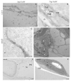
Localization of PR1 in Col-0 and rbohD leaf tissues 3 (A,C) and 7 days (B,D) after TuMV inoculation examined by immunogold electron microscopy. Control comprises Col-0 mock-inoculated leaf tissues (E) and leaf tissues without primary antibodies (F). (A) PR1 (*) in cell wall (CW), plasmodesmata (Pd) and vacuole (v) in Col-0 mesophyll 3 days after TuMV inoculation. (B) PR1 presence (*) in cell wall (CW), paramular bodies (PMBs; diamond head arrow) in apoplast area of palisade mesophyll Col-0 plants 7 days after TuMV inoculation. (C) PR1 localization (*) in cell wall (CW) and vacuoles (vs) in rbohD 3 days after TuMV inoculation. (D) PR1 deposition (*) along cell wall (CW) and in vacuoles (vs) in rbohD spongy mesophyll cell with virus cytoplasmic inclusions (CI with arrow). (E) PR1 (*) in cell wall (CW) and vacuole (v) in different type of phloem cells from mock-inoculated Col-0 plants. Se—sieve element. (F) Mesophyll from mock-inoculated Col-0 control plants without PR1 epitopes when primary antibodies were replaced by pre-immune serum. ch—chloroplast; CW—cell wall.
Figure 10
Figure 10
Localization of PR1 in rbohF and rbohD/F leaf tissues 3 (A,C) and 7 days (B,D) after TuMV inoculation examined by immunogold electron microscopy. (A) PR1 localization (*) around multivesicular bodies (MVBs) in vacuole (v) in rbohF palisade mesophyll cell. ch—chloroplast; CW—cell wall. (B) PR1 presence (*) in plasmodesmata (Pd), cell wall (CW) and paramular bodies (PMBs; white diamond head arrow) in rbohF phloem parenchyma cell. (C) PR1 (*) in and around changed (arrows) cell wall (CW) in rbohD/F spongy mesophyll cell. ga—Golgi apparatus, m—mitochondrion, v—vacuole. (D) PR1 presence (*) in paramular bodies (PMBs; diamond head arrow) and vacuole (V) in rbohD/F spongy mesophyll cell. ch—chloroplast; CW—cell wall; v—vacuole.
Figure 11
Figure 11
Quantification of PR1 antigen localization in mock- and TuMV-inoculated Arabidopsis thaliana Col-0, rbohD, rbohF and double rbohD/F mutant at 3 and 7 days after inoculation. Significant differences between classes at p < 0.05 level of significance by ANOVA with post-hoc Tukey HSD were assessed. The statistical significant values are marked by letters above chart bars.
Figure 12
Figure 12
H2O2 detection in Col-0 and rbohD mock-inoculated leaf tissues (A,D) and 3 (B,E) and 7 days (C,F) post-TuMV inoculation examined under TEM. (A) Electron-dense deposits of cerium perhydroxide (*) in the cell wall and along the trans Golgi network (tgn) and vesicles in the cytoplasm of Col-0 spongy mesophyll cell. CW—cell wall; m—mitochondrion. (B) H2O2 (*) around multivesicular bodies (MVBs) in Col-0 epidermis. Pd—plasmodesmata; CW—cell wall. (C) H2O2 (*) in cell wall (CW), intercellular space (Ints) and near paramural bodies (PMB; diamond arrowhead) in Col-0 mesophyll cells. (D) H2O2 (*) in the apoplast between cell wall and plasmalemma in rbohD mock-treated xylem cells. XP—xylem parenchyma. (E) H2O2 (*) in vacuoles (vs), vesicular structures and endoplasmic reticulum (er) in cytoplasm in rbohD phloem parenchyma cells. (F) H2O2 (*) in phloem parenchyma cells with cytoplasmic inclusions (Cis; white arrow) of TuMV in rbohD sieve element (Se).
Figure 13
Figure 13
H2O2 detection in rbohF and rbohD/F mock-inoculated leaf tissues (A,D) and 3 (B,E) and 7 days (C,F) post-TuMV inoculation examined under TEM. (A) Cerium perhydroxide precipitates (*) in cell wall and apoplast between cell wall and plasmalemma in rbohF mock-inoculated xylem tracheary elements (X). XP—xylem parenchyma. (B) H2O2 (*) along membranous structures in necrotized (Ne) palisade mesophyll cell with destructed chloroplasts in rbohF. (C) H2O2 (*) in dark necrotized (Ne) protoplast of phloem cells and in the cytoplasm of non-necrotized phloem parenchyma cells in rbohF. CW—cell wall. (D) H2O2 staining (*) along the cell wall (CW) and endoplasmic reticulum (er) in rbohD/F mock-inoculated spongy mesophyll cells. ga—Golgi apparatus. (E) H2O2 (*) along the cell wall (CW) and membranous structures of necrotized (Ne) rbohD/F spongy mesophyll cell. MVBs—multivesicular bodies; CW—cell wall. (F) H2O2 (*) in plasmodesmata (Pd) and in cytoplasm around in rbohD/F mesophyll cell.
Figure 14
Figure 14
Corrected total electron density (CTED) of cerium (IV) perhydroxide precipitate in mock- and TuMV-inoculated Arabidopsis thaliana Col-0, rbohD, rbohF and double mutant rbohD/F at 3 and 7 dpi. Significant differences between classes at p < 0.05 level of significance by ANOVA with post-hoc Tukey HSD were assessed. The statistical significant values are marked by letters above chart bars.

References

    1. Revers F., García J.A. Molecular biology of potyviruses. Adv. Virus Res. 2015;92:101–199. - PubMed
    1. Jenner C.E., Tomimura K., Ohshima K., Hughes S.L., Walsh J. Mutations in Turnip mosaic virus P3 and cylindrical inclusion proteins are separately required to overcome two Brassica napus resistance genes. Virology. 2002;300:50–59. doi: 10.1006/viro.2002.1519. - DOI - PubMed
    1. Liu X.P., Lu W.C., Liu Y.K., Li J.L. A study of TuMV strain differentiation of cruciferous vegetables from ten provinces of China. Chin. Sci. Bull. 1990;35:1734–1739.
    1. Yoon J.Y., Green S.K., Opena R.T. Inheritance of resistance to Turnip mosaic virus in Chinese cabbage. Euphytica. 1993;69:103–108. doi: 10.1007/BF00021732. - DOI
    1. Kehoe M.A., Coutts B.A., Jones R.A. Resistance phenotypes in diverse accessions, breeding lines, and cultivars of three mustard species inoculated with Turnip mosaic virus. Plant Dis. 2010;94:1–9. doi: 10.1094/PDIS-12-09-0841. - DOI - PubMed

MeSH terms

Supplementary concepts

LinkOut - more resources