

Neuroimaging of Basal Ganglia in Neurometabolic Diseases in Children
- PMID: 33198265
- PMCID: PMC7697699
- DOI: 10.3390/brainsci10110849
Neuroimaging of Basal Ganglia in Neurometabolic Diseases in Children
Abstract
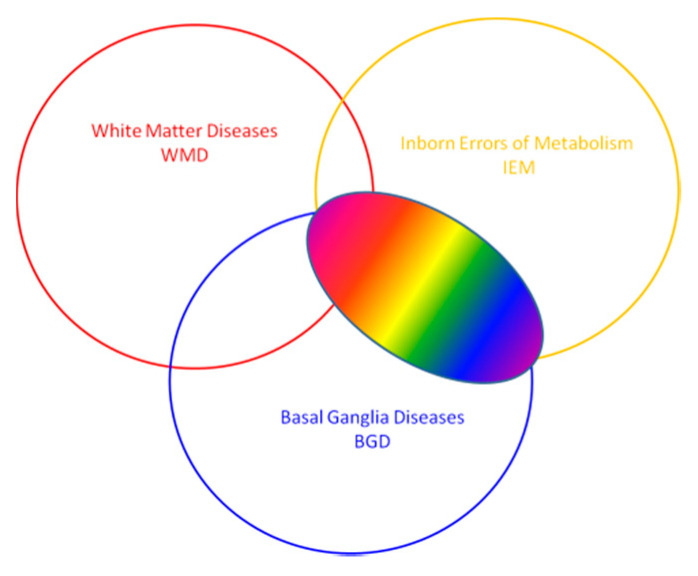
Diseases primarily affecting the basal ganglia in children result in characteristic disturbances of movement and muscle tone. Both experimental and clinical evidence indicates that the basal ganglia also play a role in higher mental states. The basal ganglia can be affected by neurometabolic, degenerative diseases or other conditions from which they must be differentiated. Neuroradiological findings in basal ganglia diseases are also known. However, they may be similar in different diseases. Their assessment in children may require repeated MRI examinations depending on the stage of brain development (mainly the level of myelination). A large spectrum of pathological changes in the basal ganglia in many diseases is caused by their vulnerability to metabolic abnormalities and chemical or ischemic trauma. The diagnosis is usually established by correlation of clinical and radiological findings. Neuroimaging of basal ganglia in neurometabolic diseases is helpful in early diagnosis and monitoring of changes for optimal therapy. This review focuses on neuroimaging of basal ganglia and its role in the differential diagnosis of inborn errors of metabolism.
Keywords: basal ganglia abnormalities; children; neurometabolic disease.
Conflict of interest statement
The authors declare no conflict of interest.
Figures
Similar articles
-
Biotin-thiamine-responsive basal ganglia disease: catastrophic consequences of delay in diagnosis and treatment.Neurol Res. 2017 Feb;39(2):117-125. doi: 10.1080/01616412.2016.1263176. Epub 2016 Dec 1. Neurol Res. 2017. PMID: 27905264
-
Magnetic resonance imaging of the brain in glutaric acidemia type I: a review of the literature and a report of four new cases with attention to the basal ganglia and imaging technique.Invest Radiol. 2003 Aug;38(8):489-96. doi: 10.1097/01.rli.0000080405.62988.f6. Invest Radiol. 2003. PMID: 12874515 Review.
-
Clinical course, early diagnosis, treatment, and prevention of disease in glutaryl-CoA dehydrogenase deficiency.Neuropediatrics. 1996 Jun;27(3):115-23. doi: 10.1055/s-2007-973761. Neuropediatrics. 1996. PMID: 8837070
-
Neurometabolic Disorders of the Newborn.Top Magn Reson Imaging. 2018 Aug;27(4):179-196. doi: 10.1097/RMR.0000000000000176. Top Magn Reson Imaging. 2018. PMID: 30086107 Review.
-
Neuroradiological and neurophysiological indices for neurometabolic disorders.Eur J Pediatr. 1994;153(7 Suppl 1):S90-3. doi: 10.1007/BF02138785. Eur J Pediatr. 1994. PMID: 7957395 Review.
Cited by
-
Inherited neurometabolic diseases and the importance of imaging-based classification systems.Radiol Bras. 2022 May-Jun;55(3):VII-VIII. doi: 10.1590/0100-3984.2022.55.3e2-en. Radiol Bras. 2022. PMID: 35795600 Free PMC article. No abstract available.
-
Decoding Post-Viral Fatigue: The Basal Ganglia's Complex Role in Long-COVID.Neurol Int. 2024 Mar 28;16(2):380-393. doi: 10.3390/neurolint16020028. Neurol Int. 2024. PMID: 38668125 Free PMC article. Review.
-
MR Neuroimaging in Pediatric Inborn Errors of Metabolism.Diagnostics (Basel). 2022 Mar 30;12(4):861. doi: 10.3390/diagnostics12040861. Diagnostics (Basel). 2022. PMID: 35453911 Free PMC article. Review.
-
Eye of the Tiger Sign in Pantothenate Kinase-Associated Neurodegeneration.Case Rep Radiol. 2021 May 7;2021:6633217. doi: 10.1155/2021/6633217. eCollection 2021. Case Rep Radiol. 2021. PMID: 34040814 Free PMC article.
-
Expanding the Epidemiological and Phenotypic Spectrum of MEGDEL Syndrome: The First Case Report From Egypt.Clin Med Insights Pediatr. 2025 Aug 14;19:11795565251348345. doi: 10.1177/11795565251348345. eCollection 2025. Clin Med Insights Pediatr. 2025. PMID: 40821445 Free PMC article.
References
Publication types
LinkOut - more resources
Full Text Sources
