. 2020 Nov 16;11(1):63.
doi: 10.1186/s13293-020-00337-0.

Sex-dependent alterations in the physiology of entorhinal cortex neurons in old heterozygous 3xTg-AD mice

Affiliations

Sex-dependent alterations in the physiology of entorhinal cortex neurons in old heterozygous 3xTg-AD mice

Dany Arsenault et al. Biol Sex Differ. .

Abstract

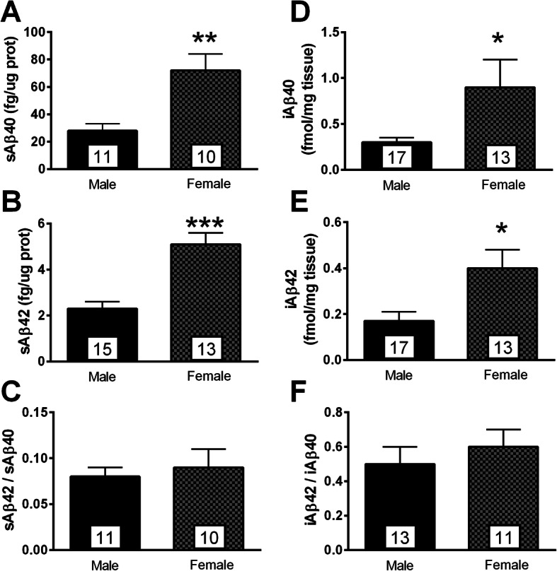
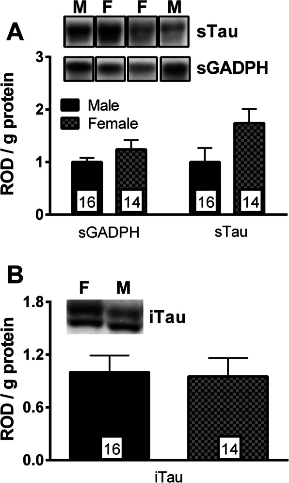
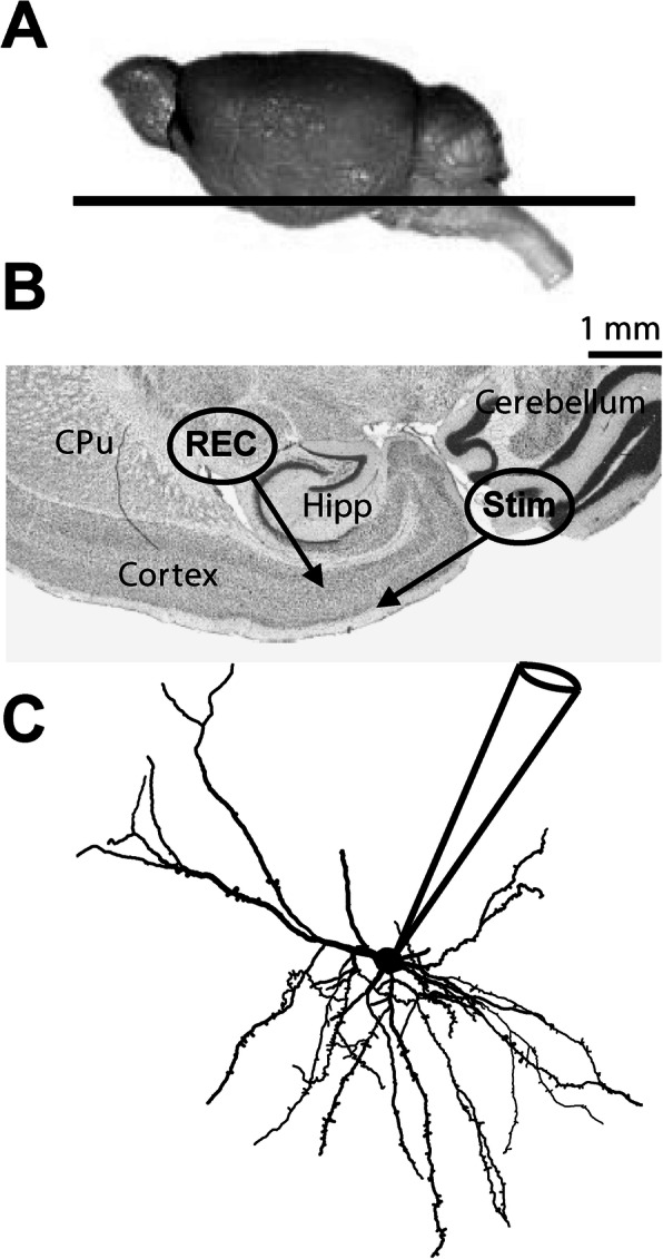
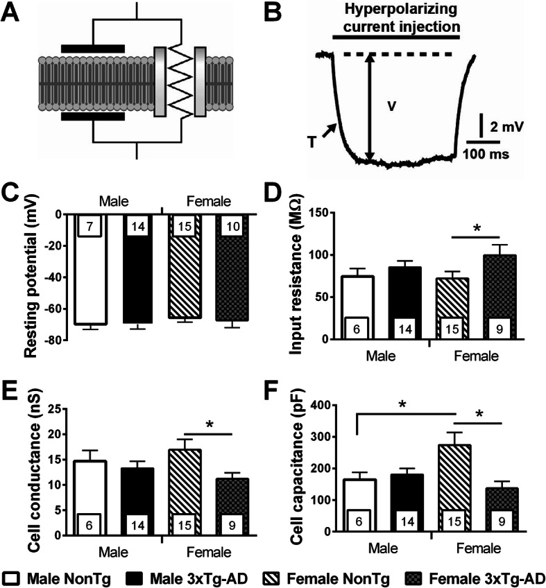
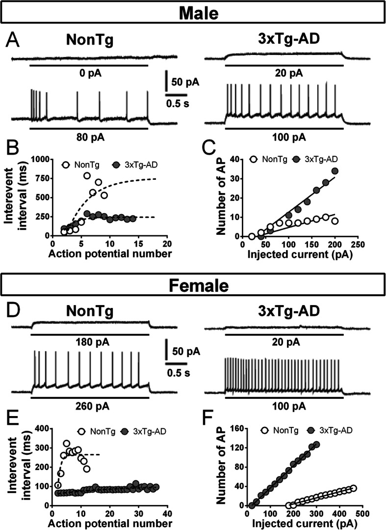
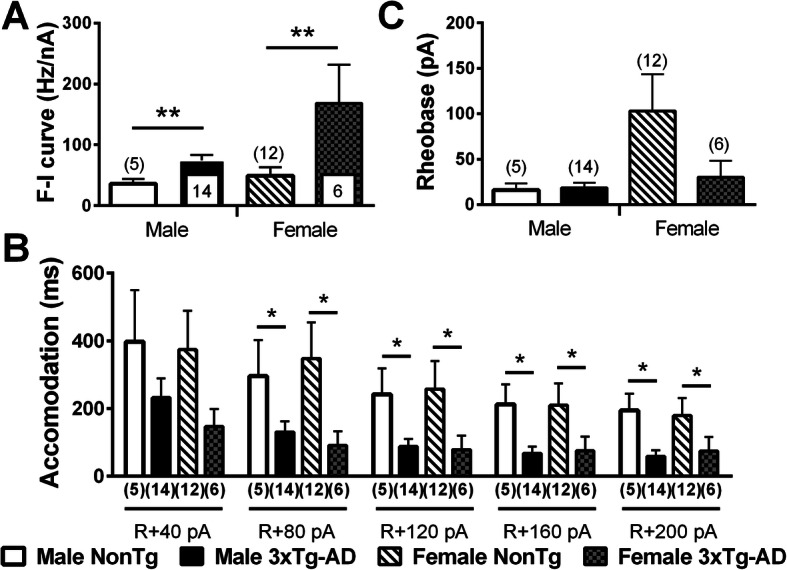
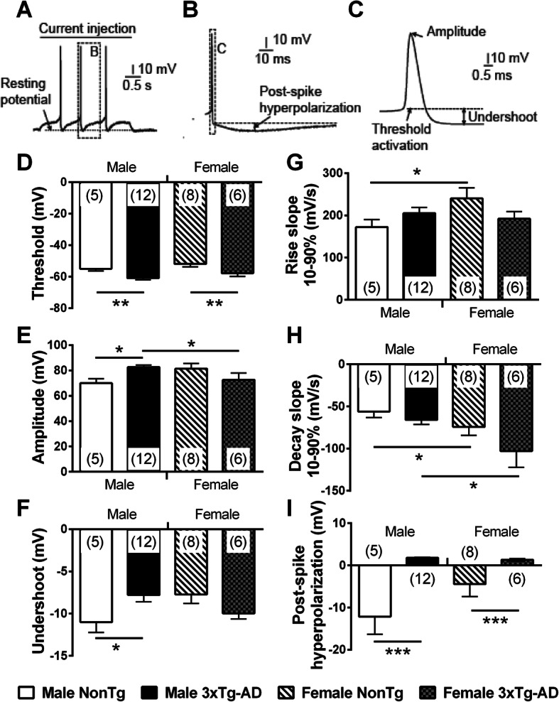
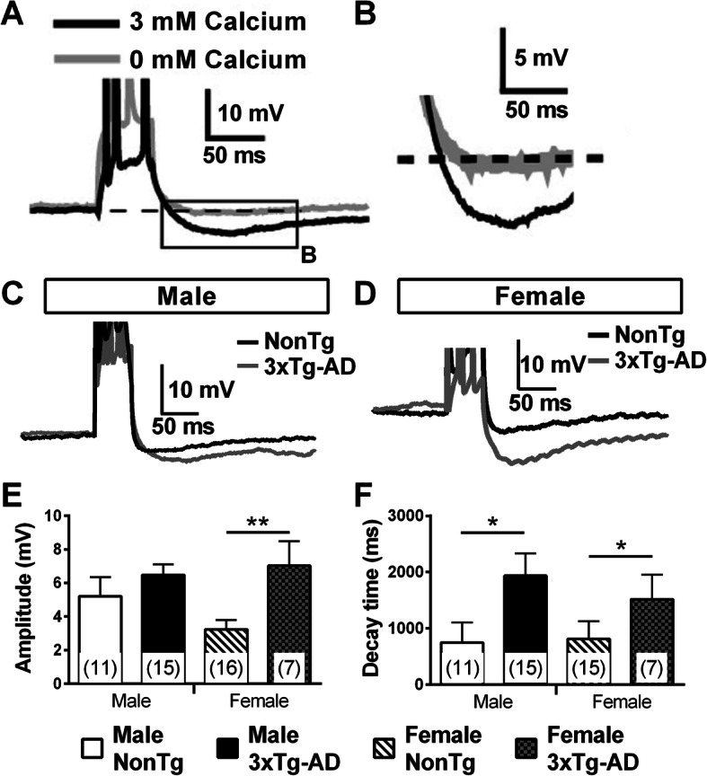
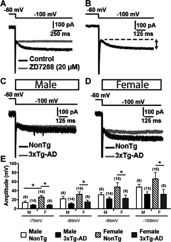
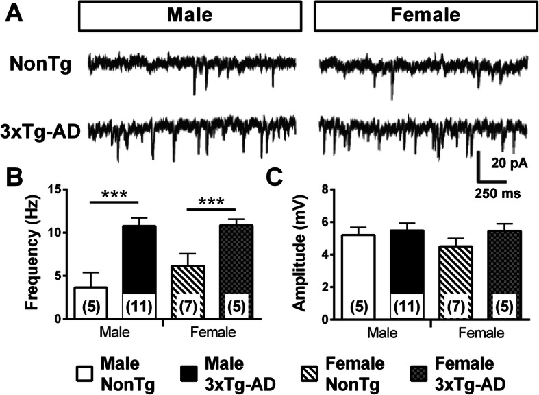
While the higher prevalence of Alzheimer's disease (AD) in women is clear, studies suggest that biological sex may also influence AD pathogenesis. However, mechanisms behind these differences are not clear. To investigate physiological differences between sexes at the cellular level in the brain, we investigated the intrinsic and synaptic properties of entorhinal cortex neurons in heterozygous 3xTg-AD mice of both sexes at the age of 20 months. This brain region was selected because of its early association with AD symptoms. First, we found physiological differences between male and female non-transgenic mice, providing indirect evidence of axonal alterations in old females. Second, we observed a transgene-dependent elevation of the firing activity, post-burst afterhyperpolarization (AHP), and spontaneous excitatory postsynaptic current (EPSC) activity, without any effect of sex. Third, the passive properties and the hyperpolarization-activated current (Ih) were altered by transgene expression only in female mice, whereas the paired-pulse ratio (PPR) of evoked EPSC was changed only in males. Fourth, both sex and transgene expression were associated with changes in action potential properties. Consistent with previous work, higher levels of Aβ neuropathology were detected in 3xTg-AD females, whereas tau deposition was similar. In summary, our results support the idea that aging and AD neuropathology differentially alter the physiology of entorhinal cortex neurons in males and females.

Keywords: 3xTg-AD mice; Aging; Alzheimer; Electrophysiology; Entorhinal cortex.


Conflict of interest statement

The authors declare that they have no competing interests.

Figures

Fig. 1
Fig. 1
Aβ pathology was more pronounced in 20-month-old heterozygous 3xTg-AD females. a, b Levels of both sAβ40 and sAβ42 were higher in transgenic females compared to males. c The sAβ42/sAβ40 ratio was not influenced by sex. d, e In insoluble fractions, amounts of iAβ40 and iAβ42 were more elevated in transgenic females. f The sex of animals did not modulate the iAβ42/iAβ40 ratio in insoluble fractions. Statistical comparisons were performed using Welch’s t test (a, d, and e) or unpaired Student’s t test (b, c, and f). *p < 0.05, **p < 0.01, ***p < 0.001
Fig. 2
Fig. 2
Sex did not influence tau pathology in 20-month-old 3xTg-AD mice. a Amounts of GADPH and tau in soluble fractions were similar between males and females. b No difference between the sexes was found in the tau levels and the proportion of phosphorylated tau at serine 396/404 in insoluble fractions. Statistical comparisons were performed using unpaired Student’s t test. Abbreviations: ROD, relative optical value; GADPH, glyceraldehyde-3-phosphate dehydrogenase (used as control)
Fig. 3
Fig. 3
Tissue preparation for electrophysiological recordings and dietary treatment. a Side view of the mouse brain. The black line represents the 250 μm horizontal section used in this study. b Horizontal mouse brain section stained with hematoxylin nuclear counterstain. Whole-cell recordings (REC) were made in the deep layer of EC. c Whole-cell patch-clamp recordings of deep-layer EC neurons. Abbreviations: CPu, caudate putamen (striatum); EC, Entorhinal cortex; Hipp, hippocampus; Rec, patch clamp cell recording region; Stim, electrically stimulated region
Fig. 4
Fig. 4
Transgene expression changed the passive properties of EC deep-layer neurons only in 20-month-old females. a Electrical representation of a cell membrane. b To quantify passive properties, different intensities of hyperpolarizing current were injected into a neuron in current clamp: voltage variation (V) and time constant (T) were measured after each injection. The panel illustrated on one trace the electrical properties measured for the calculation of passive properties. c The resting potential was not influenced by transgenes or sex. The expression of AD-related transgenes increased input resistance (d) and reduced Gc (e) in neurons of female animals, but not in males. (f) CC of neurons in NonTg females was higher than these of 3xTg-AD females and NonTg males. The number of mice included in each group was 4 for NonTg males, 5 for 3xTg-AD males, 8 for NonTg females, and 4 for 3xTg-AD females. Statistical comparisons were performed using unpaired Student’s t tests (c, d, and e) or Welch’s t tests (f). Abbreviations: CC, cell capacitance; Gc, cell conductance. *p < 0.05
Fig. 5
Fig. 5
Examples of electrophysiological recordings showing the firing properties of EC neurons, accordingly to the sex and the genotype. a The upper trace illustrates the maximum current injected in a neuron without reaching its excitation threshold. The lower trace represents the firing pattern obtained for a current injection of 80 pA greater than that of the upper trace in the same neuron. The protocol used is a 3-s depolarizing current injection generating a voltage response. Left recording shows the firing of a neuron from a NonTg male while a cell from a transgenic male is illustrated in the one at right. b Interevent interval between action potential of the recordings presented in the panel a. The firing accommodation corresponds to the difference between interevent interval at the beginning and the end of the train. 3xTg-AD neurons showed a lower firing adaptivity compared to NonTg cells. c The relationship between firing rate and injected current (F-I curves) from NonTg or 3xTg-AD neurons of males is illustrated in the graph on the right of the panel. The steepness of F-I slopes was increased by transgene expression in males. d, e, and f Same as a, b, and c, but it is for females. Transgene expression influenced similarly the firing activity and the firing accommodation in neurons of female mice
Fig. 6
Fig. 6
Transgene expression increased firing activity and reduced firing accommodation in both males and females aged of 20 months. Transgene expression increased F-I slopes (a) and reduced firing accommodation (b) in 3xTg-AD mice of both sexes. c The rheobase was not influenced by 3xTg-AD expression or sex and was estimated from the F-I plot using graphical methods. The number of mice included in each group was 5 for NonTg males, 14 for 3xTg-AD males, 12 for NonTg females, and 6 for 3xTg-AD females. Statistical comparisons were performed using two-way ANOVA (a, b) or Welch’s t test (c). Abbreviations: F-I, firing rate versus injected current. *p < 0.05, **p < 0.01
Fig. 7
Fig. 7
AP properties are differently influenced by sex and transgene expression in EC from 20-month-old mice. a An example of a recorded EC neuron following an injection of a 3-s depolarizing current. In this typical trace, the injected current triggered three APs. b Representation of a post-spike hyperpolarization (zoomed from the dashed square in a). Post-spike hyperpolarization was calculated from the difference between the voltage undershoot after the AP (the dashed line) and the voltage peak of post-spike. c Representation of AP characteristics quantified in this study (zoomed from the dashed square in the panel b). Undershoot was the difference between stabilized voltage after the AP and activation threshold. d AP threshold was significantly decreased with transgene expression in both sexes. e Amplitude of AP was higher in neurons from male 3xTg-AD, compared to those from male NonTg and female 3xTg-AD. f Transgene expression reduced undershoots only in the male. The rising slope was lower in NonTg males (g) whereas decay slope was higher in both NonTg and 3xTg-AD male mice (h). Transgene expression reduced post-spike hyperpolarization in both sexes. The number of mice included in each group was 5 for NonTg males, 12 for 3xTg-AD males, 8 for NonTg females, and 6 for 3xTg-AD females. Statistical comparisons were performed using two-way ANOVA (d, h, and i) or unpaired Student’s t test (e, f, and g). Abbreviations: AP, action potential; EC, entorhinal cortex. *p < 0.05, **p < 0.01, ***p < 0.001
Fig. 8
Fig. 8
Transgene expression modulated the post-burst AHP potential in 20-month-old mice. a An example of a recorded EC neuron following an injection of a 50-ms depolarizing current. Post-burst AHP potential is estimated in relation with the resting potential (the dashed line) and is abolished when calcium is removed from the extracellular solution. b Representation of a post-burst hyperpolarization (zoomed from the square in a). c An example of recordings illustrating post-burst hyperpolarization in neurons from male (c) or female (d) expressing or not 3xTg-AD transgenes. Transgene expression increased the amplitude of post-burst AHP only in females (e) and elevated the decay time in both sexes (f). The number of mice included in each group was 5 for NonTg males, 5 for 3xTg-AD males, 9 for NonTg females, and 4 for 3xTg-AD females. Statistical comparisons were performed using two-way ANOVA (f) or unpaired Student’s t test (e). *p < 0.05, **p < 0.01
Fig. 9
Fig. 9
Transgene expression reduced Ih current in a sex-dependent manner in mice aged of 20 months. a An example of recorded neuron following a voltage step, from − 60 to − 100 mV. Application of ZD7288 (20 μM), an antagonist of the hyperpolarized-activated current Ih [57], in the same neuron showed its slow and persistent activation in EC. b Ih was measured by subtracting the current before and after its slow and persistent activation, as illustrated by the line with two arrows. Illustrations of Ih currents generated by a voltage step, from − 60 to − 100 mV in males (c) and females (d), both transgenic or NonTg animals. e Ih current generated by hyperpolarizing voltage steps was decreased by 3xTg-AD expression in females, whereas the reduction was not significant in males. The number of mice included in each group was 4 for NonTg males, 5 for 3xTg-AD males, 8 for NonTg females, and 4 for 3xTg-AD females. Statistical comparisons were performed using unpaired Student’s t test (− 80 to − 100 mV) or Welch’s t test (− 70 mV). *p < 0.05
Fig. 10
Fig. 10
Transgene expression increased spontaneous excitatory postsynaptic current (sEPSC) in both sexes. a Examples of intracellular sEPSC recordings (voltage clamped at − 60 mV). b Frequency of sEPSC was higher in neurons from 20-month-old 3xTg-AD mice for both sexes. c sEPSC amplitude was not affected by sex or transgene expression. The number of mice included in each group was 3 for NonTg males, 5 for 3xTg-AD males, 5 for NonTg females, and 4 for 3xTg-AD females. Statistical comparisons were performed using two-way ANOVA. ***p = 0.001
Fig. 11
Fig. 11
Sex-dependent alteration of paired-pulse ratio from intracortical synaptic transmission by 3xTg-AD expression in mice aged of 20 months. a, b Evoked excitatory and inhibitory input has been discriminated by generating postsynaptic currents at different imposed voltages. The excitatory inputs produced a depolarizing current that increases with hyperpolarization of resting potential, whereas inhibitory inputs generated both hyperpolarizing (− 50 mV and -60 mV) and depolarizing currents (− 70 mV), depending on whether imposed voltage was under or over the reversal potential of Cl ions, estimated at − 63 mV. There are examples of eEPSC recordings for a paired electrical stimulation (interval of 100 ms) in males (c) and females (d), both transgenic or NonTg animals. e The P2/P1 ratio was decreased by transgene expression in male, but not in female mice. f The application of the GABAa receptor antagonist picrotoxin (100 μM) and the NMDA receptor antagonist D-APV (100 μM) did not affect the amplitude or the kinetic of eEPSC, showing that these receptors did not play a significant part in it. The number of mice included in each group was 3 for NonTg males, 4 for 3xTg-AD males, 4 for NonTg females, and 4 for 3xTg-AD females. Statistical comparisons were performed using unpaired Student’s t test. Abbreviations: eEPSC, evoked postsynaptic current. *p < 0.05

References

    1. Mielke MM, Ferretti MT, Iulita MF, Hayden K, Khachaturian AS. Sex and gender in Alzheimer’s disease – does it matter? Alzheimers Dement. 2018;14:1101–1103. - PubMed
    1. Jack CR, Wiste HJ, Weigand SD, Knopman DS, Vemuri P, Mielke MM, et al. Age, sex, and APOE ε4 effects on memory, brain structure, and β-amyloid across the adult life span. JAMA Neurol. 2015;72:511. - PMC - PubMed
    1. Mielke MM, Vemuri P, Rocca WA. Clinical epidemiology of Alzheimer’s disease: assessing sex and gender differences. Clin Epidemiol. 2014;6:37–48. - PMC - PubMed
    1. Jack CR, Wiste HJ, Weigand SD, Therneau TM, Knopman DS, Lowe V, et al. Age-specific and sex-specific prevalence of cerebral β-amyloidosis, tauopathy, and neurodegeneration in cognitively unimpaired individuals aged 50–95 years: a cross-sectional study. Lancet Neurol. 2017;16:435–444. - PMC - PubMed
    1. Snyder HM, Asthana S, Bain L, Brinton R, Craft S, Dubal DB, et al. Sex biology contributions to vulnerability to Alzheimer’s disease: a think tank convened by the Women’s Alzheimer’s Research Initiative. Alzheimers Dement. 2016;12:1186–1196. - PMC - PubMed

Publication types

Substances