. 2021 Jan 29:538:14-23.
doi: 10.1016/j.bbrc.2020.10.087. Epub 2020 Nov 6.

History of the COVID-19 pandemic: Origin, explosion, worldwide spreading

Affiliations

History of the COVID-19 pandemic: Origin, explosion, worldwide spreading

Sara Platto et al. Biochem Biophys Res Commun. .

Abstract

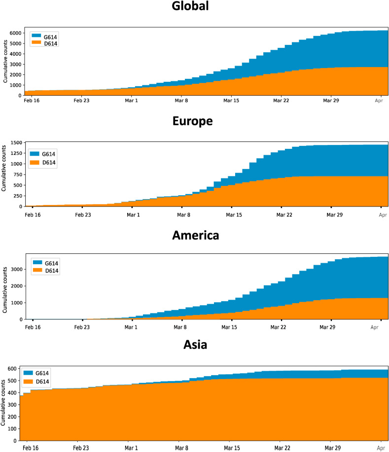
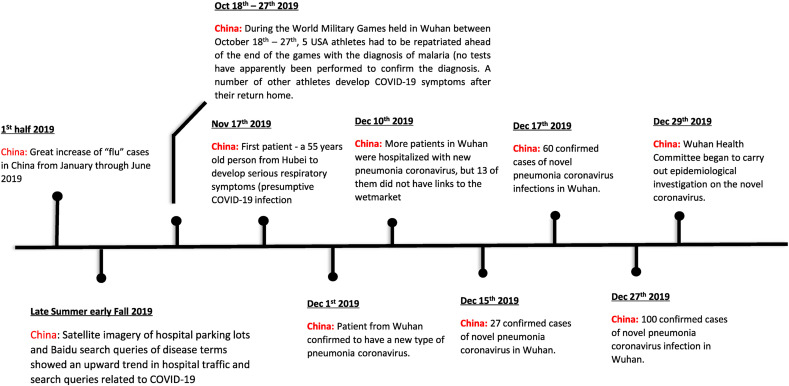
The SARS-CoV-2 virus of the COVID-19 pandemic, that is presently devastating the entire world, had been active well before January of this year, when its pathogenic potential exploded full force in Wuhan. It had caused the onset of small disease outbreaks in China, and probably elsewhere as well, which failed to reach epidemic potential. The distant general origin of its zoonosis can be traced back to the ecosystem changes that have decreased biodiversity, greatly facilitating the contacts between humans and the animal reservoirs that carry pathogens, including SARS-CoV-2. These reservoirs are the bats. The transition between the limited outbreaks that had occurred through 2019 and the epidemic explosion of December-January was made possible by the great amplification of the general negative conditions that had caused the preceding small outbreaks. In the light of what we have now learned, the explosion was predictable, and could have happened wherever the conditions that had allowed it, could be duplicated. What could not have been predicted was the second transition, from epidemic to pandemic. Research has now revealed that the globalization of the infection appears to have been caused by a mutation in the spike protein of the SARS-CoV-2, that has dramatically increased its transmissibility.

Keywords: Adenosine analogs; Antiviral therapy; RNA dependent RNA polymerase; Remdesivir.


Conflict of interest statement

Declaration of competing interest The authors declare that they don’t have conficts of interest.

Figures

Fig. 1
Fig. 1
Structure of the trimeric Spike protein of COVID-19. A: primary structure colored by domain. SS, signal sequence; S2′, protease cleavage site; FP, fusion peptide; HR1, heptad repeat 1; CH, central helix; CD, connector domain; HRE, heptad repeat 2; TM, transmembrane domain; CT, cytoplasmic tail; RDB, receptor binding domain; arrows indicate protease cleavage sites. B side and top views of the structure of the protein with a single RBD in the up conformation. The two RBD down protomers are shown in white or gray. The RBD protomer is shown in ribbons colored as shown in A. Modified from D Wrapp et al., 2020 [[17], [21]].
Fig. 2
Fig. 2
Amino acid sequence of the Spike protein of human SARS-CoV-2 and related coronaviruses.The amino acid sequence of the Spike protein (red bar in A) is expanded in B, and shows a polybasic site (RRAR) at the junction of the two subunits S1and S2 of the protein. The alignment against related coronaviruses (C) shows that the polybasic site RRAR is unique to SARS-CoV-2. The three adjacent predicted O-linked glycans are also unique to SARS-CoV-2 Sequences shown are from NCBI GenBank, accession codes MN908947, MN996532, AY278741, KY417146andMK211376. The pangolin coronavirus sequences are a consensus generated from SRR10168377and SRR10168378(NCBI BioProject PRJNA573298. Modified from K. G. Andersen et al., 2020 [[17], [21]] where additional details are found. (For interpretation of the references to color in this figure legend, the reader is referred to the Web version of this article.)
Fig. 3
Fig. 3
Proteolytic cleavage of peptides containing the S1/S2 cleavage site of SARS-CoV-2 and SARS-CoV. A number of proteases are likely to be involved in Spike protein processing, furin and PC1, trypsin, type II transmembrane serine protease matripase (the transmembrane serine protease TMPRSS2 has not been tested), cathepsins L and B. Furin cleaves SARS-CoV-2, but not SARS-CoV. In addition to furin, other proteases cleave SARS-CoV-2 much more efficiently than SARS-CoV. The only protease that cleaves SARS-CoV more efficiently than SARS-CoV-2 is cathepsin L. Clearly, the acquisition of the polybasic site insert PRRA by COVID-17 broadens the repertoire of activating proteases. Modified from Jaimes et al. (21).
Fig. 4
Fig. 4
Daily cumulative count of the relative amount of the wild type Spike protein of SARS-CoV-2 with the wild type D614 and of the mutant G614 in different world regions. After the mutant protein enters a region, it soon becomes the dominating variant. Modified from B. Korber et al. [22].
Fig. 5
Fig. 5
The D614G mutation in the Spike protein of SARS-CoV-2. Top: cryo-EM structure of the S1 (brown) and S2 (orange) subunits of the Spike protein. Residues 581–676, a C-terminal region of the S1 domain involved in S2 interaction is colored green. Aspartic acid 614 is shown in light green. Bottom: the area indicated with a black square is shown magnified. Residues within 5.5 A of D614 are shown in a ball and stick representation (see text Modified from L.Zhang et al.) , (For interpretation of the references to color in this figure legend, the reader is referred to the Web version of this article.)
Fig. 6
Fig. 6
A timeline of the of the possible COVID-19 infection in China prior to the Wuhan explosion.

Similar articles

Cited by

References

    1. Platto S., Xue T., Carafoli E. COVID-19: an announced pandemic. Cell Death Dis. 2020;11:799–812. - PMC - PubMed
    1. Deslandes V. Berti, Tandjaoui-Lambotte J., Alloui C., Carbonelle E., Zahar J.R., Brichler S., Cohen Y. SARS-CoV-2 was already spreading in France in late December 2019. Int. J. Antimicrob. Agents. 2020;55 doi: 10.1016/j.ijantimicag.2020.106006. Epub 2020 May 3. - DOI - PMC - PubMed
    1. Platto S., Zhou J., Yanqing W., Huo W., Carafoli E. Biodiversity loss and COVID-19 pandemic: the role of bats in the origin and the spreading of the disease. Biochem. Biophys. Res. Commun. 2020 doi: 10.1016/j.bbrc.2020.10.028. PMID: 33092787; PMCID: PMVC7566801. - DOI - PMC - PubMed
    1. Luis A.D., Hayman D.T.S., O’Shea T.J., Cryan P.M., Gilbert A.T., Pulliam J.R.C., Mills J.N., Timonin M.E., Willis C.K.R., Cunningham A.A., Fooks A.R., Rupprecht C.E., Wood J.L.N., Webb C.T. A comparison of bats and rodents as reservoirs of zoonotic viruses: are bats special ? Proc. Royal Soc. B. 2013;280 doi: 10.1098/rspb.2012.2753. Print 2013 Apr 7. - DOI - PMC - PubMed
    1. Forster P., Forster L., Renfrew C., Forster M. Phylogenetic network analysis of SARS-CoV-2 genomes. Proc. Natl. Acad. Sci. U.S.A. 2020;117:9241–9243. - PMC - PubMed

Substances