Review
. 2021 Mar;27(3):207-219.
doi: 10.1016/j.molmed.2020.10.004. Epub 2020 Nov 13.

PD-L1 and B7-1 Cis-Interaction: New Mechanisms in Immune Checkpoints and Immunotherapies

Affiliations
Review

PD-L1 and B7-1 Cis-Interaction: New Mechanisms in Immune Checkpoints and Immunotherapies

Christopher D Nishimura et al. Trends Mol Med. 2021 Mar.

Abstract

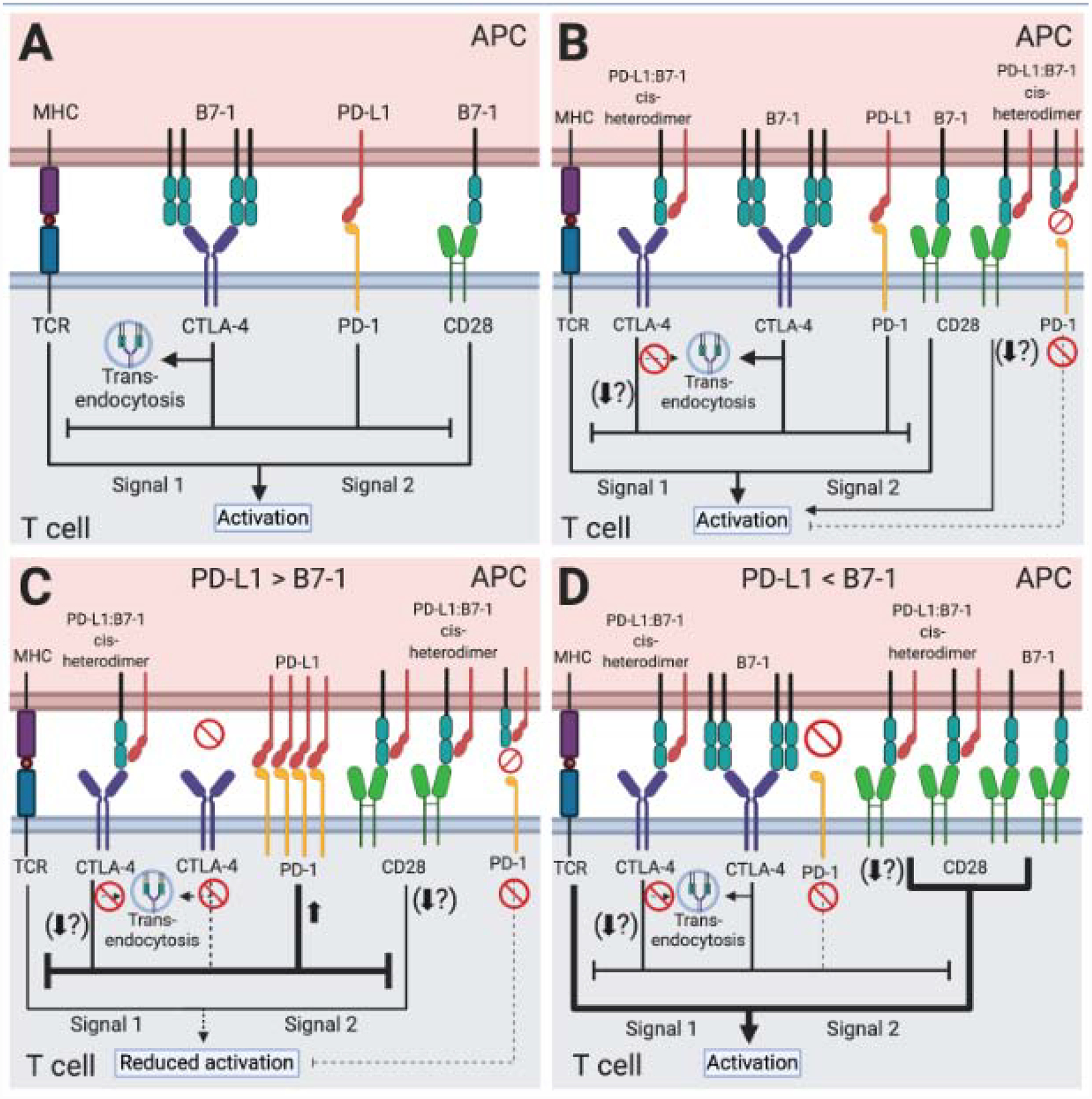
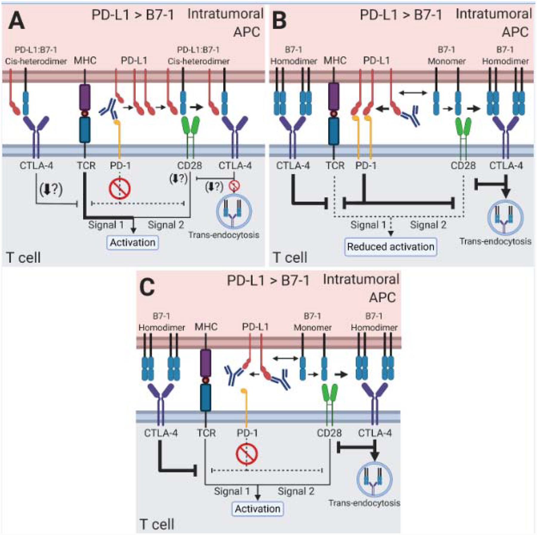
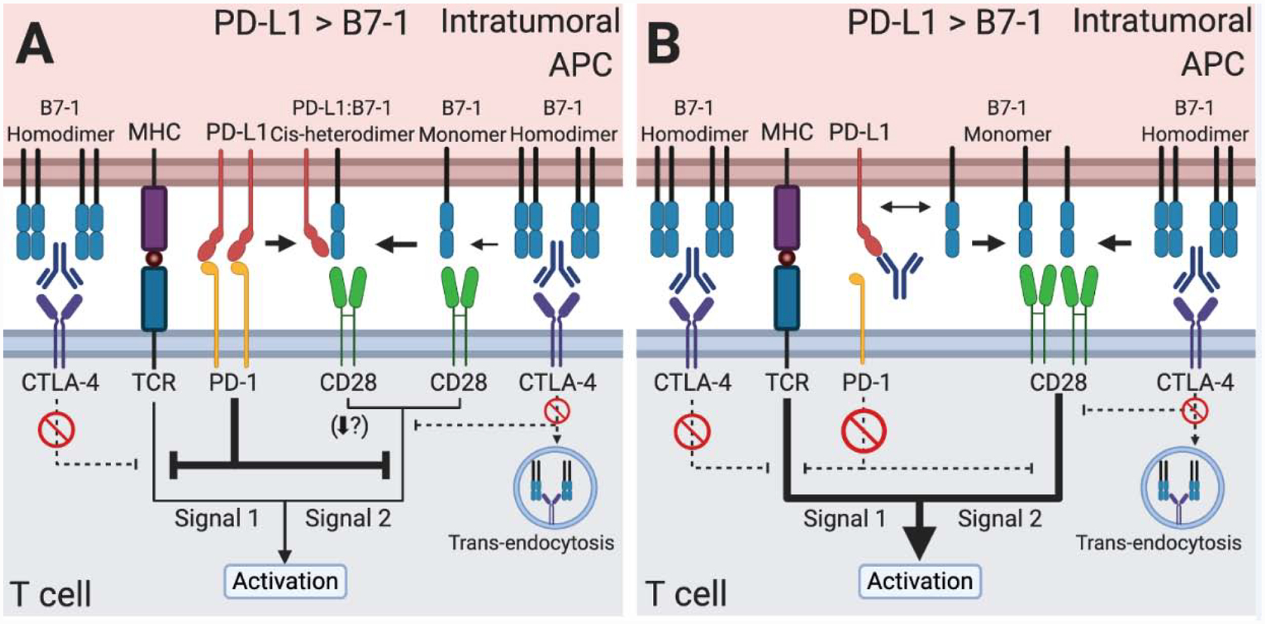
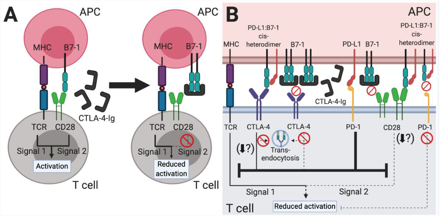
Immune checkpoints negatively regulate immune cell responses. Programmed cell death protein 1:programmed death ligand 1 (PD-1:PD-L1) and cytotoxic T lymphocyte-associated protein 4 (CTLA-4):B7-1 are among the most important immune checkpoint pathways, and are key targets for immunotherapies that seek to modulate the balance between stimulatory and inhibitory signals to lead to favorable therapeutic outcomes. The current dogma of these two immune checkpoint pathways has regarded them as independent with no interactions. However, the newly characterized PD-L1:B7-1 ligand-ligand cis-interaction and its ability to bind CTLA-4 and CD28, but not PD-1, suggests that these pathways have significant crosstalk. Here, we propose that the PD-L1:B7-1 cis-interaction brings novel mechanistic understanding of these pathways, new insights into mechanisms of current immunotherapies, and fresh ideas to develop better treatments in a variety of therapeutic settings.

Keywords: B7-1; CD28; CTLA-4; PD-1; PD-L1; cis-interaction; immunotherapy.


Conflict of interest statement

Conflicts of interest

The authors declare no competing financial interests.

Figures

Figure 1.
Figure 1.. PD-L1 and B7–1 immune checkpoint signaling pathways.
T cell activation requires two signals. Engagement of the peptide-MHC complex on APCs with the TCR complex provides signal one. Costimulatory ligand-receptor interactions such as B7–1:CD28 provide signal two. Immune checkpoint interactions like PD-L1:PD-1 and B7–1:CTLA-4 regulate this response. (A) Although both inhibit T cell activation, originally the PD-L1:PD-1 pathway and the B7–1:CTLA-4 pathway were considered completely independent with no interactions. (B) The PD-L1:B7–1 cis-interaction demonstrates crosstalk between these pathways. The PD-L1:B7–1 cis-heterodimer binds CTLA-4, reduces B7–1 trans-endocytosis, and potentially alters other CTLA-4-mediated cell-intrinsic mechanisms of inhibition. It also binds CD28; while most studies demonstrate no alteration in functional outcomes, one study shows decreased T cell activation. The PD-L1:B7–1 heterodimer does not bind to PD-1. By sequestering B7–1, the PD-L1:B7–1 cis-heterodimer indirectly reduces CTLA-4 inhibition. Although not detailed in this figure, the roles of B7–2 and PD-L2 should be further examined in the context of PD-L1:B7–1 engagement in T cell activation. (C) If PD-L1 outnumbers B7–1, PD-L1:B7–1 cis-heterodimers reduce free B7–1. The PD-L1:B7–1 cis-heterodimer binds to CD28 which most, but not all, studies suggest does not alter functional outcomes as described above. It also binds to CTLA-4, reduces CTLA-4-mediated trans-endocytosis, and possibly other CTLA-4 cell-intrinsic inhibitory mechanisms and outcomes. Finally, excess PD-L1 binds to PD-1, inducing strong inhibitory signals. Altogether, this reduces T cell activation. (D) If B7–1 outnumbers PD-L1, PD-L1:B7–1 cis-heterodimers reduce free PD-L1. The heterodimer prevents PD-1:PD-L1 binding. It reduces CTLA-4:B7–1 trans-endocytosis, possibly other CTLA-4 inhibitory mechanisms. Most studies show that it does not alter B7–1:CD28 functional outcomes, although one study demonstrates that it reduces T cell activation. While B7–1 homodimers may interact with CTLA-4, B7–1 monomers and PD-L1:B7–1 cis-heterodimers continue transducing CD28 costimulatory signals. Collectively, this generates a net-stimulatory effect.
Figure 2.
Figure 2.. New considerations of the PD-L1:B7–1 cis-interaction in anti-PD-1/PD-L1 monotherapies to treat cancer.
Intratumoral APCs exhibit high PD-L1 expression, outnumbering B7–1. B7–2 and PD-L2 (not shown) likely also play roles in the therapies described below, and their contribution should be explored in future studies. (A) Anti-PD-1 mAb monotherapy blocks PD-1 binding. Excess PD-L1 sequesters B7–1. The PD-L1:B7–1 cis-heterodimer induces CD28 costimulatory signaling. CTLA-4 sequesters the PD-L1:B7–1 cis-heterodimer from CD28, reducing the CD28 costimulatory signal. (B) Anti-PD-L1 mAb monotherapy only targeting the PD-L1:B7–1 cis-interaction frees B7–1 from PD-L1. B7–1 can bind to either CD28 or CTLA-4. B7–1 is sequestered and trans-endocytosed by CTLA-4, reducing its binding to CD28. PD-1 binding increases due to freed PD-L1. The PD-1 pathway inhibits the CD28 costimulatory signal. Together this reduces T cell activation. (C) Anti-PD-L1 mAb monotherapy targeting both PD-1 binding and PD-L1:B7–1 cis-interaction eliminates the PD-1 inhibitory pathway and frees B7–1 from PD-L1. Free B7–1 binds to CD28 to induce a costimulatory signal. Additionally, CTLA-4 may sequester the B7–1 and mediates trans-endocytosis. But the combination of reduced PD-1 and increased CD28 signaling leads to T cell activation. If high CTLA-4 levels are present (not shown), the increased sequestration of B7–1 may reduce T cell activation.
Figure 3.
Figure 3.. The PD-L1:B7–1 cis-interaction in anti-CTLA-4 monotherapy and the optimal anti-CTLA-4 and anti-PD-L1 combination therapy to treat cancer.
(A) Anti-CTLA-4 mAb monotherapy prevents CTLA-4 sequestration and trans-endocytosis of B7–1. The B7–1 heterodimerizes with PD-L1. The PD-L1:B7–1 cis-heterodimer induces CD28 costimulatory signaling. Given a high enough level of PD-L1 to remain able to bind PD-1, PD-1 signaling may prevent optimal T cell activation. (B) Combination therapy of anti-CTLA-4 mAb and anti-PD-L1 mAb targeting both PD-1 binding and the PD-L1:B7–1 cis-interaction blocks PD-1 inhibitory signaling, CTLA-4 binding of B7–1, and CTLA-4-mediated trans-endocytosis. This combination maximizes B7–1 levels and induces a strong CD28 costimulatory signal, strengthening the overall T cell activation signal. B7–2 and PD-L2 (not shown) may be important in these therapies as well.
Figure 4.
Figure 4.. The PD-L1:B7–1 cis-interaction offers new mechanistic insight into CTLA-4-Ig therapy.
(A) Based on our original understanding of immune checkpoint signaling pathways (Figure 1A), CTLA-4-Ig therapy functions by decreasing CD28-mediated T cell costimulation. The fusion protein mimics the native CTLA-4 protein and binds to B7–1 and B7–2 (not shown) on APCs, thus preventing a CD28:B7–1 or B7–2 interaction. (B) Our new understanding of the PD-L1:B7–1 cis-interaction shows additional immune checkpoint signaling pathways which underlie CTLA-4-Ig therapy. As CTLA-4-Ig sequesters most free B7–1 and B7–2 (not shown), the B7–1 receptors CD28 and CTLA-4 do not transduce activating or inhibitory signals, respectively. PD-L1 is unaffected and may continue to bind PD-1, inhibiting signals one and two of T cell activation. PD-L2 (not shown) may also continue to inhibit T cell activation. Although likely sparse, PD-L1:B7–1 cis-heterodimers may continue to modulate CTLA-4, CD28, and PD-1 signaling. The PD-L1:B7–1:CTLA-4 interaction reduces B7–1 trans-endocytosis and potentially other CTLA-4 functional outcomes as well. Most studies indicate that it mediates continued CD28 costimulation, although one study demonstrated that it reduced T cell activation. As the PD-L1:B7–1 cis-heterodimer cannot bind PD-1, it does not further inhibit T cell activation through this pathway. Taken together, this therapy is highly immunosuppressive, but the preformed PD-L1:B7–1 cis-heterodimer may continue to interact with CD28 and provide enough activation over the long-term to mediate low levels of T cell responses.

References

    1. Ledford H, et al. (2018) Cancer immunologists scoop medicine Nobel prize. Nature 562, 20–21 - PubMed
    1. Mease PJ, et al. (2017) Efficacy and safety of abatacept, a T-cell modulator, in a randomised, double-blind, placebo-controlled, phase III study in psoriatic arthritis. Ann Rheum Dis 76, 1550–1558 - PMC - PubMed
    1. Brunner HI, et al. (2018) Subcutaneous Abatacept in patients with polyarticular-course juvenile idiopathic arthritis: results from a phase III open-label study. Arthritis Rheumatol 70, 1144–1154 - PMC - PubMed
    1. van Nimwegen JF, et al. (2020) Abatacept treatment for patients with early active primary Sjögren’s syndrome: a single-centre, randomised, double-blind, placebo-controlled, phase 3 trial (ASAP-III study). Lancet Rheumatol 2, e153–e163 - PubMed
    1. Kliwinski C, et al. (2005) Prophylactic administration of abatacept prevents disease and bone destruction in a rat model of collagen-induced arthritis. J Autoimmun 25, 165–171 - PubMed

Publication types