. 2021 Jan-Jun:296:100085.
doi: 10.1074/jbc.RA120.015824. Epub 2020 Nov 23.

The KH domain facilitates the substrate specificity and unwinding processivity of DDX43 helicase

Affiliations

The KH domain facilitates the substrate specificity and unwinding processivity of DDX43 helicase

Manisha Yadav et al. J Biol Chem. 2021 Jan-Jun.

Abstract

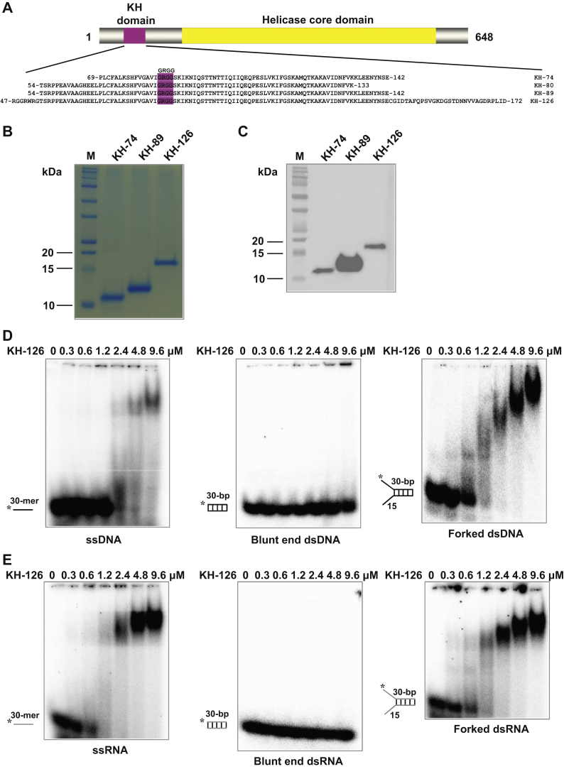
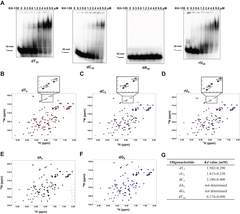
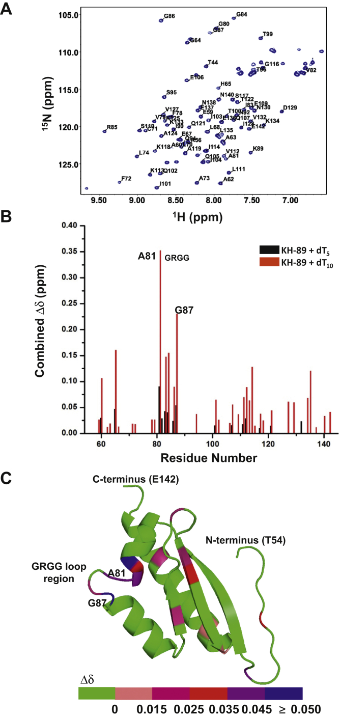
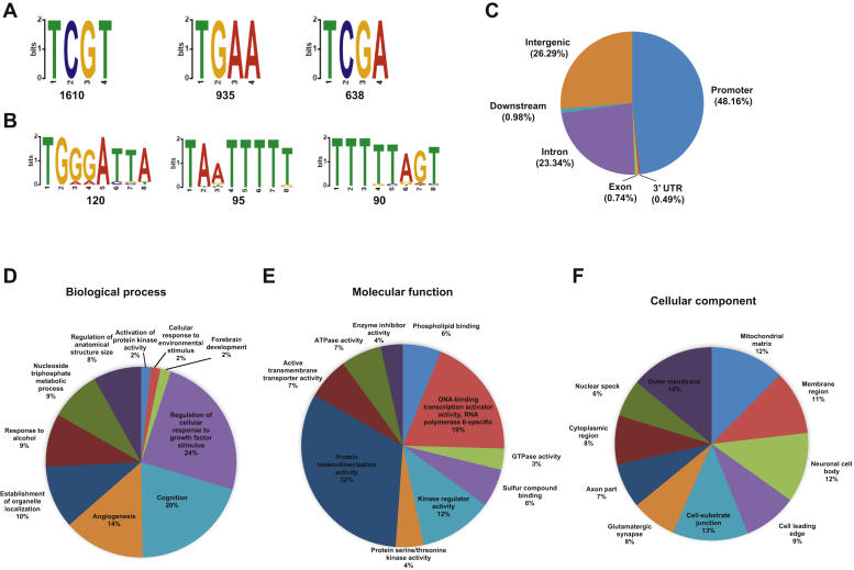
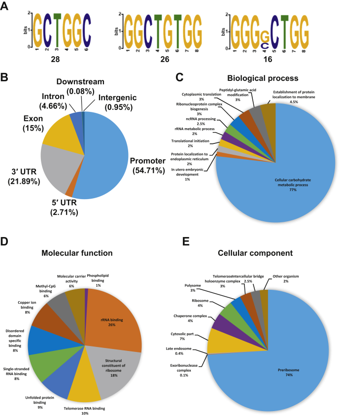
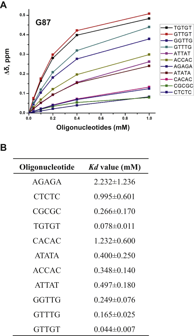
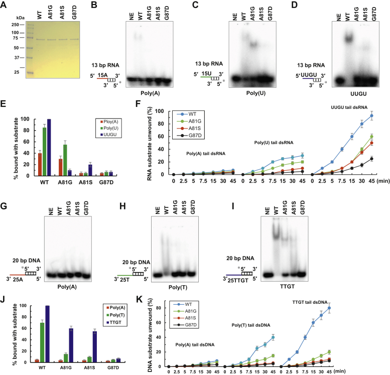
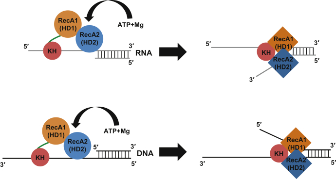
The K-homology (KH) domain is a nucleic acid-binding domain present in many proteins. Recently, we found that the DEAD-box helicase DDX43 contains a KH domain in its N-terminus; however, its function remains unknown. Here, we purified recombinant DDX43 KH domain protein and found that it prefers binding ssDNA and ssRNA. Electrophoretic mobility shift assay and NMR revealed that the KH domain favors pyrimidines over purines. Mutational analysis showed that the GXXG loop in the KH domain is involved in pyrimidine binding. Moreover, we found that an alanine residue adjacent to the GXXG loop is critical for binding. Systematic evolution of ligands by exponential enrichment, chromatin immunoprecipitation-seq, and cross-linking immunoprecipitation-seq showed that the KH domain binds C-/T-rich DNA and U-rich RNA. Bioinformatics analysis suggested that the KH domain prefers to bind promoters. Using 15N-heteronuclear single quantum coherence NMR, the optimal binding sequence was identified as TTGT. Finally, we found that the full-length DDX43 helicase prefers DNA or RNA substrates with TTGT or UUGU single-stranded tails and that the KH domain is critically important for sequence specificity and unwinding processivity. Collectively, our results demonstrated that the KH domain facilitates the substrate specificity and processivity of the DDX43 helicase.

Keywords: CLIP-seq; ChIP-seq; DDX43; KH domain; NMR; SELEX; helicase processivity; substrate specificity.


Conflict of interest statement

Conflict of interest The authors declare that they have no conflicts of interest with the contents of this article.

Figures

Figure 1
Figure 1
Purification and characterization of DDX43 KH-domain proteins.A, the schematic representation of four DDX43 KH-domain fragments. The KH domain and GRGG loop are indicated in red and helicase core domain in yellow. B, the SDS-PAGE analysis of recombinant DDX43 KH-domain proteins eluting from a Sephacryl S-100 HR column. C, the Western blot analysis for proteins shown in panel B with an anti-His antibody (SC-8036, Santa Cruz). DE, the representative EMSA images of increasing DDX43 KH-domain protein (126 aa, 0–9.6 μM) binding with 0.5 nM of indicated DNA (D) and RNA (E) substrates. See Table S2 for substrate sequence. DNA is in black and RNA in gray. EMSA, electrophoretic mobility shift assay; HR, high-resolution.
Figure 2
Figure 2
DDX43 KH domain prefers to bind pyrimidines.A, the representative images of electrophoretic mobility shift assays (EMSA) performed by incubating 0.5 nM of indicated oligonucleotide with increasing DDX43 KH-domain proteins (126 aa, 0–9.6 μM) at RT for 30 min. DNA is in black and RNA in gray. BF, 1H-15N HSQC spectrum of DDX43 KH-domain protein (89 aa, 200 μM) with indicated oligonucleotide (ranged 40–1000 μM). Chemical shifts are indicated by arrows in the enlarged image in panels BD. G, affinities of DDX43-KH domain (89 aa) with indicated oligo determined by NMR spectroscopy. HSQC, heteronuclear single quantum coherence.
Figure 3
Figure 3
The conserved GRGG loop region is involved in nucleotide binding.A, a 600-MHz 1H-15N HSQC spectrum of DDX43 KH-89 protein with some of the peak assignments. B, combined chemical shift changes caused by dT5 and dT10 binding to DDX43-KH domain (89 aa) as a function of amino acid. C, a model structure of DDX43 KH domain (89 aa) generated by Phyre 2. Potential amino acids involved in dT5 binding are indicated by colors according to their combined Δδ. HSQC, heteronuclear single quantum coherence.
Figure 4
Figure 4
The alanine adjacent to the GRGG loop is critical for nucleic acid binding.AB, the representative EMSA images of increasing protein concentration (0–9.6 μM) of DDX43 KH domain (89 aa, including WT, A81G, and A81S) binding with 0.5 nM of ssDNA (A) or ssRNA (B). CD, peak assignments of NMR spectra for KH-A81G (C) and A81S (D) and overlap of WT. E, combined chemical shift changes caused by dT5 binding to the KH-domain protein (89 aa, including WT, A81G, and A81S) as a function of the amino acid residue number. EMSA, electrophoretic mobility shift assay.
Figure 5
Figure 5
Identification of DDX43 KH domain bound DNA by the SELEX and ChIP-seq methods.A, top three motif sequences obtained by the SELEX method. B, top three motif sequences obtained by the ChIP-Seq method. C, distribution of DDX43 KH-domain peaks across the human genome. DF, distribution of gene ontology terms among the annotated sequences for biological process (D), molecular function (E), and cellular component (F). ChIP-seq, chromatin immunoprecipitation sequence; SELEX, systematic evolution of ligands by exponential enrichment.
Figure 6
Figure 6
Identification of DDX43 KH domain bound RNA by the CLIP-seq method.A, top three motif sequences obtained by the ChIP-Seq method. B, distribution of DDX43 KH-domain peaks across the human genome. CE, distribution of gene ontology terms among the annotated sequences for biological process (C), molecular function (D), and cellular component (E). ChIP-seq, chromatin immunoprecipitation sequence; CLIP-seq, cross-linking immunoprecipitation sequence.
Figure 7
Figure 7
Affinity of DDX43 KH domain with the oligonucleotide determined by NMR spectroscopy.A, combined chemical shift change (Δδ) plotted as a function of different oligonucleotide concentrations (0–1 mM) at 200 μM DDX43 KH-89 protein for residue G87. B, affinities of DDX43 KH domain (89 aa) with indicated oligonucleotides determined by NMR spectroscopy.
Figure 8
Figure 8
Mutated alanine (A81) or glycine (G87) affects DDX41 in binding and unwinding processivity.A, the SDS-PAGE analysis of purified DDX43 full-length proteins (WT and mutants, 1 ug each). BE, the representative images of EMSA performed by incubating DDX43 full-length proteins (2 μM) with 0.5 nM of 13-bp duplex RNA with 5′ tail of polyA (B), polyU (C), UUGU repeats (D), and their quantitative analysis (E). F, the quantitative analysis of helicase assays of DDX43 proteins (9.6 μM) on 0.5 nM of 13-bp duplex RNA with 5′ tail of polyA, polyU, or UUGU repeats as a function of time (0–45 min). GJ, the representative images of EMSA performed by incubating DDX43 full-length proteins (2 μM) with 0.5 nM of 20-bp duplex DNA with 3′ tail of polyA (G), polyT (H), TTGT repeats (I), and their quantitative analysis (J). K, the quantitative analysis of helicase assays of DDX43 proteins (9.6 μM) on 0.5 nM of 20-bp duplex DNA with 3′ tail of polyA, polyT, or TTGT repeats as a function of time (0–45 min). The triangle indicates heat-denatured RNA or DNA substrate control. Data are presented as the mean ± SD, n = 3. EMSA, electrophoretic mobility shift assay; NE, no enzyme.
Figure 9
Figure 9
Proposed models for the role of the KH domain in DDX43 helicase unwinding dsRNA (upper) and dsDNA (bottom). In initial recognition and binding, the KH domain is mainly responsible for ssRNA/ssDNA binding. The helicase core domain, especially the RecA2 domain, might also be involved in nucleic acid binding. The binding of ATP and Mg causes conformational changes of two RecA domains. During unwinding, the KH domain binds with ssRNA/ssDNA (either strand) to stabilize RecA1/2 interacting with the fork junction and also prevent ssRNA/ssDNA reannealing. This, in turn, accelerates the unwinding processivity of helicase domains. RecA1 (HD1), RecA2 (HD2), and KH domains are shown in orange, blue, and dark red, respectively, and linkers are in green.

Similar articles

Cited by

References

    1. Jarmoskaite I., Russell R. RNA helicase proteins as chaperones and remodelers. Annu. Rev. Biochem. 2014;83:697–725. - PMC - PubMed
    1. Linder P., Jankowsky E. From unwinding to clamping - the DEAD box RNA helicase family. Nat. Rev. Mol. Cell Biol. 2011;12:505–516. - PubMed
    1. Lohman T.M., Bjornson K.P. Mechanisms of helicase-catalyzed DNA unwinding. Annu. Rev. Biochem. 1996;65:169–214. - PubMed
    1. Pyle A.M. Translocation and unwinding mechanisms of RNA and DNA helicases. Annu. Rev. Biophys. 2008;37:317–336. - PubMed
    1. Fairman-Williams M.E., Guenther U.P., Jankowsky E. SF1 and SF2 helicases: family matters. Curr. Opin. Struct. Biol. 2010;20:313–324. - PMC - PubMed

Publication types

MeSH terms