. 2020 Nov 16;4(1):e202000795.
doi: 10.26508/lsa.202000795. Print 2021 Jan.

Structure of the helicase core of Werner helicase, a key target in microsatellite instability cancers

Affiliations

Structure of the helicase core of Werner helicase, a key target in microsatellite instability cancers

Joseph A Newman et al. Life Sci Alliance. .

Abstract

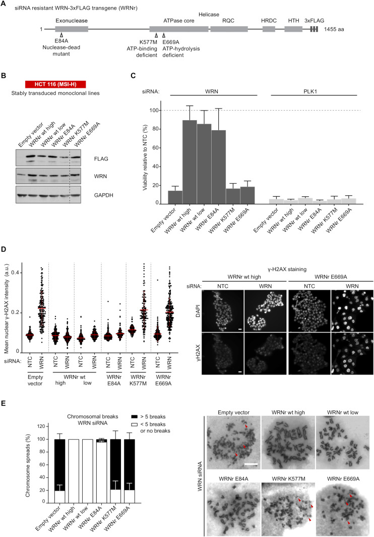
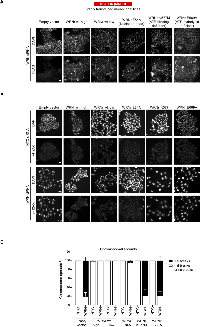
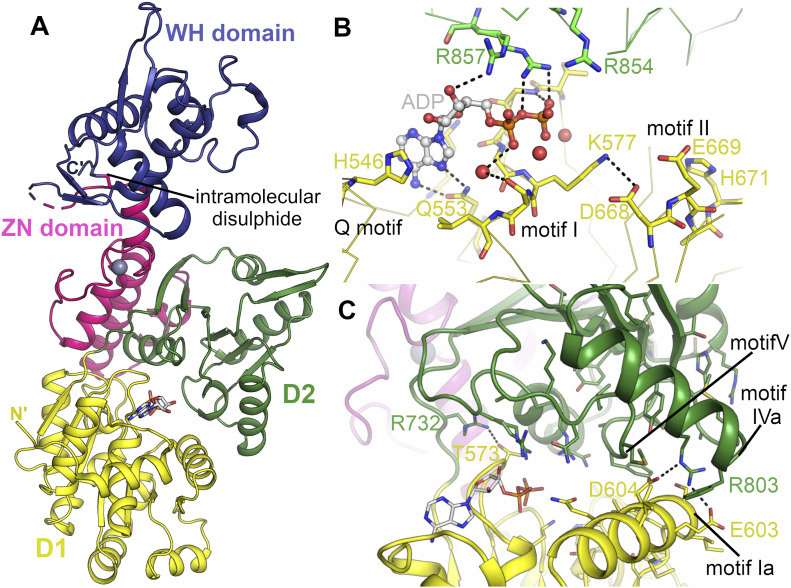
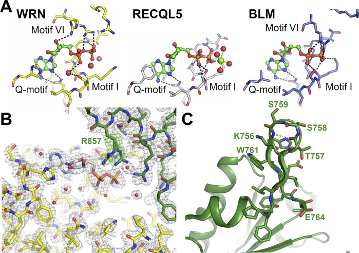
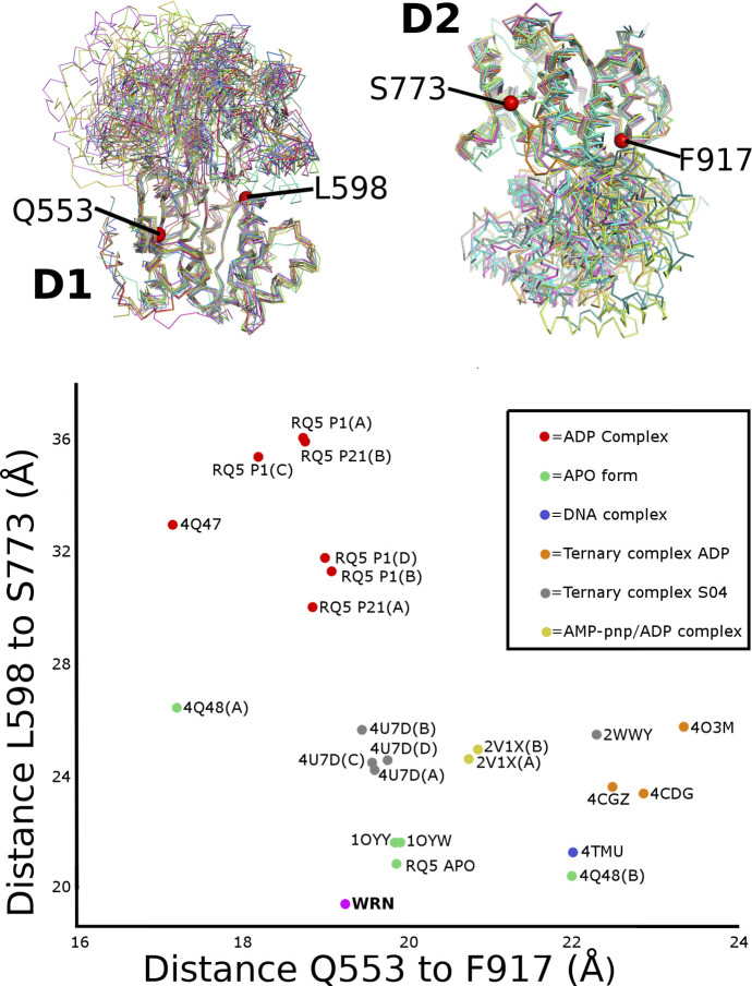
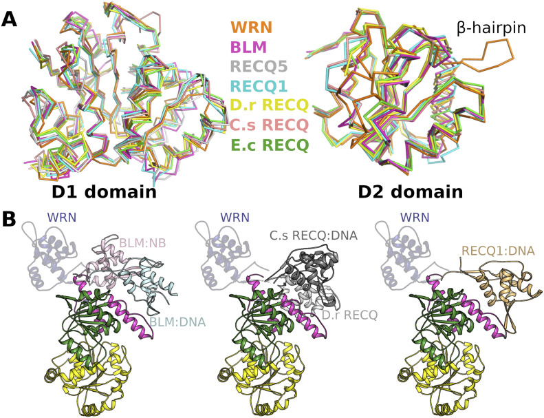
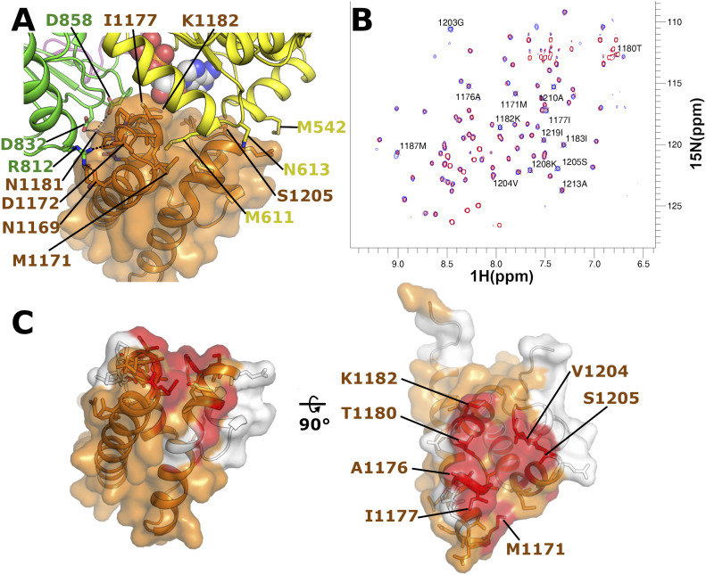
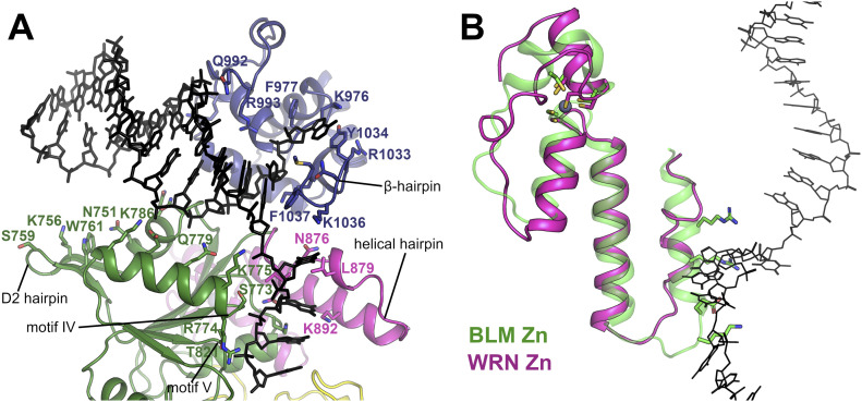
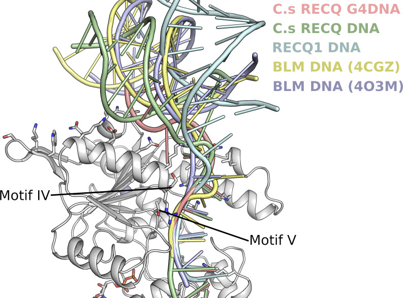
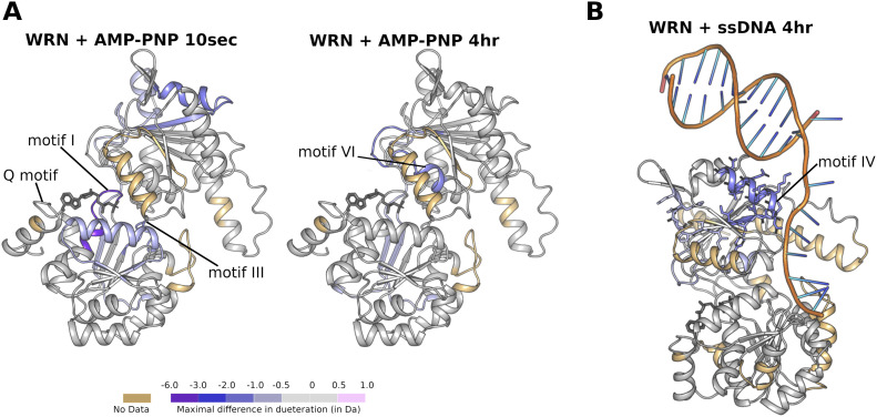
Loss of WRN, a DNA repair helicase, was identified as a strong vulnerability of microsatellite instable (MSI) cancers, making WRN a promising drug target. We show that ATP binding and hydrolysis are required for genome integrity and viability of MSI cancer cells. We report a 2.2-Å crystal structure of the WRN helicase core (517-1,093), comprising the two helicase subdomains and winged helix domain but not the HRDC domain or nuclease domains. The structure highlights unusual features. First, an atypical mode of nucleotide binding that results in unusual relative positioning of the two helicase subdomains. Second, an additional β-hairpin in the second helicase subdomain and an unusual helical hairpin in the Zn2+ binding domain. Modelling of the WRN helicase in complex with DNA suggests roles for these features in the binding of alternative DNA structures. NMR analysis shows a weak interaction between the HRDC domain and the helicase core, indicating a possible biological role for this association. Together, this study will facilitate the structure-based development of inhibitors against WRN helicase.


Conflict of interest statement

The authors declare that they have no conflict of interest.

Figures

Figure 1.
Figure 1.. ATP hydrolysis by WRN is essential for viability and genome integrity in MSI-H CRC cells.
(A) Schematic representing the WRN domain structure. Location of nuclease-dead and ATPase-inactivating mutations (Walker A and B mutants) in siRNA-resistant WRN expression constructs containing a C-terminal 3xFLAG tag (WRNr) are indicated. (B) Monoclonal HCT 116 (MSI-H) cell clones were isolated after transduction with an empty vector control and WRNr wild-type or mutant transgenes. Immunoblotting of cell lysates with anti-FLAG and anti-WRN antibodies was used to determine the expression of the WRNr wild-type and mutant forms along with total WRN protein levels. Two WRNr wild-type clones (high and low) were selected to cover the expression range of WRNr mutant variants. (C) HCT 116 cells expressing WRNr transgenes were transfected with either non-targeting control or WRN siRNAs. Viability measurements were performed 7 d after siRNA transfection, and the data are represented relative to non-targeting control siRNA. Data information: In (C), viability data are shown as mean ± SD of three biological repeat experiments. (D) Immunofluorescence analysis of γ-H2AX was performed 72 h after siRNA transfection. The mean nuclear γH2AX intensity (a.u.) was quantified after siRNA transfection. Data points shown (n ≥ 120 cells per condition) are derived from a single representative experiment that is consistent with a biological repeat experiment. Scale bar, 20 μM. (E) Mitotic chromosome spread analysis was performed 72 h after siRNA transfection. At the 66 h time-point, cells were treated with 6 h of Nocodazole (1.5 μM) before spreading to enrich for mitotic stages. As a reference, some chromosome breaks are highlighted by red arrowheads. Each mitotic spread was categorized into less than five breaks or more than five breaks (n ≥ 28 mitotic spreads per condition). Data values and error bars presented here are the mean and the SD, respectively, from biological repeats (n = 2). Scale bar, 10 μM. Source data are available for this figure.
Figure S1.
Figure S1.
WRN expression and DNA damage in engineered cell lines. (A) To monitor cellular expression and nuclear accumulation of the WRNr transgenes, an anti-FLAG immunofluorescence analysis was performed in monoclonal cell lines expressing wild-type and mutant variants of WRNr. Scale bar, 20 μM. (B) Representative images of the immunofluorescence analysis of γ-H2AX that was performed 72 h after siRNA transfection. Scale bar, 20 μM. (C) Mitotic chromosome spread analysis was performed on each of the transgenic cell lines 72 h post-siRNA transfection that included a 6 h nocodazole (1.5 μM) treatment. Quantification was performed under a microscope where each mitotic spread was categorized into less than five (<5) breaks or more than five (>5) breaks (n ≥ 28 mitotic spreads). Data values and error bars presented here are the mean and the SD, respectively, from biological repeats (n = 2).
Figure 2.
Figure 2.. Structure of WRN helicase catalytic core and the nucleotide-binding site.
(A) Overall structure of WRN helicase catalytic core with domains coloured individually. (B) Close-up view of the WRN nucleotide-binding site with conserved helicase motifs and key residues labelled. (C) Close-up view of the contact formed and interface between the D1 and D2 domains with key residues and motifs labelled.
Figure S2.
Figure S2.
Details of nucleotide binding and comparisons with other human RecQ helicases. (A) Comparison of the nucleotide binding modes of WRN (left), RECQL5 (centre), and BLM (right) helicases. The nucleotide in the WRN structure is bound with extensive contacts from motif VI, whereas contacts from motif I are less extensive than in other RecQ helicase structures. (B) WRN electron density maps, the final 2Fo-1Fc electron density map is shown in grey in the vicinity of the nucleotide-binding site contoured at 1.2σ. A Fo-Fc difference map is show in green contoured at 2.7σ. This map was calculated with the side chain of R857 omitted from the model used for map calculation. (C) Close-up view of the WRN-specific β-hairpin insertion within the D2 domain, the hydrogen bond formed from the type II′ β-turn is show in yellow dashes.
Figure S3.
Figure S3.. Global comparison of RecQ family relative domain positioning of D1 and D2.
The upper panel shows a structural superposition of selected RecQ family structures on the basis of the D1 domain (left) or D2 domain (right). Invariant points used to define the positioning of the two domains are depicted as red spheres with numbering according to the WRN sequence. The lower panel shows a 2D vector diagram of distances between the vector pairs. The WRN structure occupies a distinct position at the bottom of the plot with a conformation even more compact that the APO form structures.
Figure 3.
Figure 3.. Comparison of WRN with other members of the RecQ family.
(A) Comparison of current RecQ helicase structures superposed on the basis of the D1 domain (left) and D2 domain (right). (B) Comparison of the relative positioning of the winged hHelix domain with respect to the helicase core in various RecQ family structures, alignments were performed on the basis of the D2 domain with BLM-nanobody (NB) complex versus BLM-DNA complex shown on the left, D.r RecQ versus C.s RecQ-DNA complex in the centre and RecQ1–DNA complex shown on the right. The WRN Winged Helix domain is shown throughout in semi-transparent blue.
Figure 4.
Figure 4.. Examination of possible contacts between the WRN HRDC domain and helicase core.
(A) Structural model of the possible WRN HRDC domain–helicase core interaction interface created by positioning the isolated WRN HRDC structure into its expected position based on the BLM helicase structure. (B) Superposition of 2D 15N SoFast HMQC spectra of 30 μM 15N-labelled WRN HRDC in absence (blue) or presence (red) of 250 μM unlabelled WRN helicase (residues 531–950). Resonances with a strong intensity decrease are highlighted. (C) Mapping of the interaction site between the WRN HRDC domain and the helicase core by NMR. Residues whose resonance is strongly affected by the interaction with the helicase domain are highlighted in red. Residues which could not be assigned are highlighted in white. All residues within 4 Å of a helicase core residue in our model are shown in stick format. The left-hand panel shows the HRDC domain in the same orientation as in section A, whereas the right-hand panel shows an orthogonal view with the interface.
Figure 5.
Figure 5.. A model of WRN bound to DNA containing a 3′ overhang.
(A) Overview of the WRN DNA model with predicted DNA contacting residues and motifs labelled. (B) Comparison of the zinc-binding domain in BLM (green) and WRN (pink) helicases and its contacts to the 3′ DNA overhang (shown in black stick format), the WRN Zn-binding domain features an extended linker helix, alternate positioning of Zn coordinating residues, and coil conformation of the N-terminal arm of the helical hairpin. Side chain residues from the helical hairpin that form contacts to DNA in the BLM structure are shown in stick format for reference.
Figure S4.
Figure S4.. Comparison of current RecQ family DNA complex structures.
The double-stranded part of the DNA is positioned variably with respect to the D2 domain, whereas the single-stranded 3′ overhang can be seen to converge an makes conserved interactions with helicase motifs IV and V.
Figure 6.
Figure 6.. Hydrogen deuterium exchange MS measurements of WRN in solution.
(A) Comparative HDX (DAMP-PNP–Dunbound) of WRN in complex with the ATP analogue AMP-PNP mapped onto the WRN structure. Protection can be seen for nucleotide-binding features in D1 (left), whilst protection for nucleotide contacting residues from D2 are observed over longer time scales (right). (B) Comparative HDX of WRN (DssDNA–Dunbound) in complex with single-stranded DNA, mapped on to the WRN DNA-binding model. Protection can be seen for residues in the D2 hairpin and motif IV.

References

    1. Bachrati CZ, Hickson ID (2003) RecQ helicases: Suppressors of tumorigenesis and premature aging. Biochem J 374: 577–606. 10.1042/bj20030491 - DOI - PMC - PubMed
    1. Huang S, Lee L, Hanson NB, Lenaerts C, Hoehn H, Poot M, Rubin CD, Chen DF, Yang CC, Juch H, et al. (2006) The spectrum of WRN mutations in Werner syndrome patients. Hum Mutat 27: 558–567. 10.1002/humu.20337 - DOI - PMC - PubMed
    1. Saintigny Y, Makienko K, Swanson C, Emond MJ, Monnat RJ Jr. (2002) Homologous recombination resolution defect in werner syndrome. Mol Cell Biol 22: 6971–6978. 10.1128/mcb.22.20.6971-6978.2002 - DOI - PMC - PubMed
    1. Crabbe L, Verdun RE, Haggblom CI, Karlseder J (2004) Defective telomere lagging strand synthesis in cells lacking WRN helicase activity. Science 306: 1951–1953. 10.1126/science.1103619 - DOI - PubMed
    1. Poot M, Yom JS, Whang SH, Kato JT, Gollahon KA, Rabinovitch PS (2001) Werner syndrome cells are sensitive to DNA cross-linking drugs. FASEB J 15: 1224–1226. 10.1096/fj.00-0611fje - DOI - PubMed

Publication types

MeSH terms