. 2021 Jan 25;134(5):jcs254268.
doi: 10.1242/jcs.254268.

CMV-encoded GPCR pUL33 activates CREB and facilitates its recruitment to the MIE locus for efficient viral reactivation

Affiliations

CMV-encoded GPCR pUL33 activates CREB and facilitates its recruitment to the MIE locus for efficient viral reactivation

Benjamin A Krishna et al. J Cell Sci. .

Abstract

Human cytomegalovirus (HCMV) establishes life-long latent infection in hematopoietic progenitor cells and circulating monocytes in infected individuals. Myeloid differentiation coupled with immune dysregulation leads to viral reactivation, which can cause severe disease and mortality. Reactivation of latent virus requires chromatin reorganization and the removal of transcriptional repressors in exchange for transcriptional activators. While some factors involved in these processes are identified, a complete characterization of the viral and cellular factors involved in their upstream regulation remains elusive. Herein, we show the HCMV-encoded G protein-coupled receptor (GPCR), UL33, is expressed during latency. Although this viral GPCR is not required to maintain latent infection, our data reveal UL33-mediated signaling is important for efficient viral reactivation. Additionally, UL33 signaling induces cellular cyclic AMP response element binding protein (CREB1, referred to here as CREB) phosphorylation, a transcription factor that promotes reactivation when recruited to the major immediate early (MIE) enhancer/promoter. Finally, targeted pharmacological inhibition of CREB activity reverses the reactivation phenotype of the UL33 signaling-deficient mutant. In sum, our data reveal UL33-mediated signaling functions to activate CREB, resulting in successful viral reactivation.

Keywords: CMV; GPCR; Latency reactivation; UL33.


Conflict of interest statement

Competing interestsThe authors declare no competing or financial interests.

Figures

Fig. 1.
Fig. 1.
UL33 is expressed during latent HCMV infection. (A,B) Kasumi-3 cells were infected with HCMV TB40/EmCherry (WT; MOI=1.0) in conditions favoring latency. Following 7 days in culture, cells were treated either with vehicle control DMSO (–TPA; open bars) or TPA (+TPA; checked bars) to induce viral reactivation for 2 days. Cells were then collected, total RNA isolated, and (A) US28 or UL33 was quantified relative to GAPDH or (B) the ratio of US28 or UL33 was quantified relative to UL123 and normalized to cellular GAPDH by RT-qPCR. Each data point (circles) is the mean of three technical replicates. Data are mean±s.d. of three biological replicates. Statistical significance was calculated using the two-tailed, unpaired Student's t-test with Benjamini, Krieger and Yekutieli false discovery rate.
Fig. 2.
Fig. 2.
pUL33 mutant viruses grow with wild-type kinetics in lytically infected fibroblasts. (A) Schematic of recombinant viruses. BAC-derived TB40/EmCherry (wild type, WT) was used to generate TB40/EmCherry-UL33-3xF (UL33-3xF) by adding three FLAG epitopes to the C-terminus (denoted by the checkered box), which was then used to delete the entire UL33 ORF [TB40/EmCherry-UL33Δ (UL33Δ)]. Also using UL33-3xF as a backbone, the UL33 G protein-coupling domain (‘DRY’ motif) was replaced with three alanines (denoted by the asterisk), resulting in TB40/EmCherry-UL33AAA-3xF (UL33AAA). TRL, terminal repeat long; UL, unique long; IRL, internal repeat long; IRS, internal repeat short; US, unique short; TRS, terminal repeat short. (B) NuFF-1 fibroblasts were mock infected or infected with the indicated viruses (MOI=0.5). At 96 h post infection, cell lysates (30 µg) were probed for pUL33 expression via the 3xF epitope, pp65, p-CREB, t-CREB and cellular actin. Molecular weight markers (in kDa) are shown to the left of the blot. Representative blots shown. (C) Multistep growth analyses were performed in NuFF-1 fibroblasts infected with the indicated viruses (MOI=0.01) over a 16-day time course. Viral supernatants were collected every 4 days, and viral titers were quantified by TCID50 using naïve NuFF-1 fibroblasts. Representative experiment shown. Samples were analyzed in triplicate. Data are mean±s.d. of the technical replicates.
Fig. 3.
Fig. 3.
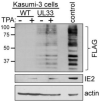
pUL33 is expressed during HCMV latency. Kasumi-3 cells were infected (MOI=1.0) with wild type (WT) or UL33-3xF (UL33) for 7 days, then treated either with DMSO (–TPA) or TPA (+TPA) for an additional 2 days. Cells were then collected, and lysates (60 µg) were assessed for pUL33 expression by immunoblot using an antibody against the C-terminal triple FLAG (3xF) epitope. IE2 served as a control to distinguish latent versus reactivated infections, and cellular actin was probed as a loading control. Lysate from UL33-3xF-lytically infected fibroblasts (MOI=0.5, 96 h, 30 µg) is shown as a positive control for pUL33 expression. Molecular weight markers (in kDa) are shown to the left of the blot. n=3, representative blots shown.
Fig. 4.
Fig. 4.
Disruption of the UL33 G protein-coupling domain attenuates HCMV reactivation. (A) Kasumi-3 cells were infected (MOI=1.0) with the indicated viruses. At 7 dpi, half of each infected population was cultured for an additional 2 days with vehicle (DMSO; –TPA, open bars) or TPA (+TPA, checked bars). Cells were then co-cultured with naïve NuFF-1 fibroblasts to quantify the fold change in the frequency of infectious centers [relative to wild type (WT) –TPA; open gray bar] by ELDA. (B) Kasumi-3 cells were infected (MOI=1.0) as indicated over a 7-day time course in conditions favoring latency. At the indicated times, cells were harvested and cell-associated viral genomes were quantified by qPCR using primers directed at the MIE locus. Viral genome copies are shown relative to those in WT-infected cells (gray bars) at each time point. (C) CD34+ HPCs were infected as indicated (MOI=2.0; 7 days). Cells were then co-cultured with fibroblasts in medium favoring latency (pre-reactivation; open bars) or in culture conditions favoring reactivation (reactivation; checked bars). Fold change in the frequency of infectious centers was quantified by ELDA and is shown relative to WT, pre-reactivation (open gray bar). (A-C) Each data point (circles) is the mean of three technical replicates. Data are mean±s.d. of three biological replicates. Statistical significance was calculated using two-way ANOVA followed by Tukey's post-hoc analysis (*P<0.05; **P<0.01; ****P<0.005; ns, not significant).
Fig. 5.
Fig. 5.
pUL33-mediated signaling induces UL123 transcription in THP-1 cells. (A) THP-1 cells transduced with the indicated lentiviral vectors were treated with DMSO (–DOX) or +DOX, and harvested 24 h post treatment. Lysates (30 μg) were probed with a FLAG antibody to detect pUL33 and pUL33AAA, each with a C-terminal 3xFLAG (3xF) tag. Cell lysate (10 μg) from NuFF-1 fibroblasts lytically infected with TB40/EmCherry-UL33-3xF (UL33-3xF; MOI=0.5, 96 h post infection) is shown as a control for pUL33 expression (control). Cellular actin is shown as a loading control. Molecular weight markers (in kDa) are shown to the left of the blot. Representative blots are shown (n=3). (B) THP-1-pSLIK, (C) THP-1-pSLIK-UL33-3xF, or (D) THP-1-pSLIK-UL33AAA-3xF cells were infected with the indicated viruses (MOI=1.0; 7 days) and maintained under conditions favoring latency. (B-D) At 7 dpi, half of each infected population was cultured for an additional 2 days with vehicle (DMSO) to maintain latency (–TPA, black bars), or with TPA to induce reactivation (+TPA, blue bars) in the absence or presence of DOX. Total RNA was harvested and UL123 was measured by RT-qPCR relative to cellular GAPDH. Each data point (circles) is the mean of three technical replicates. Data are mean±s.d. of three biological replicates, and statistical significance was calculated using two-way ANOVA followed by Tukey's post-hoc analysis (*P<0.05; **P<0.01; ***P<0.001).
Fig. 6.
Fig. 6.
pUL33 activates CREB and enhances its recruitment to the MIE enhancer/promoter. (A) THP-1-pSLIK (empty vector, EV), THP-1-pSLIK-UL33-3xF (UL33), or THP-1-pSLIK-UL33AAA-3xF (UL33AAA) cells were treated with vehicle (DMSO; –DOX) or DOX (+DOX) for 24 h. Lysates (30 µg) were analyzed by immunoblot using antibodies directed at phosphorylated CREB (p-CREB), total CREB (t-CREB), and cellular actin as a loading control. Representative blots are shown (n=3). p-CREB expression relative to t-CREB was quantified by densitometry from two independent experiments and indicated below the blots. (B) Kasumi-3 cells (MOI=1.0; 7 days) were infected with the indicated viruses. At 7 dpi, half of each infected population was cultured for an additional 2 days with vehicle (DMSO; –TPA, open bars) or TPA (+TPA, checked bars). Binding of p-CREB to the MIE enhancer/promoter was quantified by ChIP using an anti-p-CREB antibody. Co-precipitated MIE enhancer/promoter was quantified by qPCR, and data are shown as fold change relative to input for WT –TPA cultures (open gray bar). Each data point (circles) is the mean of three technical replicates. Data are mean±s.d. of three biological replicates, and the statistical significance was calculated using two-way ANOVA followed by Tukey's post-hoc analysis (*P<0.05; ***P<0.001).
Fig. 7.
Fig. 7.
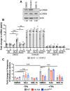
Pharmacological stimulation of p-CREB rescues the reactivation deficiency of UL33Δ- and UL33AAA-infected Kasumi-3 cells treated with TPA. (A) Kasumi-3 cells were treated for 24 h with vehicle control (DMSO), forskolin (20 µM) or 666-15 (1.0 µM). Lysates were harvested for immunoblot and assessed for p-CREB and t-CREB expression. Cellular actin is shown as a loading control. p-CREB expression was quantified by densitometry relative to t-CREB, indicated below the blots. Representative blots are shown (n=3). (B) THP-1-pSLIK (empty vector; EV), THP-1-pSLIK-UL33-3xF (UL33) and THP-1-pSLIK-UL33AAA-3xF (UL33AAA) cells were treated with vehicle (DMSO; –DOX) or DOX (+DOX) in the absence (DMSO) or presence of forskolin (20 µM) or 666-15 (1.0 µM) for 24 h, as indicated. Total RNA was harvested, and Bcl-2 (black) and COX-2 (light gray) were measured by RT-qPCR and plotted as ΔΔCt relative to EV +DMSO/–DOX. (C) Kasumi-3 cells (MOI=1.0) were infected with wild type (WT, gray), UL33Δ (red) and UL33AAA (blue). At 7 dpi, half of each infected population was cultured for an additional 2 days with vehicle (DMSO, –TPA; open bars) or TPA (+TPA; checked bars) in the presence of vehicle control (DMSO), forskolin (20 µM) or 666-15 (1.0 µM). Cells were then co-cultured with naïve NuFF-1 cells to quantify the fold change in the frequency of infectious centers (relative to WT –TPA/DMSO; open gray bar) by ELDA. (B,C) Each data point (circles) is the mean of three technical replicates. Data are mean±s.d. *P<0.05; **P<0.01; ***P<0.001; n.s., not significant (two-way ANOVA and Tukey's post-hoc analysis).

Similar articles

Cited by

References

    1. Adamson, C. S. and Nevels, M. M. (2020). Bright and early: inhibiting human cytomegalovirus by targeting major immediate-early gene expression or protein function. Viruses 12, 110 10.3390/v12010110 - DOI - PMC - PubMed
    1. Arend, K. C., Ziehr, B., Vincent, H. A. and Moorman, N. J. (2016). Multiple transcripts encode full-length human cytomegalovirus IE1 and IE2 proteins during lytic infection. J. Virol. 90, 8855-8865. 10.1128/JVI.00741-16 - DOI - PMC - PubMed
    1. Bate, S. L., Dollard, S. C. and Cannon, M. J. (2010). Cytomegalovirus seroprevalence in the United States: the national health and nutrition examination surveys, 1988-2004. Clin. Infect. Dis. 50, 1439-1447. 10.1086/652438 - DOI - PMC - PubMed
    1. Bechtel, J. T. and Shenk, T. (2002). Human cytomegalovirus UL47 tegument protein functions after entry and before immediate-early gene expression. J. Virol. 76, 1043-1050. 10.1128/JVI.76.3.1043-1050.2002 - DOI - PMC - PubMed
    1. Beisser, P. S., Vink, C., Van Dam, J. G., Grauls, G., Vanherle, S. J. V. and Bruggeman, C. A. (1998). The R33 G protein-coupled receptor gene of rat cytomegalovirus plays an essential role in the pathogenesis of viral infection. J. Virol. 72, 2352-2363. 10.1128/JVI.72.3.2352-2363.1998 - DOI - PMC - PubMed

Publication types

MeSH terms

Substances