. 2020 Dec 1;117(48):30649-30660.
doi: 10.1073/pnas.2007206117. Epub 2020 Nov 16.

Thymus-derived B cell clones persist in the circulation after thymectomy in myasthenia gravis

Affiliations

Thymus-derived B cell clones persist in the circulation after thymectomy in myasthenia gravis

Ruoyi Jiang et al. Proc Natl Acad Sci U S A. .

Abstract

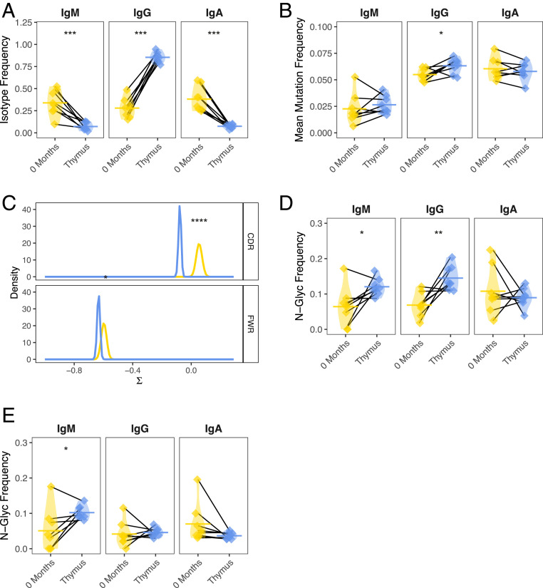
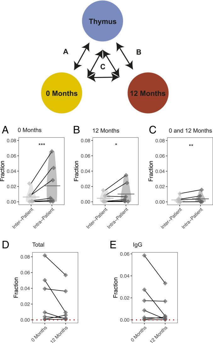
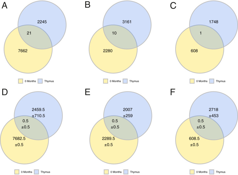
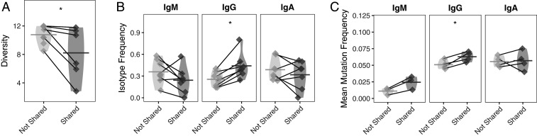
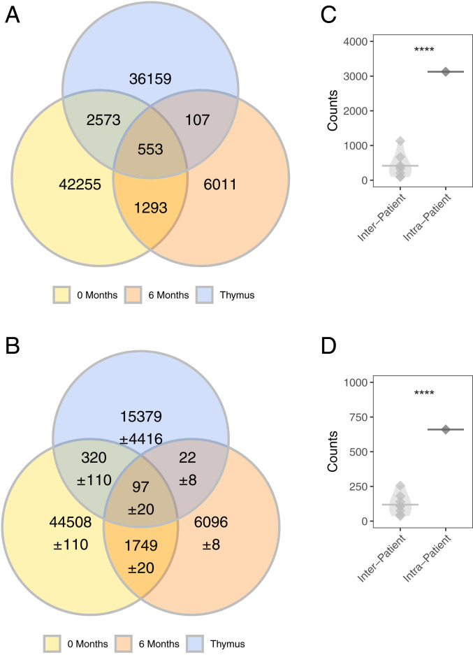
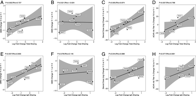
Myasthenia gravis (MG) is a neuromuscular, autoimmune disease caused by autoantibodies that target postsynaptic proteins, primarily the acetylcholine receptor (AChR) and inhibit signaling at the neuromuscular junction. The majority of patients under 50 y with AChR autoantibody MG have thymic lymphofollicular hyperplasia. The MG thymus is a reservoir of plasma cells that secrete disease-causing AChR autoantibodies and although thymectomy improves clinical scores, many patients fail to achieve complete stable remission without additional immunosuppressive treatments. We speculate that thymus-associated B cells and plasma cells persist in the circulation after thymectomy and that their persistence could explain incomplete responses to resection. We studied patients enrolled in a randomized clinical trial and used complementary modalities of B cell repertoire sequencing to characterize the thymus B cell repertoire and identify B cell clones that resided in the thymus and circulation before and 12 mo after thymectomy. Thymus-associated B cell clones were detected in the circulation by both mRNA-based and genomic DNA-based sequencing. These antigen-experienced B cells persisted in the circulation after thymectomy. Many circulating thymus-associated B cell clones were inferred to have originated and initially matured in the thymus before emigration from the thymus to the circulation. The persistence of thymus-associated B cells correlated with less favorable changes in clinical symptom measures, steroid dose required to manage symptoms, and marginal changes in AChR autoantibody titer. This investigation indicates that the diminished clinical response to thymectomy is related to persistent circulating thymus-associated B cell clones.

Keywords: B cells; adaptive immune cell receptor repertoire sequencing; autoimmune disease; myasthenia gravis; thymectomy.


Conflict of interest statement

Competing interest statement: K.C.O. has received research support from Ra Pharma and is a consultant and equity shareholder of Cabaletta Bio. K.C.O. is the recipient of a sponsored research subaward from the University of Pennsylvania, the primary financial sponsor of which is Cabaletta Bio. S.H.K. receives consulting fees from Northrop Grumman. R.J.N. has received research support from Alexion Pharmaceuticals, Genentech, Grifols, and Ra Pharma. H.J.K. has served as an advisor to Alnylam Pharmaceuticals, Ra Pharmaceuticals, and UCB Pharmaceuticals, and is Chief Exectutive Officer and Chief Marketing Officer of ARC Biotechnology, LLC, based on US Patent 8,961,98.

Figures

Fig. 1.
Fig. 1.
The thymus-associated B cell repertoire is distinct from the paired circulating repertoire. AIRR-seq was performed to generate B cell repertoires for eight MGTX trial-derived thymus specimens and paired PBMCs collected at baseline. Key features of antigen-experience B cells in each of the respective repertoires were then compared. (A) Overall constant region (isotype) usage frequencies are quantified for thymus compared to the circulating compartment per patient. Horizontal bars show the average frequency of constant region usage across patients. Frequencies belonging to the same patient are paired with a black line. (B) Individual SHM frequencies for thymus compared to circulating B cells are presented. Horizontal bars show the average SHM frequency for a given sample status (i.e., thymus or circulation). Frequencies belonging to the same patient are paired with a black line. (C) BASELINe probability distribution functions (PDF) are shown for thymus and circulating repertoires, with density shown for a range of selection strengths (∑) on the x axis. PDFs were determined via convolution of subject-specific PDFs for each status to generate a single aggregate status. (D) Individual frequencies for the occurrence of glycosylation (N[X][S/T]) sites are quantified for thymus compared to circulating B cells and (E) for the inferred germline of these sequences. Horizontal bars show the average glycosylation frequency for a given cluster. Frequencies belonging to the same patient are paired with a black line. Statistical differences are shown only when significant (****P < 0.0001; ***P < 0.001; **P < 0.01; *P < 0.05).
Fig. 2.
Fig. 2.
B cell clones are shared between thymus and circulation before and 12 mo after thymectomy. An mRNA-based sequencing approach was used to evaluate how B cells clones are shared between the thymus and circulation before and 12 mo after thymectomy. The schematic diagram shows the corresponding computation of clonal sharing associated with each subpanel below. Clonal sharing is quantified as the number of shared clones divided by the number of clones in the corresponding thymus repertoire (or background thymus repertoire). This is shown for the sharing of clones found in the thymus with A clones found in prethymectomy circulation, (B) the postthymectomy circulation samples, and also (C) clones shared between the pre- and postthymectomy circulation samples. The change in sharing per patient is also shown before and after thymectomy (D) for both the total repertoire and (E) for the repertoire filtered only for IgG-switched V(D)J sequences. Quantification of clonal sharing from the same patient is paired with a gray line. Statistical differences are shown only when significant (***P < 0.001; **P < 0.01; *P < 0.05).
Fig. 3.
Fig. 3.
B cell clones are shared between thymus and circulation before a thymectomy. A gDNA-based sequencing approach was used to confirm that B cells clones are shared between the thymus and circulation before thymectomy. The observed number of shared clones is shown for patient THY6 (A), patient THY7 (B), and patient THY2 (C). The computed background sharing is shown as Venn-diagrams for THY6 (D), patient THY7 (E), and patient THY2 (F). Background sharing was computed by changing the identity of the thymus used to assess for clonal sharing: That is, the observed sharing of clones from thymus samples derived from different patients with the circulation and computing an average. Uncertainty was computed as the SD of values from the corresponding background sharing and are shown in DF using the “±” symbol.
Fig. 4.
Fig. 4.
Features of clones shared between the thymus and prethymectomy circulation are distinct from circulating clones that are not shared. (A) Simpson’s diversity of shared prethymectomy and thymus clones in the circulation. Lower values of diversity correspond to greater clonal expansion; that is, circulating clones shared with the thymus are more clonally expanded than their nonshared circulating counterparts. Diversity values belonging to the same patient are paired with a gray line. (B) Usage of different isotypes among shared and nonshared clones is shown for IgM, IgG, and IgA for the circulating repertoire. Horizontal bars show the average isotype usage frequency for clones that are either shared or not shared. Frequencies belonging to the same patient are paired with a gray line. (C) Average SHM frequencies among shared and nonshared clones are shown for IgM, IgG, and IgA for the circulating repertoire. Horizontal bars show the average SHM frequency for a given cluster. Frequencies belonging to the same patient are paired with a gray line. Statistical differences are shown only when significant (*P < 0.05).
Fig. 5.
Fig. 5.
Confirmation of B cell clonal sharing with the circulation in specimens (THY-Y) independent of the MGTX trial. (A) Observed number of clones shared between the prethymectomy, postthymectomy circulation, and thymus shown as Venn diagrams. (B) Computed background sharing between the prethymectomy, postthymectomy circulation and thymus are shown as Venn diagrams. Background sharing was computed by permuting the identity of the thymus used to assess for clonal sharing and computing an average; this approach is identical to the approach used to compute clonal sharing for thymus samples from the MGTX trial. Uncertainty was computed as the SD of the background clonal sharing and shown using the “±” symbol. Statistical testing of observed counts of shared clones between circulation and thymus repertoire (C) prethymectomy and (D) postthymectomy. Statistical differences are shown only when significant (****P < 0.0001).
Fig. 6.
Fig. 6.
Correlation between clinical outcome measurements and circulating thymus-associated B cells. Correlation of the log fold-change in clonal sharing between the thymus and circulation as assessed with a Bray–Curtis index (on the x axis) and with (A) the change in QMG score between years 1 and 2 after thymectomy, (B) the change in QMG score between baseline and year 1 after thymectomy, (C) the change in steroid dose between years 1 and 2 after thymectomy, and (D) the percentage change in AChR autoantibody titer between years 1 and 2 after thymectomy (on the y axis). The same analysis is shown for the log fold-change in IgG clonal sharing (EH), respectively. AChR autoantibody titer was not collected for THY7 (D and H).

Comment in

  • Thymectomy and myasthenia gravis.
    Lisak RP, Richman DP. Lisak RP, et al. Proc Natl Acad Sci U S A. 2020 Dec 22;117(51):32195-32196. doi: 10.1073/pnas.2022901117. Epub 2020 Dec 3. Proc Natl Acad Sci U S A. 2020. PMID: 33273117 Free PMC article. No abstract available.

References

    1. Vincent A., Unravelling the pathogenesis of myasthenia gravis. Nat. Rev. Immunol. 2, 797–804 (2002). - PubMed
    1. Gilhus N. E., et al. , Myasthenia gravis—Autoantibody characteristics and their implications for therapy. Nat. Rev. Neurol. 12, 259–268 (2016). - PubMed
    1. Lindstrom J. M., Engel A. G., Seybold M. E., Lennon V. A., Lambert E. H., Pathological mechanisms in experimental autoimmune myasthenia gravis. II. Passive transfer of experimental autoimmune myasthenia gravis in rats with anti-acetylcholine recepotr antibodies. J. Exp. Med. 144, 739–753 (1976). - PMC - PubMed
    1. Oda K., Korenaga S., Ito Y., Myasthenia gravis: Passive transfer to mice of antibody to human and mouse acetylcholine receptor. Neurology 31, 282–287 (1981). - PubMed
    1. Sterz R., et al. , Effector mechanisms in myasthenia gravis: End-plate function after passive transfer of IgG, Fab, and F(ab′)2 hybrid molecules. Muscle Nerve 9, 306–312 (1986). - PubMed

Publication types

MeSH terms