. 2020 Dec 1;117(48):30775-30786.
doi: 10.1073/pnas.2016959117. Epub 2020 Nov 16.

The intracellular Ca2+ release channel TRPML1 regulates lower urinary tract smooth muscle contractility

Affiliations

The intracellular Ca2+ release channel TRPML1 regulates lower urinary tract smooth muscle contractility

Caoimhin S Griffin et al. Proc Natl Acad Sci U S A. .

Abstract

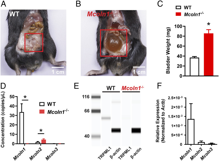
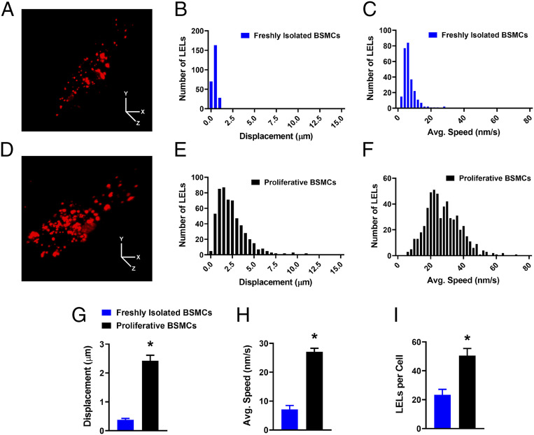
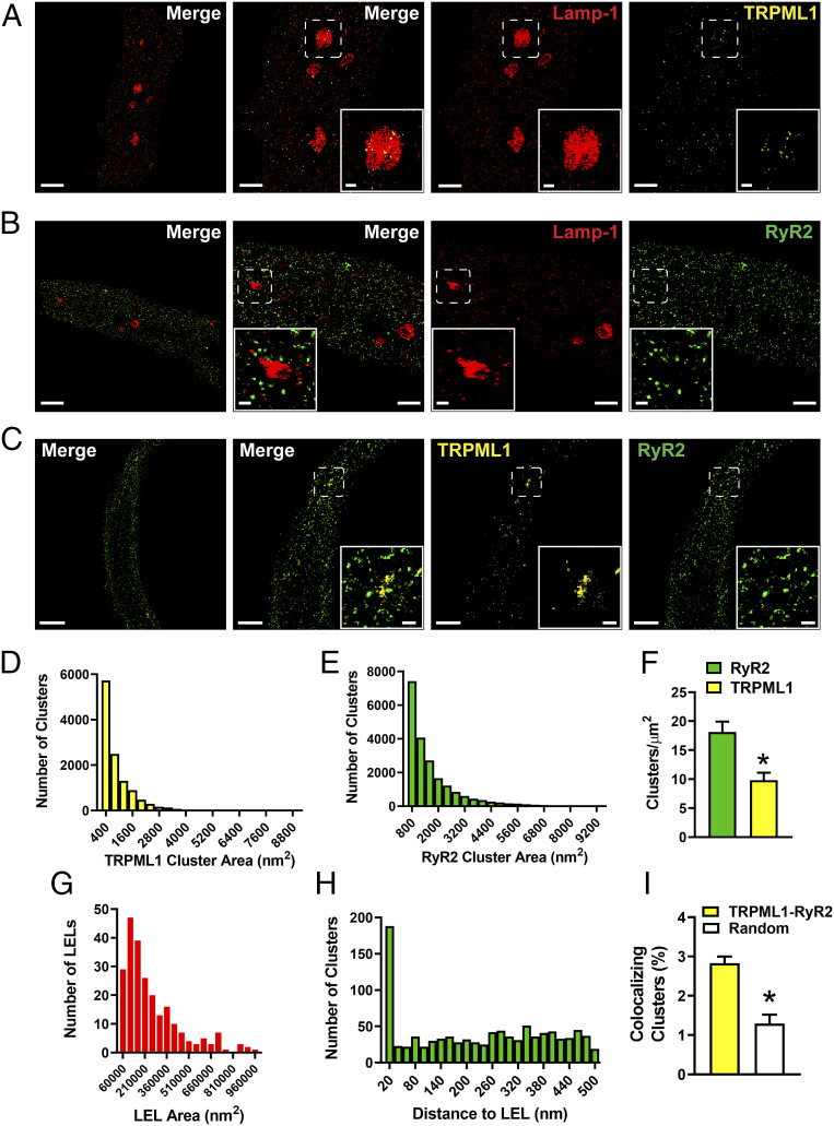
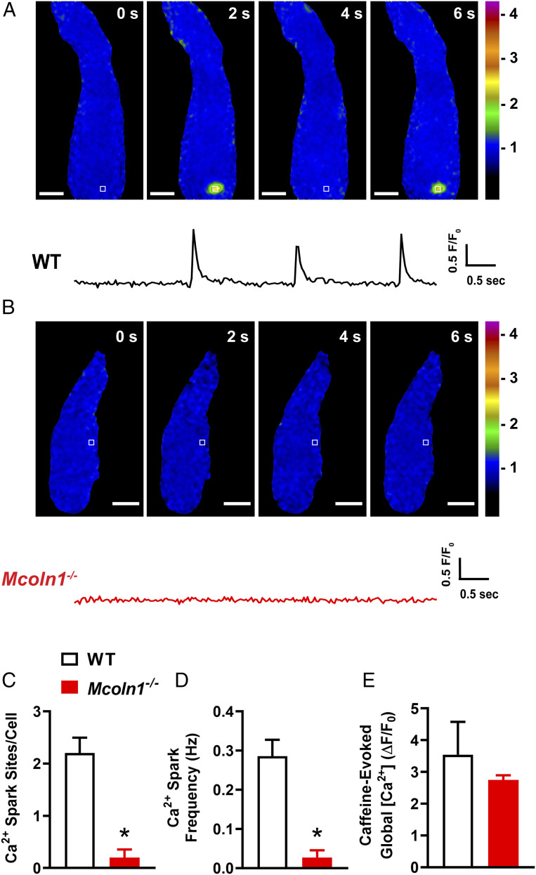
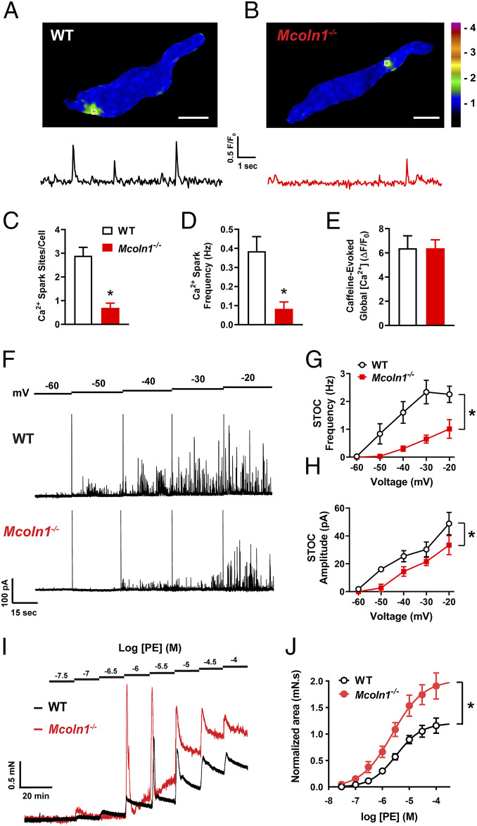
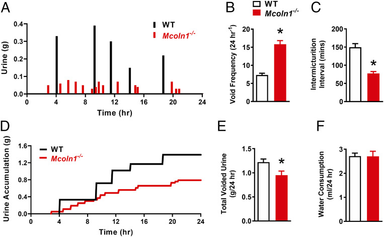
TRPML1 (transient receptor potential mucolipin 1) is a Ca2+-permeable, nonselective cation channel that is predominantly localized to the membranes of late endosomes and lysosomes (LELs). Intracellular release of Ca2+ through TRPML1 is thought to be pivotal for maintenance of intravesicular acidic pH as well as the maturation, fusion, and trafficking of LELs. Interestingly, genetic ablation of TRPML1 in mice (Mcoln1-/- ) induces a hyperdistended/hypertrophic bladder phenotype. Here, we investigated this phenomenon further by exploring an unconventional role for TRPML1 channels in the regulation of Ca2+-signaling activity and contractility in bladder and urethral smooth muscle cells (SMCs). Four-dimensional (4D) lattice light-sheet live-cell imaging showed that the majority of LELs in freshly isolated bladder SMCs were essentially immobile. Superresolution microscopy revealed distinct nanoscale colocalization of LEL-expressing TRPML1 channels with ryanodine type 2 receptors (RyR2) in bladder SMCs. Spontaneous intracellular release of Ca2+ from the sarcoplasmic reticulum (SR) through RyR2 generates localized elevations of Ca2+ ("Ca2+ sparks") that activate plasmalemmal large-conductance Ca2+-activated K+ (BK) channels, a critical negative feedback mechanism that regulates smooth muscle contractility. This mechanism was impaired in Mcoln1-/- mice, which showed diminished spontaneous Ca2+ sparks and BK channel activity in bladder and urethra SMCs. Additionally, ex vivo contractility experiments showed that loss of Ca2+ spark-BK channel signaling in Mcoln1-/- mice rendered both bladder and urethra smooth muscle hypercontractile. Voiding activity analyses revealed bladder overactivity in Mcoln1-/- mice. We conclude that TRPML1 is critically important for Ca2+ spark signaling, and thus regulation of contractility and function, in lower urinary tract SMCs.

Keywords: calcium signaling; endolysosomes; ion channels; lower urinary tract; superresolution microscopy.


Figures

Fig. 1.
Fig. 1.
Bladder abnormalities in Mcoln1−/− mice and TRPML subtype expression in Mcoln1−/− BSMCs. (A and B) Representative images of the typical bladder anatomy observed in WT mice (A) and the hyperdistended/hypertrophic bladder phenotype observed in all Mcoln1−/− mice (B). (C) Summary data illustrating the differences in whole-bladder weight (in milligrams) between WT and Mcoln1−/− mice (n = 8 animals/group; *P < 0.05). (D) ddPCR analysis of the expression of Mcoln1, Mcoln2, and Mcoln3 mRNA (copies/µL PCR) in BSM from WT and Mcoln1−/− mice (n = 4 animals/group; *P < 0.05). (E) Representative Wes analysis results for TRPML1 protein in BSM. A single band (∼60 kDa) was detected in lysates of BSM from WT mice, but was absent in lysates of BSM from Mcoln1−/− mice (n = 4 animals/group). β-Actin was used as a loading control. (F) qRT-PCR analysis of Mcoln1, Mcoln2, and Mcoln3 mRNA expression levels (normalized to Actb) in FACS-enriched homogeneous populations of BSMCs (n = 3 animals/group). All data are presented as means ± SEM.
Fig. 2.
Fig. 2.
LELs are immobile in freshly isolated BSMCs. (A) Representative lattice light-sheet image of a freshly isolated BSMC stained with LysoTracker Red. (Scale bars, 10 µm.) (B and C) Histogram showing the distributions of LEL total displacement (B) and average speed (C) within freshly isolated BSMCs, recorded over a 10-min period. A total of 262 LELs (n = 12 cells) were tracked and analyzed. (D) Representative lattice light-sheet image of a cultured, proliferative BSMC stained with LysoTracker Red. (Scale bars, 10 µm.) (E and F) Histogram showing the distributions of LEL total displacement (E) and average speed (F) within proliferative BSMCs, recorded over a 10-min period. A total of 555 LELs (n = 11 cells) were tracked and analyzed. Summary data showing the mean values for LEL (G) displacement and (H) average speed, and the (I) cellular density of LELs in both freshly isolated and proliferative BSMCs (freshly isolated BSMCs, n = 12 cells from n = 4 mice; proliferative BSMCs, n = 11 cells from n = 4 mice; *P < 0.05). All data are shown as mean ± SEM.
Fig. 3.
Fig. 3.
Nanoscale colocalization of TRPML1 with RyR2 and Lamp-1 in native BSMCs. (AC) Representative superresolution localization maps of isolated BSMCs coimmunolabeled for Lamp-1 and TRPML1 (A), Lamp-1 and RyR2 (B), or TRPML1 and RyR2 (C). (Scale bars, 2 µm.) Representative of n = 8 to 10 cells isolated from n = 3 to 4 animals. The second panel shows a magnified view of the region enclosed in the white boxes. (Scale bars, 1 µm.) Insets show magnified views of the indicated regions of interest. (Scale bars, 0.2 µm.) (D and E) Histograms showing the distribution of the surface areas of individual protein clusters for TRPML1 (D) and RyR2 (E) (TRPML1, n = 11,862 clusters; RyR2, n = 21,030 clusters). (F) TRPML1 and RyR2 protein cluster density (n = 12 cells, n = 4 animals per group; *P < 0.05). (G) Histogram showing that individual Lamp-1 ovoid metastructures are between 62,000 and 1,000,000 nm2 in size (mean area = 270,779 ± 13,888 nm2, n = 240). (H) Nearest-neighbor analysis showing the distribution of distances between the center of RyR2 protein clusters and the outer edge of Lamp-1–positive LELs (n = 1,019 RyR2 protein clusters). (I) Object-based analysis comparing the fraction of TRPML1 and RyR2 colocalizing clusters with a simulated random distribution of clusters (TRPML1–RyR2, 2.83% ± 0.17%; random, 1.29% ± 0.23%; n = 8 cells from three animals; *P < 0.05). All data are presented as means ± SEM.
Fig. 4.
Fig. 4.
BSMCs from Mcoln1−/− mice lack spontaneous Ca2+ sparks. (A) Representative confocal time course images of a Ca2+ spark site (in seconds) in a Fluo-4 AM loaded BSMC from a WT mouse, and trace showing changes in fractional fluorescence (F/F0) in a defined region of interest (white box). (Scale bar, 10 µm.) (B) Representative confocal time course images (in seconds) and complimentary trace recorded from a BSMC isolated from a Mcoln1−/− mouse showing lack of Ca2+ spark activity. (Scale bar, 10 µm.) (C and D) Summary data showing (C) the number of spontaneous Ca2+ spark sites per cell and (D) Ca2+ spark frequency in BSMCs isolated from WT and Mcoln1−/− mice (WT, n = 20 cells from n = 4 mice; Mcoln1−/−, n = 20 cells from n = 4 mice; *P < 0.05). (E) Total SR Ca2+ store load in BSMCs from WT and Mcoln1−/− mice, assessed by imaging changes in global intracellular [Ca2+] in response to administration of caffeine (10 mM) (WT, n = 7 cells from n = 4 mice; Mcoln1−/−, n = 7 cells from n = 4 mice). All data are presented as means ± SEM.
Fig. 5.
Fig. 5.
Reduced spontaneous BK channel activity in BSMCs isolated from Mcoln1−/− mice. (A) Representative traces of STOCs recorded in the perforated-patch configuration from voltage-clamped (−40 and −20 mV) BSMCs isolated from WT and Mcoln1−/− mice. (B and C) Summary data for mean STOC frequency (B) and amplitude (C) over a range of membrane potentials (−60 to −20 mV) in BSMCs isolated from WT and Mcoln1−/− mice (WT, n = 9 cells from n = 5 mice; Mcoln1−/−, n = 7 cells from n = 4 mice; *P < 0.05). (D) Representative traces showing increases in STOC frequency following application of the TRPML activator ML-SA1 (3 µM) in BSMCs isolated from WT, but not Mcoln1−/− mice. BSMCs were voltage clamped at −30 mV. (E) Summary data showing the effects of ML-SA1 on STOC frequency (WT, n = 9 cells from n = 5 mice; Mcoln1−/−, n = 8 cells from n = 4 mice; *P < 0.05). All data are presented as means ± SEM.
Fig. 6.
Fig. 6.
BSM contractility is enhanced in Mcoln1−/− mice. (A) Representative isometric tension recordings showing spontaneous contractions intrinsic to WT and Mcoln1−/− BSM strips. (B and C) Summary data for mean contraction (B) amplitude and (C) frequency (n = 11 muscle strips from n = 6 animals for both groups; *P < 0.05). (D) Representative isometric tension traces showing BSM contractions induced by the cumulative addition of increasing concentrations (1 nM to 10 µM) of the muscarinic receptor agonist CCh in WT (black) and Mcoln1−/− (red) mice. (E) Concentration–response data plotting the mean amplitude of CCh-induced contractions of BSM strips from WT and Mcoln1−/− mice, normalized to the high-K+ response (WT, n = 8 muscle strips from n = 4 mice; Mcoln1−/−, n = 9 muscle strips from n = 5 mice; *P < 0.05). All data are presented as means ± SEM.
Fig. 7.
Fig. 7.
USMCs from Mcoln1−/− mice show impaired Ca2+ sparks and STOC activity, leading to USM hypercontractility. (A) Representative confocal image of a Ca2+ spark site (Scale bar, 10 µm.) in a Fluo-4 AM loaded USMC from a WT mouse, and trace showing changes in fractional fluorescence (F/F0) in a defined region of interest (white box). (B) Representative confocal image (Scale bar, 10 µm.) and complimentary trace recorded from a USMC isolated from a Mcoln1−/− mouse showing reduced Ca2+ spark activity. (C and D) Summary data showing (C) the number of spontaneous Ca2+ spark sites per cell and (D) Ca2+ spark frequency in USMCs isolated from WT and Mcoln1−/− mice (WT, n = 19 cells from n = 4 mice; Mcoln1−/−, n = 20 cells from n = 4 mice; *P < 0.05). (E) Total SR Ca2+ store load in USMCs from WT and Mcoln1−/− mice, assessed by imaging changes in global intracellular [Ca2+] in response to administration of caffeine (10 mM) (WT, n = 7 cells from n = 4 mice; Mcoln1−/−, n = 8 cells from n = 4 mice). (F) Representative traces of STOCs recorded in the perforated-patch configuration from voltage-clamped (−60 to −20 mV) USMCs isolated from WT and Mcoln1−/− mice. (G and H) Summary data for mean STOC frequency (G) and amplitude (H) over a range of membrane potentials (−60 to −20 mV) in WT and Mcoln1−/− mice (n = 6 cells from n = 4 mice in both groups; *P < 0.05). (I) Representative isometric tension traces showing USM contractions induced by the cumulative addition of increasing concentrations (30 nM to 100 µM) of the α1-AR agonist phenylephrine (PE) in WT (black) and Mcoln1−/− (red) mice. (J) Concentration–response data plotting mean amplitudes of PE-induced contractions of USM ring preparations from WT and Mcoln1−/− mice, normalized to the high-K+ response (WT, n = 6 from n = 6 mice; Mcoln1−/−, n = 6 from n = 6 mice; *P < 0.05). All data are presented as means ± SEM.
Fig. 8.
Fig. 8.
Mcoln1−/− mice show hyperactive voiding. (A) Representative voiding micturogram displaying the micturition activity of WT (black) and Mcoln1−/− (red) mice over a 24-h period. (B and C) Summary data showing the daily average void frequency (B) and intermicturition interval (C) for WT and Mcoln1−/− mice (WT, n = 6; Mcoln1−/−, n = 6; *P < 0.05). (D) Representative trace, complimentary to the micturogram in A, showing urine accumulation in WT (black) and Mcoln1−/− (red) mice. (E and F) Summary data showing the total daily voided urine volume (E) and water consumption (F) for WT and Mcoln1−/− mice (WT, n = 6; Mcoln1−/−, n = 6; *P < 0.05). All data are presented as means ± SEM.

Comment in

  • Igniting Ca2+ sparks with TRPML1.
    Sergeant GP, Hollywood MA, Thornbury KD. Sergeant GP, et al. Proc Natl Acad Sci U S A. 2020 Dec 29;117(52):32836-32838. doi: 10.1073/pnas.2022896117. Epub 2020 Dec 1. Proc Natl Acad Sci U S A. 2020. PMID: 33262276 Free PMC article. No abstract available.

References

    1. Montell C., The TRP superfamily of cation channels. Sci. STKE 2005, re3 (2005). - PubMed
    1. Sun M., et al. , Mucolipidosis type IV is caused by mutations in a gene encoding a novel transient receptor potential channel. Hum. Mol. Genet. 9, 2471–2478 (2000). - PubMed
    1. Venkatachalam K., Wong C. O., Zhu M. X., The role of TRPMLs in endolysosomal trafficking and function. Cell Calcium 58, 48–56 (2015). - PMC - PubMed
    1. Lindvall J. M., Blomberg K. E., Wennborg A., Smith C. I., Differential expression and molecular characterisation of Lmo7, Myo1e, Sash1, and Mcoln2 genes in Btk-defective B-cells. Cell. Immunol. 235, 46–55 (2005). - PubMed
    1. Samie M. A., et al. , The tissue-specific expression of TRPML2 (MCOLN-2) gene is influenced by the presence of TRPML1. Pflugers Arch. 459, 79–91 (2009). - PMC - PubMed

Publication types

MeSH terms