. 2020 Dec 1;117(48):30577-30588.
doi: 10.1073/pnas.2013012117. Epub 2020 Nov 16.

Exo1 recruits Cdc5 polo kinase to MutLγ to ensure efficient meiotic crossover formation

Affiliations

Exo1 recruits Cdc5 polo kinase to MutLγ to ensure efficient meiotic crossover formation

Aurore Sanchez et al. Proc Natl Acad Sci U S A. .

Abstract

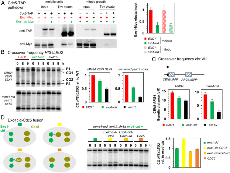
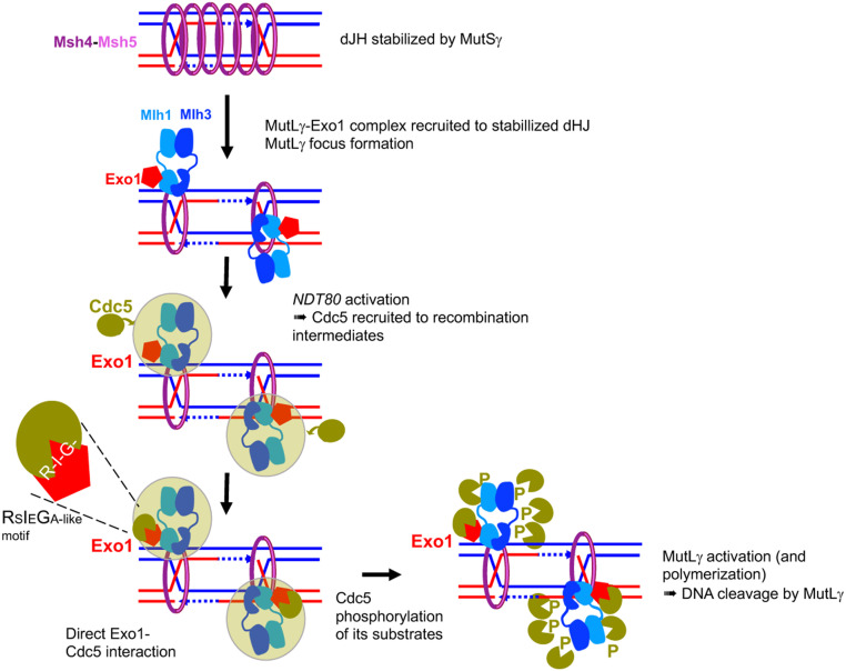
Crossovers generated during the repair of programmed meiotic double-strand breaks must be tightly regulated to promote accurate homolog segregation without deleterious outcomes, such as aneuploidy. The Mlh1-Mlh3 (MutLγ) endonuclease complex is critical for crossover resolution, which involves mechanistically unclear interplay between MutLγ and Exo1 and polo kinase Cdc5. Using budding yeast to gain temporal and genetic traction on crossover regulation, we find that MutLγ constitutively interacts with Exo1. Upon commitment to crossover repair, MutLγ-Exo1 associate with recombination intermediates, followed by direct Cdc5 recruitment that triggers MutLγ crossover activity. We propose that Exo1 serves as a central coordinator in this molecular interplay, providing a defined order of interaction that prevents deleterious, premature activation of crossovers. MutLγ associates at a lower frequency near centromeres, indicating that spatial regulation across chromosomal regions reduces risky crossover events. Our data elucidate the temporal and spatial control surrounding a constitutive, potentially harmful, nuclease. We also reveal a critical, noncatalytic role for Exo1, through noncanonical interaction with polo kinase. These mechanisms regulating meiotic crossovers may be conserved across species.

Keywords: MutL; crossovers; meiosis; polo kinase; recombination.


Conflict of interest statement

The authors declare no competing interest.

Figures

Fig. 1.
Fig. 1.
Mlh3 forms foci on yeast pachytene meiotic chromosomes, distinct from ZMM foci. (A) Crystal structure of the C-terminal region of Saccharomyces cerevisiae Mlh1–Mlh3 heterodimer showing the position of the internal tags in Mlh3. The Mlh1 and Mlh3 regions are colored in light and dark blue, respectively. The Mlh1 binding motif for Exo1 and the endonuclease site of Mlh3 are colored in red and yellow, respectively. (B) Comparison of Mlh3–Myc18 and Zip3 foci. (C) Comparison of Mlh3–Myc18 and Msh4-HA foci. (D and E) Quantification of Mlh3, Zip3, and Msh4 foci from B and C. (F and G): Mlh3-myc foci in zip3∆ (F) and msh4∆ (G). (H) Quantification of Mlh3-Myc foci in zip3∆ and msh4∆ mutants from B, F, and G; 51 nuclei examined in each. (I) Colocalization quantification of Mlh3 with Zip3 or Msh4 foci. The percent of Mlh3 foci colocalizing is indicated. (J) Quantification of Mlh3 foci in pachytene-selected nuclei of wild-type (same as in B) and ndt80∆; 51 nuclei examined in each. (BJ) All experiments at 4 h in meiosis, except ndt80∆ (6 h in meiosis). Scale bars: 5 μm. See also SI Appendix, Fig. S1.
Fig. 2.
Fig. 2.
Mlh3 associates with meiotic DSB hotspots and its distribution is influenced locally by specific chromosome features. (A) Mlh3–Myc levels at the three indicated meiotic DSB hotspots and one axis-associated site relative to a negative control site (NFT1) assessed by ChIP and qPCR during meiotic time courses. Values are the mean ± SEM from three (four in wild-type) independent experiments at the time-point of maximum enrichment. The full corresponding time courses are in SI Appendix, Fig. S2. The cartoon illustrates the position of sites analyzed by qPCR relative to the meiotic chromosome structure. (B) ChIP-seq binding of Mlh3 (at 5.5 h in meiosis) compared to the binding of the Mer3 and Zip4 (33) ZMMs, to DSBs (Spo11 oligos) (82), and to axis sites (Red1 ChIP-seq) (72). Normalized data are smoothed with a 200-bp window. (C) Average ChIP-seq signal at the indicated features. Same data as in B). The Mer3 and Zip4 ChIP-seq signals are aligned on the Spo11 hotspots midpoints from ref. , and Mlh3 ChIP-seq signal on the pCUP1-IME1 Spo11 hotspots midpoints (this study). (Bottom) ChIP-seq signal is aligned on the Red1 peaks summits from ref. . (D) ZMM and Mlh3 signals per DSB vary with the proximity to a centromere or a telomere. The ChIP-seq signal of each protein divided by their corresponding Spo11 oligo signal (Spo11 signal from ref. for Mer3 and Zip4, and pCUP1-IME1 Spo11 signal for Mlh3) was computed on the width plus 1 kb on each side of the strongest corresponding 2,000 Spo11 hotspots at the indicated chromosome regions: Interstitial (1,805 and 1,829 hotspots); 0 to10 kb from a centromere (29 and 28 hotspots); 10 to 20 kb from a centromere (50 and 46 hotspots); 0 to 40 kb from a telomere (117 and 97 hotspots). For each region, the corresponding interstitial control was computed by randomly selecting groups of interstitial hotspot regions with the same median Spo11 oligo level; this step was repeated 10,000 times. Statistical differences (Mann–Whitney Wilcoxon test) between different regions and their interstitial control are indicated. (E) Examples of DSB (pCUP1-IME1 Spo11 oligo) and Mlh3 binding in an interstitial region and two pericentromeric regions. The normalized signal smoothed with a 200-bp window size is indicated. (F) The ZMM and Mlh3 signals per DSB vary with the timing of DNA replication. Same legend as in D for the indicated chromosomal regions, except that early and late regions were directly compared to each other. See also SI Appendix, Figs. S2–S5.
Fig. 3.
Fig. 3.
MutLγ forms a complex with Exo1 and transiently interacts with MutSγ in vivo. (A) Affinity pull-down of Mlh3-Flag from cells at 4 h in meiosis. Silver-stained gel of pulled-down proteins. Table: Mass-spectrometry analysis of selected proteins reproducibly identified in all replicates and not in the controls (no tag strain). The number of peptides of four independent experiments is shown (two with benzonase treatment, two without). Detail of pulled-down proteins in Dataset S3. (B) Coimmunoprecipitation by Exo1-TAP or Msh4-TAP from cells at 4 h in meiosis analyzed by Western blot. The asterisk indicates the uncleaved Msh4-TAP, of which the protein A region is weakly recognized by the anti-HA antibody. The tobacco etch virus (Tev)-cleaved Msh4-TAP is no longer recognized by the anti-HA. (C) Exo1-TAP pull-down throughout meiosis. The graph indicates the ratio of immunoprecipitated Mlh1 and Mlh3 relative to Exo1 in the Tev eluates at the indicated times in meiosis. Values are mean ± SD of two independent experiments. (D) Coimmunoprecipitation between Pms1-Flag and Mlh3-Myc (Upper) or between Mlh3-Flag and Mlh3–Myc (Lower) from cells at 4 h in meiosis analyzed by Western blot. Asterisks indicate a nonspecific cross-hybridizing bands. See also SI Appendix, Fig. S6.
Fig. 4.
Fig. 4.
The Cdc5 kinase interacts with both MutLγ and Exo1 bound on recombination sites. (A) Coimmunoprecipitation by Cdc5-TAP of Mlh1-HA, Mlh3-Myc, and Exo1-Myc from pCUP1-IME1 synchronized cells at 5.5 h in meiosis analyzed by Western blot. (B) Mlh3-Myc and Cdc5-TAP association with the indicated DSB hotspots as revealed by ChIP and qPCR at the indicated times of a pCUP1-IME1 synchronized meiotic time course. Same normalization as in Fig. 2. Values are the average of three (Mlh3-Myc) or four (Cdc5-TAP) independent experiments ± SEM (C) Coimmunoprecipitation by Cdc5-TAP of Mlh1-HA and Mlh3-Myc is independent of Exo1. Same conditions as in A. (D) Coimmunoprecipitation by Cdc5-TAP of Exo1 is independent of Exo1 interaction with MutLγ. Same conditions as in A. (E) Direct, phosphorylation-independent interaction between recombinant Exo1 and Cdc5 proteins. Western blot showing the pull-down of purified Cdc5-PM (phosphomimetic; see Materials and Methods) by Exo1-Flag, in the presence or absence of CDK1 or λ-phosphatase. See also SI Appendix, Fig. S7.
Fig. 5.
Fig. 5.
Cdc5 directly interacts with Exo1 through a noncanonical site. (A) Delineation of the Exo1 motif responsible for interaction with Cdc5 PBD by two-hybrid assays. The GAL4-BD fusions with indicated Exo1 fragments were tested in combination with a GAL4–AD–Cdc5–PBD fusion; “+” indicates an interaction. Exo1-cid: Cdc5 interaction-deficient. (B) Conservation of the Exo1 region interacting with Cdc5 PBD and illustration of the Exo1-cid mutation. The Dbf4 motif interacting with Cdc5 PBD (47) is indicated (consensus from 17 Saccharomycetaceae family species; see Materials and Methods). (C) Modeling of Dbf4 and Exo1 motifs on the crystal structure of Cdc5 PBD. (i) Crystal structure of the Cdc5 PBD bound to a Dbf4-derived peptide encompassing the RSIEGA motif (PDB ID code 6MF6) (48). The structural elements of the polo box domain are color coded. The region of the domain where phosphorylated substrates bind is labeled for reference. (ii) Model of the Cdc5–Exo1 interaction based on the crystal structure of the Cdc5–Dbf4 complex with Cdc5 shown in the same orientation and color scheme as in (i). The inset shows the residues mediating the interaction between the RSIEGA motif of Exo1 (red labels) and Cdc5 (black labels). (D) The same surface of Cdc5 used for interaction with Dbf4 is used for interaction with Exo1. The GAL4–BD fusions with indicated fragments were tested in combination with a GAL4–AD–Cdc5–PBD fusion with the indicated mutations (WHK stands for W517F H641A K643M); “+” indicates an interaction. See also SI Appendix, Figs. S7 and S8.
Fig. 6.
Fig. 6.
Cdc5 direct interaction with Exo1 promotes crossover formation. (A) Coimmunoprecipitation by Cdc5-TAP of Exo1-Myc in meiotic cells or in cells growing mitotically. Same conditions as in Fig. 4A. (Right) Quantification of Exo1-Myc levels in the Tev eluate relative to the input. Error bars represent SD of two independent experiments. (B) Meiotic crossover frequencies at the HIS4LEU2 hotspot in the exo1-cid mutant. (Left) Representative Southern blot analysis of crossovers in the indicated exo1 mutants, in an otherwise wild-type (MMS4 YEN1 SLX4) or triple nuclease mutant (mms4-md yen1∆ slx4∆) background. mms4-md stands for pCLB2-mms4. (Right) Quantification of crossovers. Values are the mean ± SEM of four independent experiments (wild-type background) or mean ± SD of two independent experiments (triple nuclease mutant), normalized to the corresponding EXO1 value. (C) Meiotic crossovers on chromosome VIII. (Left) Illustration of the fluorescent spore set-up (78). (Right) Genetic distances measured in the CEN8-ARG4 genetic interval for each indicated genotype. (D) Meiotic crossover frequencies at the HIS4LEU2 hotspot with an Exo1-cid–Cdc5 fusion protein. (Left) Scheme illustrating the different experimental setups. In each cell, proteins are expressed from the two allelic endogenous EXO1 promoters (Left) or CDC5 promoters (Right). Same legend as in B. Values are the mean ± SD of two independent experiments, normalized to the homozygous exo1-cid-Myc value.
Fig. 7.
Fig. 7.
Model of MutLγ binding to sites of recombination and activation for crossover formation. MutSγ stabilizes dHJ intermediates and forms foci on chromosomes. MutLγ, in complex with Exo1, binds dJH intermediates that have been stabilized by MutSγ and other ZMM proteins. For simplicity, MutSγ is shown embracing both DNA duplexes of the intermediates, but recent data also suggest it may embrace separately each DNA duplex (74). Once bound on the dHJ, MutLγ may form a focus when interacting with MutSγ (model shown here), or later in the process of its activation (not shown). Upon NDT80 activation, the Cdc5 kinase is induced, and interacts with the MutLγ–Exo1 complex through multiple interactions (green cloud). Among these interactions, the direct interaction of Cdc5 with Exo1 is important for MutLγ-driven crossover formation. We propose that this interaction allows Cdc5 to phosphorylate MutLγ, which activates its nuclease function and produces crossover formation. This may occur through transient MutLγ polymerization.

References

    1. Hunter N., Meiotic recombination: The essence of heredity. Cold Spring Harb. Perspect. Biol. 7, a016618 (2015). - PMC - PubMed
    1. Lam I., Keeney S., Mechanism and regulation of meiotic recombination initiation. Cold Spring Harb. Perspect. Biol. 7, a016634 (2014). - PMC - PubMed
    1. De Muyt A., et al. , BLM helicase ortholog Sgs1 is a central regulator of meiotic recombination intermediate metabolism. Mol. Cell 46, 43–53 (2012). - PMC - PubMed
    1. Allers T., Lichten M., Differential timing and control of noncrossover and crossover recombination during meiosis. Cell 106, 47–57 (2001). - PubMed
    1. Hunter N., Kleckner N., The single-end invasion: An asymmetric intermediate at the double-strand break to double-Holliday junction transition of meiotic recombination. Cell 106, 59–70 (2001). - PubMed

Publication types

MeSH terms