. 2020 Dec 1;117(48):30722-30727.
doi: 10.1073/pnas.2016858117. Epub 2020 Nov 16.

Fast recovery of disrupted tip links induced by mechanical displacement of hair bundles

Affiliations

Fast recovery of disrupted tip links induced by mechanical displacement of hair bundles

R G Alonso et al. Proc Natl Acad Sci U S A. .

Abstract

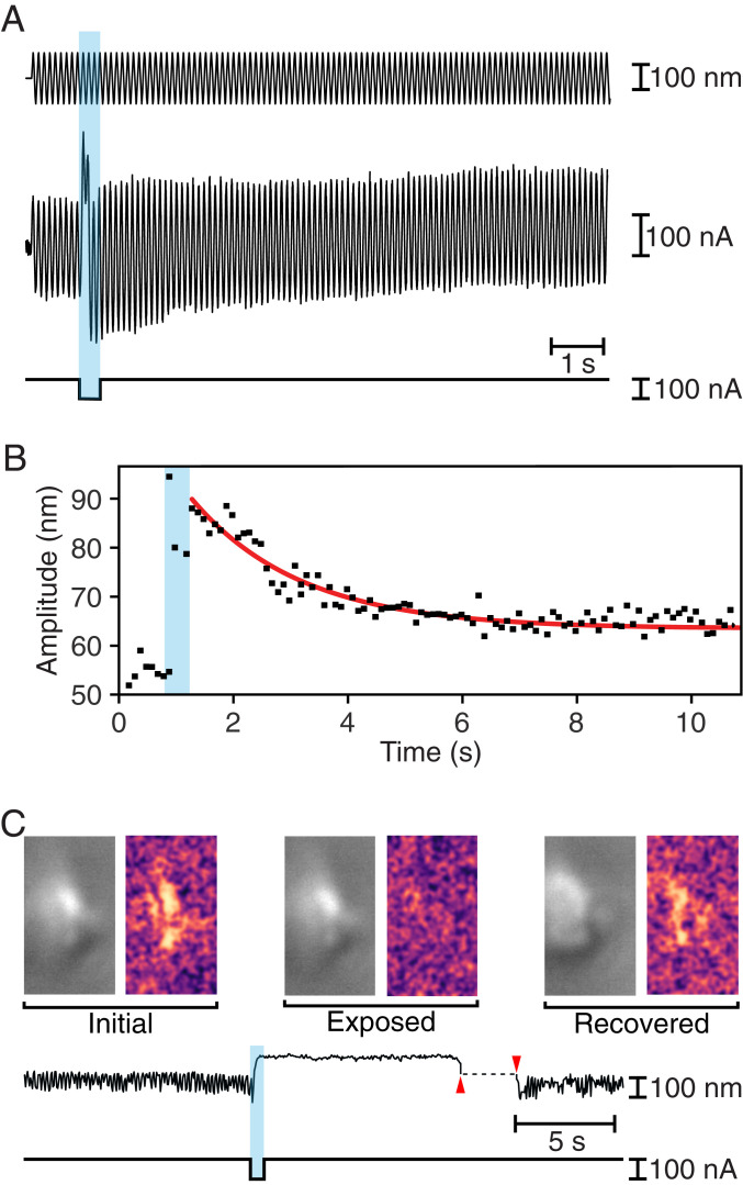
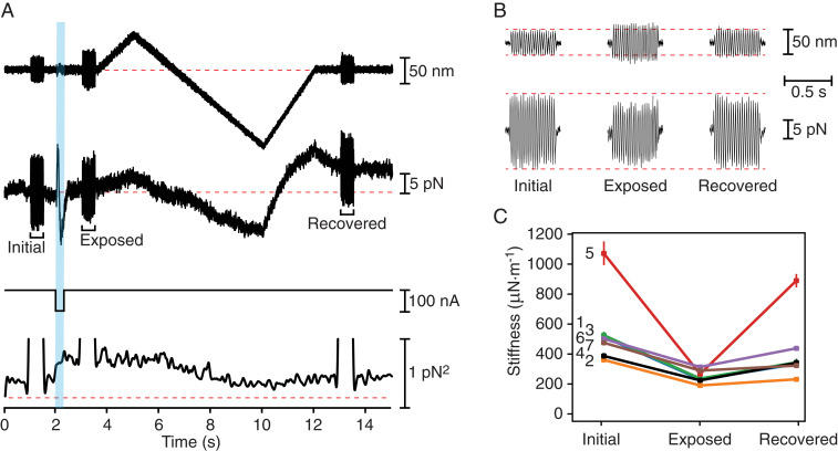
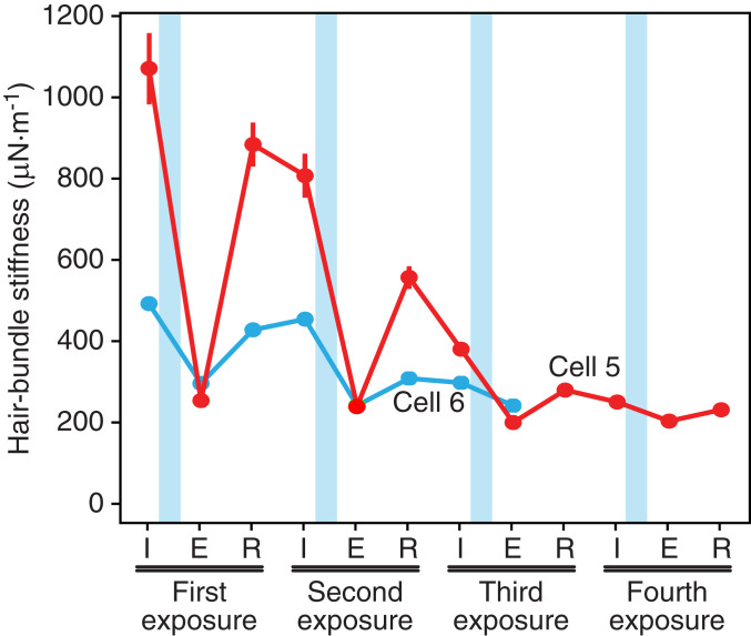
Hearing and balance rely on the capacity of mechanically sensitive hair bundles to transduce vibrations into electrical signals that are forwarded to the brain. Hair bundles possess tip links that interconnect the mechanosensitive stereocilia and convey force to the transduction channels. A dimer of dimers, each of these links comprises two molecules of protocadherin 15 (PCDH15) joined to two of cadherin 23 (CDH23). The "handshake" that conjoins the four molecules can be disrupted in vivo by intense stimulation and in vitro by exposure to Ca2+ chelators. Using hair bundles from the rat's cochlea and the bullfrog's sacculus, we observed that extensive recovery of mechanoelectrical transduction, hair bundle stiffness, and spontaneous bundle oscillation can occur within seconds after Ca2+ chelation, especially if hair bundles are deflected toward their short edges. Investigating the phenomenon in a two-compartment ionic environment that mimics natural conditions, we combined iontophoretic application of a Ca2+ chelator to selectively disrupt the tip links of individual frog hair bundles with displacement clamping to control hair bundle motion and measure forces. Our observations suggest that, after the normal Ca2+ concentration has been restored, mechanical stimulation facilitates the reconstitution of functional tip links.

Keywords: auditory system; cadherin; cochlea; hair cell; vestibular system.


Conflict of interest statement

The authors declare no competing interest.

Figures

Fig. 1.
Fig. 1.
Rapid recovery of mechanoelectrical transduction in outer hair cells from the rat’s cochlea. (A) A fluid-jet stimulator was driven at 60 Hz with two sinusoidal stimulus trains (top trace), one before and one after an iontophoretic current pulse (bottom trace) that released EDTA. The pale blue band here and in subsequent figures delineates the period of iontophoresis. The transduction current (second trace) was initially large but fell to nearly zero after iontophoresis before recovering more than one-half of its original magnitude during the second stimulus train. The variance of the transduction current (third trace) fell during iontophoresis as transduction was interrupted, but recovered partially after a second epoch of stimulation. The abscissa represents zero variance. (B) For the record shown in A, the recovery of the transduction current after the iontophoretic pulse followed an exponential relation (red line) with a time constant of 100 ms.
Fig. 2.
Fig. 2.
Rapid recovery of mechanical properties in hair bundles of the bullfrog’s sacculus. (A) While a 10-Hz sinusoidal stimulus of amplitude 100 nm (top trace) was delivered to the base of a flexible glass fiber, a pulse of iontophoretic current (bottom trace) released EDTA. The hair bundle’s movement (middle trace) increased immediately after iontophoresis, but returned toward the initial value over a few seconds. (B) For the record shown in A, the decline in the amplitude of hair bundle oscillation after the iontophoretic pulse followed an exponential relation (red line) with a time constant of 1,910 ms. (C) To the left in each pair of images in the top row are individual frames of a movie of a spontaneously oscillating hair bundle (Movie S1) representing the unperturbed bundle (Initial), the same bundle after exposure to EDTA (Exposed), and finally the bundle after transient displacement in the negative direction (Recovered). To the right are three images, each obtained by subtracting the original frame from the subsequent frame. The Initial and Recovered images reveal spontaneous hair bundle motion, which is absent in the Exposed image. The time course of the hair bundle’s position in the movie (upper trace) shows suppression of the spontaneous oscillations during iontophoretic application of EDTA (lower trace) and their recovery after the bundle was pushed in the negative direction (between the red arrowheads).
Fig. 3.
Fig. 3.
Facilitation of hair bundle recovery in the bullfrog’s sacculus by mechanical displacement. (A) In a displacement-clamp experiment, a feedback system imposed a ramp displacement on a hair bundle (first trace), moving the bundle first in the positive direction to prevent prompt recovery, and then more extensively in the negative direction. At three times during this paradigm, a 500-ms epoch of ±25-nm, 40-Hz sinusoidal stimulation was superimposed on the displacement-command signal. An iontophoretic pulse (third trace) released EDTA to break tip links. The force (second trace) necessary to clamp the bundle at the outset (Initial) diminished after exposure to iontophoretically applied EDTA (Exposure) but recovered almost completely by the experiment’s end (Recovered). The variance of the force (fourth trace) confirmed the bundle’s softening after iontophoresis and its recovery during the negative phase of the ramp. The dashed line represents the background noise. (B) Enlarged records of the hair bundle displacement (top traces) and clamp force (bottom traces) from A demonstrate that maintaining an oscillation of similar—or even greater—magnitude required less force shortly after iontophoresis. (C) Data from seven hair cells, which are numbered as in Table 1, reveal a significant decrease (P < 0.01 by a single-sided paired t test) in hair bundle stiffness after iontophoretic pulses. The stiffness then recovered significantly (P < 0.05 by the same test) following negative hair bundle displacements. The bundle whose responses are depicted in A and B is number 4; SDs are shown when they exceed the size of the data points.
Fig. 4.
Fig. 4.
Sequential disruption and recovery of tip links in the bullfrog’s sacculus. For two hair bundles also described in Table 1, successive applications of EDTA reduced the stiffness to approximately that associated with the stereociliary pivots. When subjected to ramp displacements, one bundle recovered part of its stiffness at least three times and the other twice. The data points represent the initial stiffness (I), that just after EDTA exposure (E), and that following recovery (R).

References

    1. Hudspeth A. J., Integrating the active process of hair cells with cochlear function. Nat. Rev. Neurosci. 15, 600–614 (2014). - PubMed
    1. Reichenbach T., Hudspeth A. J., The physics of hearing: Fluid mechanics and the active process of the inner ear. Rep. Prog. Phys. 77, 076601 (2014). - PubMed
    1. Ó Maoiléidigh D., Ricci A. J., A bundle of mechanisms: Inner-ear hair-cell mechanotransduction. Trends Neurosci. 42, 221–236 (2019). - PMC - PubMed
    1. Howard J., Roberts W. M., Hudspeth A. J., Mechanoelectrical transduction by hair cells. Annu. Rev. Biophys. Biophys. Chem. 17, 99–124 (1988). - PubMed
    1. Jacobs R. A., Hudspeth A. J., Ultrastructural correlates of mechanoelectrical transduction in hair cells of the bullfrog’s internal ear. Cold Spring Harb. Symp. Quant. Biol. 55, 547–561 (1990). - PubMed

Publication types

LinkOut - more resources