. 2020 Nov 16;11(1):5801.
doi: 10.1038/s41467-020-19625-9.

Risk of yellow fever virus transmission in the Asia-Pacific region

Affiliations

Risk of yellow fever virus transmission in the Asia-Pacific region

Lucy de Guilhem de Lataillade et al. Nat Commun. .

Abstract

Historically endemic to Sub-Saharan Africa and South America, yellow fever is absent from the Asia-Pacific region. Yellow fever virus (YFV) is mainly transmitted by the anthropophilic Aedes mosquitoes whose distribution encompasses a large belt of tropical and sub tropical regions. Increasing exchanges between Africa and Asia have caused imported YFV incidents in non-endemic areas, which are threatening Asia with a new viral emergence. Here, using experimental infections of field-collected mosquitoes, we show that Asian-Pacific Aedes mosquitoes are competent vectors for YFV. We observe that Aedes aegypti populations from Singapore, Taiwan, Thailand, and New Caledonia are capable of transmitting YFV 14 days after oral infections, with a number of viral particles excreted from saliva reaching up to 23,000 viral particles. These findings represent the most comprehensive assessment of vector competence and show that Ae. aegypti mosquitoes from the Asia-Pacific region are highly competent to YFV, corroborating that vector populations are seemingly not a brake to the emergence of yellow fever in the region.


Conflict of interest statement

The authors declare no competing interests.

Figures

Fig. 1
Fig. 1. Vector competence of 12 Aedes aegypti populations assessed 14 and 21 days after an infectious blood meal containing 107 ffu/mL of YFV (West-African genotype).
Batches of 20–24 mosquitoes were examined in each population for viral infection (a, d), dissemination (b, e), and transmission (c, f) by estimating respectively the proportion of mosquitoes with infected bodies (1), head (2), and saliva (3). Titrations were performed on C6/36 cells in 96-well plates. Viral particles were detected by FFA using a primary anti-YFV antibody and a secondary fluorescent-conjugated antibody. Infection rate (IR) refers to the percentage of mosquitoes having an infected body among blood-fed mosquitoes. Dissemination rate (DR) is the percentage of mosquitoes with an infected head (containing viral particles having disseminated in the general cavity after crossing successfully the midgut) among mosquitoes with an infected body. Transmission rate (TR) corresponds to the percentage of mosquitoes with infectious saliva (viral particles having successively crossed the two anatomical barriers, midgut and salivary glands) among mosquitoes with infected head. Stars indicate statistical significance of comparisons by Fisher’s exact test (two-sided test; *P ≤ 0.05, ****P ≤ 0.0001). a ****P ≤ 0.0001, *P = 0.02; b *P0.042; c ****P ≤ 0.0001; d ****P ≤ 0.0001; e ****P ≤ 0.0001. ns (non-significant) indicates a lack of statistical significance (P > 0.05). In brackets are the numbers of mosquitoes tested. dpi days post-infection. Source data are provided in Supplementary Data 1 file.
Fig. 2
Fig. 2. Vector competence of 6 Aedes albopictus populations assessed 14 and 21 days after an infectious blood meal containing 107 ffu/mL of YFV (West-African genotype).
Batches of mosquitoes were examined in each population for viral infection (a, d), dissemination (b, e), and transmission (c, f) by estimating respectively the proportion of mosquitoes with infected bodies (1), head (2), and saliva (3). Infection rate (IR) refers to the percentage of mosquitoes having an infected body among blood-fed mosquitoes. Dissemination rate (DR) is the percentage of mosquitoes with an infected head (containing viral particles having disseminated in the general cavity after crossing successfully the midgut) among mosquitoes with an infected body. Transmission rate (TR) corresponds to the percentage of mosquitoes with infectious saliva (viral particles having successively crossed the two anatomical barriers, midgut and salivary glands) among mosquitoes with infected head. Stars indicate statistical significance of comparisons by Fisher’s exact test (two-sided test; *P ≤ 0.05, **P ≤ 0.01, ****P ≤ 0.0001). a ****P ≤ 0.0001; d **P = 0.003; e *P = 0.038. ns (non-significant) indicates a lack of statistical significance (P > 0.05). In brackets are the numbers of mosquitoes tested. dpi days post-infection. Source data are provided in Supplementary Data 1 file.
Fig. 3
Fig. 3. Viral loads measured in individual mosquito saliva at 14 and 21 days after an infectious blood meal with West-African YFV.
a, b Saliva viral loads of Aedes aegpti at 14 and 21 dpi; c, d saliva viral loads of Aedes albopictus at 14 and 21 dpi. Saliva was collected for 30 min using the forced salivation technique by removing legs and wings and inserting a tip containing FBS in mosquito proboscis. Salivas were titrated on C3/36 cells and the numbers of viral particles are expressed in ffu/saliva. ns (non-significant) indicates the lack of statistical significance for comparisons using the Kruskal–Wallis test (two-sided test; P > 0.05). Bars indicate the mean. In brackets are the numbers of mosquitoes tested. dpi days post-infection. Source data are provided in Supplementary Data 1 file.
Fig. 4
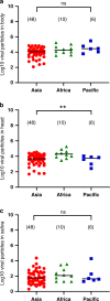
Fig. 4. Virals loads in body, head, and saliva of Aedes aegypti populations from Asia, Africa, and the Pacific region.
Mosquito (a) body, (b) head, and (c) saliva were titrated on C6/36 cells and the number of viral particles was expressed in ffu/sample. Stars indicate statistical significance of comparisons by the Kruskal–Wallis test (two-sided test; **P ≤ 0.01). b **P = 0.0095. ns (non-significant) indicates a lack of statistical significance (P > 0.05). Bars indicate the mean. In brackets are the numbers of mosquitoes tested. Red dots: samples from Asia; green triangles: samples from Africa; blue squares: samples from the Pacific region. Source data are provided in Supplementary Data 2 file.
Fig. 5
Fig. 5. Risk of Aedes aegypti-mediated YFV transmission in Asia.
a Original data from Kraemer et al. showing the probability of encountering Aedes aegypti in South-East Asia. The colors correspond to probabilities: lower (blue) or higher (red) than the median probability across the whole map (white). b Modeled vector occurrence (colored bars matching the values from the scale in a) along with mosquito transmission efficiency (gray bar) of Aedes aegypti populations tested in laboratory conditions shown in a. This map uses data published by Kraemer et al. and was generated with R v4.0.1 (package raster v3.1-5). Source data are provided in Supplementary Data 1 file.
Fig. 6
Fig. 6. Geographical distribution of the 18 mosquito sample locations (12 Aedes aegypti and 6 Aedes albopictus).
Black dots: Aedes aegypti; red dots: Aedes albopictus. The map was built using the open-source map site “https://d-maps.com/conditions.php?lang=en/”. Each dot corresponds to a sampling location.

Similar articles

Cited by

References

    1. Wilder-Smith A, et al. Epidemic arboviral diseases: priorities for research and public health. Lancet Infect. Dis. 2017;17:e101–e106. doi: 10.1016/S1473-3099(16)30518-7. - DOI - PubMed
    1. Barrett AD, Higgs S. Yellow fever: a disease that has yet to be conquered. Annu Rev. Entomol. 2007;52:209–229. doi: 10.1146/annurev.ento.52.110405.091454. - DOI - PubMed
    1. Wasserman S, Tambyah PA, Lim PL. Yellow fever cases in Asia: primed for an epidemic. Int J. Infect. Dis. 2016;48:98–103. doi: 10.1016/j.ijid.2016.04.025. - DOI - PubMed
    1. Black WC, 4th, et al. Flavivirus susceptibility in Aedes aegypti. Arch. Med. Res. 2002;33:379–388. doi: 10.1016/S0188-4409(02)00373-9. - DOI - PubMed
    1. Mutebi JP, Barrett AD. The epidemiology of yellow fever in Africa. Microbes Infect. 2002;4:1459–1468. doi: 10.1016/S1286-4579(02)00028-X. - DOI - PubMed