. 2020 Nov 16;10(1):19861.
doi: 10.1038/s41598-020-76963-w.

The architecture of corneal stromal striae on optical coherence tomography and histology in an animal model and in humans

Affiliations

The architecture of corneal stromal striae on optical coherence tomography and histology in an animal model and in humans

Pietro Emanuele Napoli et al. Sci Rep. .

Abstract

The purpose of this study was to use a portable optical coherence tomography (OCT) for characterization of corneal stromal striae (CSS) in an ovine animal model and human corneas with histological correlation, in order to evaluate their architectural pattern by image analysis. Forty-six eyes from female adult sheep (older than 2 years), and 12 human corneas, were included in our study. The eyes were examined in situ by a portable OCT, without enucleation. All OCT scans were performed immediately after death, and then the eyes were delivered to a qualified histology laboratory. In the ovine animal model, CSS were detected with OCT in 89.1% (41/46) of individual scans and in 93.4% (43/46) of histological slices. In human corneas, CSS were found in 58.3% (7/12) of cases. In both corneal types, CSS appeared as "V"- or "X"-shaped structures, with very similar angle values of 70.8° ± 4° on OCT images and 71° ± 4° on histological slices (p ≤ 0.01). Data analysis demonstrated an excellent degree of reproducibility and inter-rater reliability of measurements (p < 0.001). The present study demonstrated that by using a portable OCT device, CSS can be visualized in ovine and human corneas. This finding suggests their generalized presence in various mammals. The frequent observation, close to 60%, of such collagen texture in the corneal stroma, similar to a 'truss bridge' design, permits to presume that it plays an important structural role, aimed to distribute tensile and compressive forces in various directions, conferring resilience properties to the cornea.


Conflict of interest statement

The authors declare no competing interests.

Figures
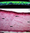
Figure 1
Figure 1
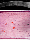
Optical coherence tomography image (A) of corneal stromal striae obtained by in situ scanning of our animal model (resembling a ‘truss bridge’ design). (B) Histological image (hematoxylin–eosin ×40) of the same cornea (the arrows indicate the “X-shaped” stromal structures) (Scale bar = 100 µm).
Figure 2
Figure 2
Quantitative analysis of angles within the geometric stromal striae. Grayscale images obtained by splitting the original picture into the green (left) and red (right) channels, which provide the best contrasts to explain how angles have been defined. Optical coherence tomography scans (top) and histological slides (bottom) are reported without (left) and with (right) the placement of markers for measurement of θA for θB angles, respectively (see text). The elements of the criss-crossing superstructure are defined by two continuous, oblique lines ("V"- or "X"-shaped) in the stromal tissue, which are indicated by the dashed lines. For the purposes of our analysis, since the vertically opposite angles (i.e. the upper and the lower) have identical (or congruent) values, only one of these was indiscriminately included in the data collection (according to the vertical angle theorem). Conversely, the lateral ones (or adjacent angles) were not considered in our computation. Of note, the various angles within the individual corneas have demonstrated to be equal, or approximately equal, in measure.
Figure 3
Figure 3
Corneal stromal striae in human cornea on optical coherence tomography imaging (top) and histology (bottom) (grayscale and color images). The arrows indicate the most easily detectable “X-shaped” linear structures (Scale bar = 100 µm).

Similar articles

Cited by

References

    1. Sturrock G. Glassy corneal striae. Albrecht Von Graefes Arch. Klin. Exp. Ophthalmol. 1973;188:245–252. doi: 10.1007/BF00410978. - DOI - PubMed
    1. Grieve K, Ghoubay D, Georgeon C, et al. Stromal striae: A new insight into corneal physiology and mechanics. Sci. Rep. 2017;7(1):13584. doi: 10.1038/s41598-017-13194-6. - DOI - PMC - PubMed
    1. Kame GY, Abo JT, Kame RT, et al. The incidence of glassy corneal striae. Int. Contact Lens Clin. 1999;26(6):147–151. doi: 10.1016/S0892-8967(00)00041-9. - DOI - PubMed
    1. Napoli PE, Nioi M, d’Aloja E, Fossarello M. Post-mortem corneal thickness measurements with a portable optical coherence tomography system: A reliability study. Sci. Rep. 2016;6:30428. doi: 10.1038/srep30428. - DOI - PMC - PubMed
    1. Nioi M, Napoli PE, Paribello F, Demontis R, De Giorgio F, Porru E, Fossarello M, d'Aloja E. Use of optical coherence tomography on detection of postmortem ocular findings: Pilot data from two cases. J. Integr. OMICS. 2018;8(1):5–7. doi: 10.5584/jiomics.v8i1.226. - DOI