. 2021 Jan;39(1):47-55.
doi: 10.1038/s41587-020-0741-7. Epub 2020 Nov 16.

A long-term study of AAV gene therapy in dogs with hemophilia A identifies clonal expansions of transduced liver cells

Affiliations

A long-term study of AAV gene therapy in dogs with hemophilia A identifies clonal expansions of transduced liver cells

Giang N Nguyen et al. Nat Biotechnol. 2021 Jan.

Abstract

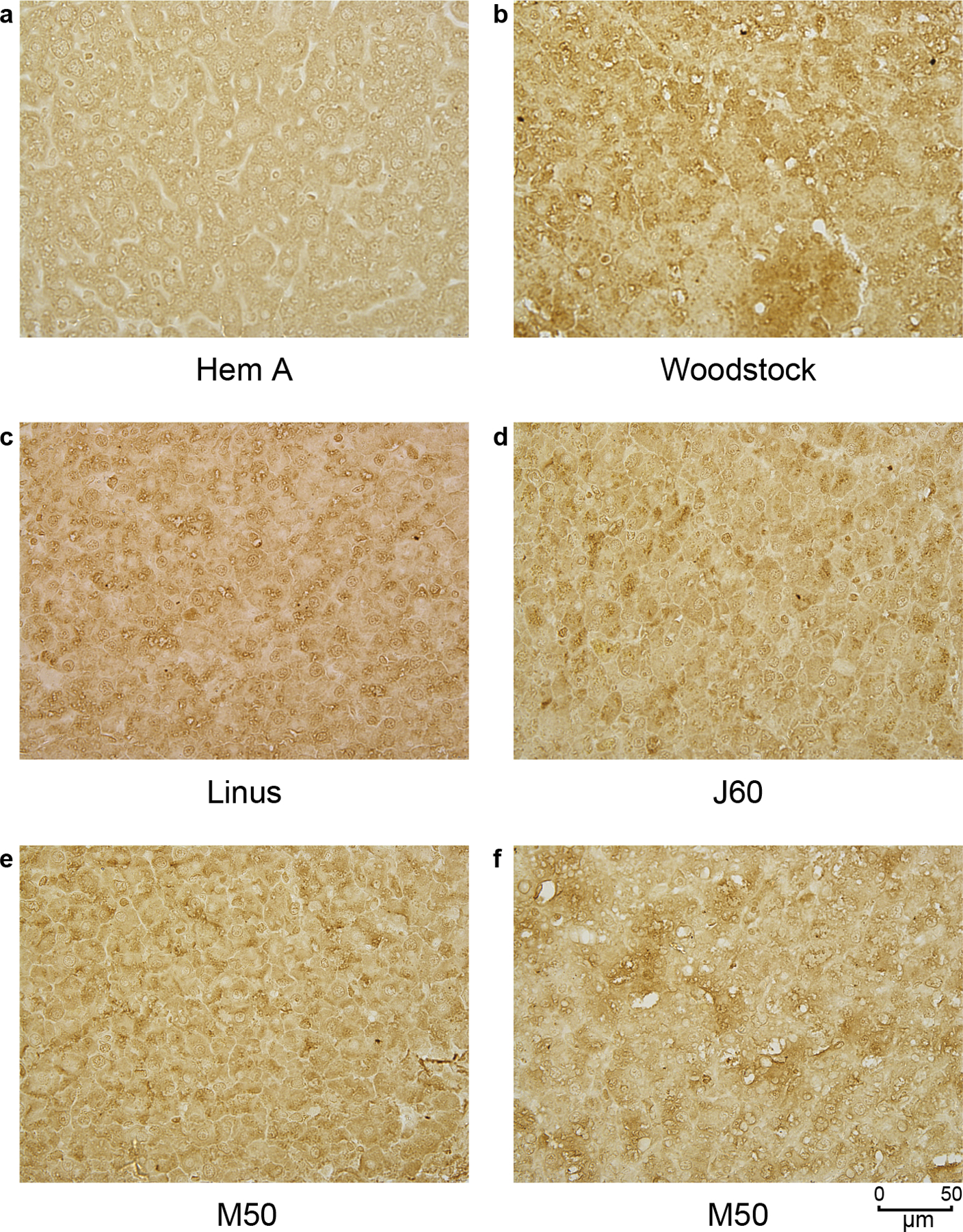
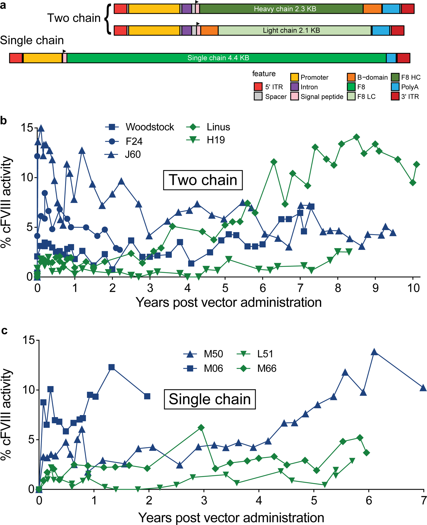
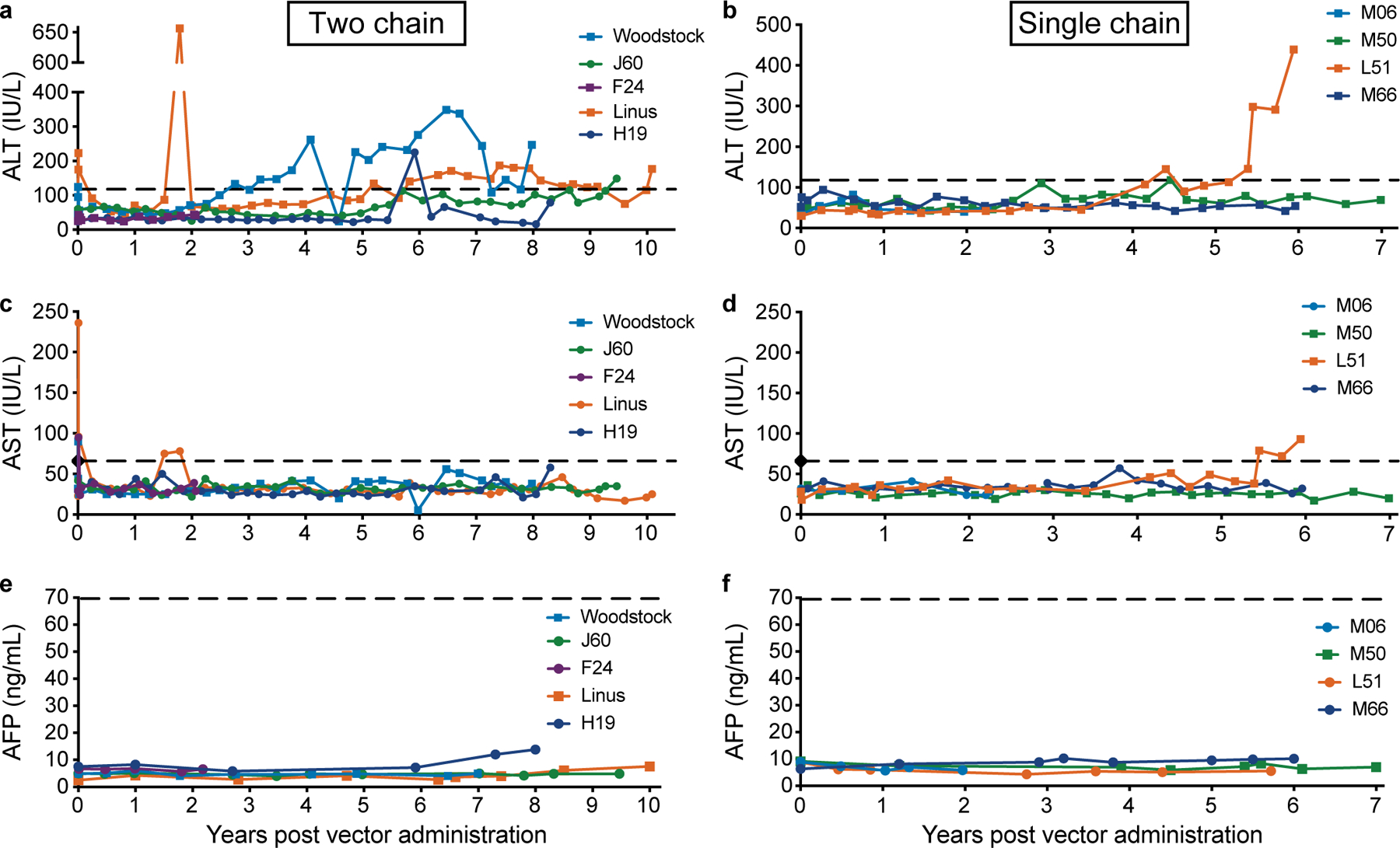
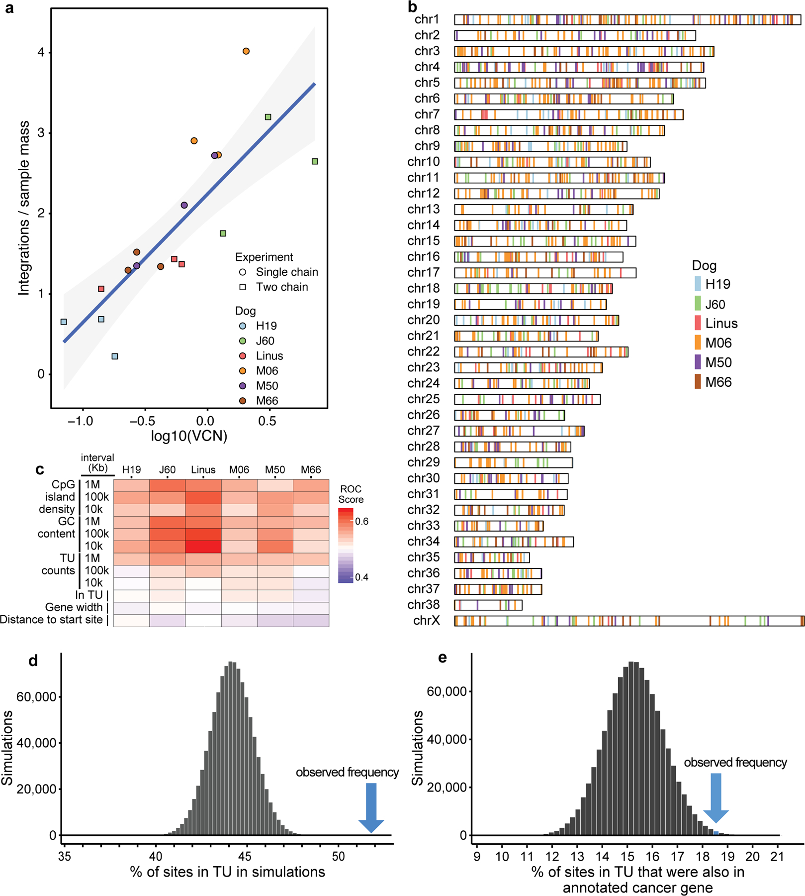
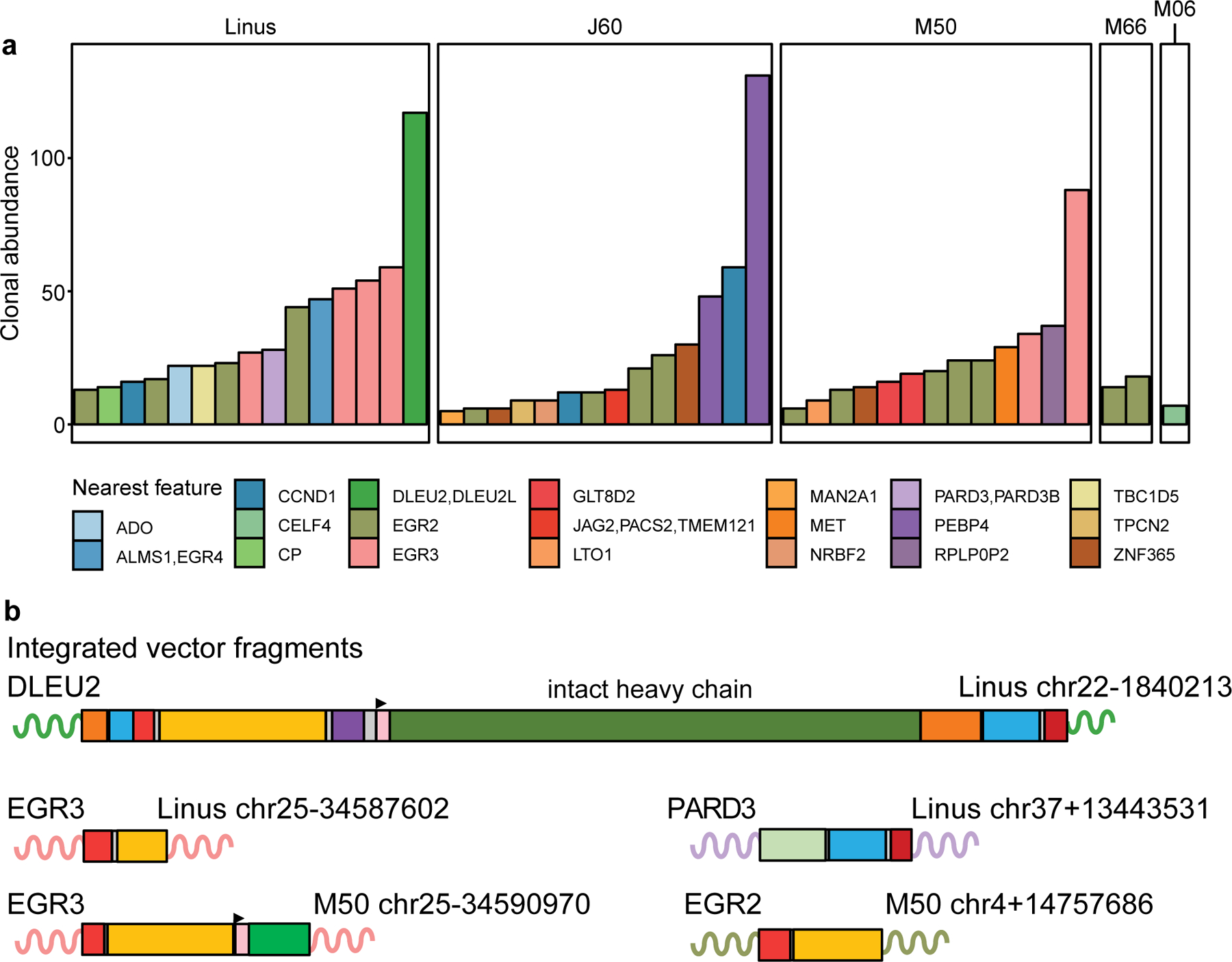
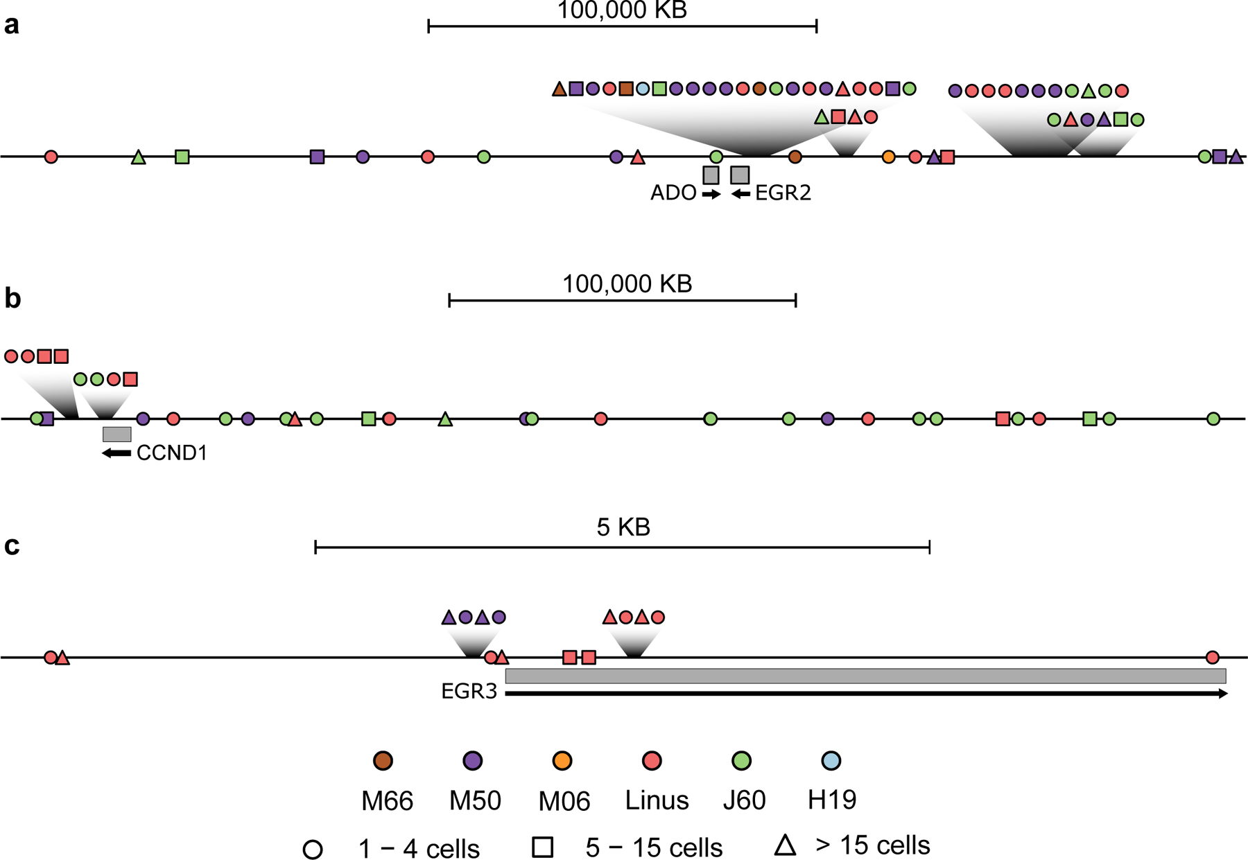
Nine dogs with hemophilia A were treated with adeno-associated viral (AAV) gene therapy and followed for up to 10 years. Administration of AAV8 or AAV9 vectors expressing canine factor VIII (AAV-cFVIII) corrected the FVIII deficiency to 1.9-11.3% of normal FVIII levels. In two of nine dogs, levels of FVIII activity increased gradually starting about 4 years after treatment. None of the dogs showed evidence of tumors or altered liver function. Analysis of integration sites in liver samples from six treated dogs identified 1,741 unique AAV integration events in genomic DNA and expanded cell clones in five dogs, with 44% of the integrations near genes involved in cell growth. All recovered integrated vectors were partially deleted and/or rearranged. Our data suggest that the increase in FVIII protein expression in two dogs may have been due to clonal expansion of cells harboring integrated vectors. These results support the clinical development of liver-directed AAV gene therapy for hemophilia A, while emphasizing the importance of long-term monitoring for potential genotoxicity.


Conflict of interest statement

Competing Interests Statement

D.E.S. receives royalites from a licensing agreement with Spark Therapeutics. D.E.S. and G.N.N. are inventors on a patent on factor VIII and hemophilia A gene therapy.

Figures

Extended Data Figure 1
Fig. 1.
Fig. 1.. Long term expression of AAV-cFVIII in hemophilia A dogs.
(a) Genetic structures of the vectors used, illustrating the two chain (TC) (top) and single chain (SC) (bottom) designs. The features of these constructs are indicated by colors shown in the legend. The TC delivery approach co-delivered the 2.3 Kb cF8 heavy chain in one AAV vector and the 2.1 Kb cF8 light chain in a second AAV vector. These vectors used the thyroxine-binding globulin (TBG) promoter and two copies of a1-microglobulin/bikunin enhancer sequences (695 bp) with an intron (175 bp) for liver-directed gene expression. The construct utilized a 263 bp SV40 polyadenylation signal (poly A). The SC delivery approach delivered the 4.4 Kb B-domain deleted cF8 driven by a minimal hepatic control region (192 bp) and human a-1 antitrypsin promoter (266 bp) (HCR-hAAT). This construct includes a 134 bp SV40 poly A. The grey regions indicate spacers or cloning sites. The TC vector was delivered by portal vein infusion while the SC vector was delivered by peripheral vein infusion. (b) Longitudinal quantification of FVIII levels after the TC delivery approach. In the two chain approach the total AAV dose was 2.5 × 10¹³ vg/kg dose for F24 (AAV8), Woodstock and J60 (AAV9)(blue). Linus (AAV8) and H19 (AAV9) were treated at the low vector dose (total vector dose of 1.2 × 10¹³ vg/kg)(green). (c) FVIII levels after the SC delivery approach. AAV8 delivery of the SC approach in hemophilia A dogs was at the vector dose of 4 × 10¹³ vg/kg (M50, M06)(blue) and at a vector dose of 2 × 10¹³ vg/kg (L51, M66)(green).
Fig. 2.
Fig. 2.. Assessment of liver function after AAV-cFVIII administration.
ALT (a and b), AST (c and d) and AFP (e and f) were monitored after AAV administration and throughout the study. In each case the x-axis shows time in years, the y-axis shows the liver analyte measured in blood. a), c) and e) Dogs treated with the two chain strategy. b), d), and f) Dogs treated with the single chain strategy. The upper limit of normal for ALT (120 IU/L), AST (65 IU/L) and AFP (70 ng/ml) are shown as dashed lines. Clinically, increase in liver enzyme levels in dogs are defined by veterinary clinical pathologists as mild (<5 times the upper limit of normal), moderate (5–10 times the upper limit of normal) and marked (>10 times the upper limit of normal) .
Fig. 3.
Fig. 3.. Distributions of sites of AAV vector integration in the treated dogs.
a) Correlation of the VCN value and the numbers of integration sites recovered from dog liver DNA specimens. Liver DNA samples (n=3 biologically independent samples) analyzed for each dog are shown as individual points. The gray envelope shows a 95% confidence interval. b) Distribution of AAV vector integration sites in the dog chromosomes. c) Distribution of AAV vector integration sites in the dog genome relative to genomic annotation. Associations are calculated using the ROC area method. Values of the ROC area vary between 0 (negatively associated; blue) and 1 (positively associated; red). TU indicates transcription unit. Several length chromosomal intervals were used for comparison. d) Enrichment of integration sites in dog transcription units. The dark bars show the frequency of appearance of randomly selected sites in transcription units (TU), summarized over 106 random simulations. The blue arrow shows the observed frequency of AAV vector integration sites in TUs. e) Enrichment of integration sites within dog transcription units that are also cancer-associated genes. A list of human cancer-associated genes was used to annotate the dog genome.
Fig. 4.
Fig. 4.. Clonal expansion of cells harboring AAV vectors.
a) Summary of notable expanded clones in five of the six dogs studied. No expanded clones were detected in H19. Shown are most abundant 15 clones per dog represented by five or more cells sampled. The genes affected are shown by the color code. Data on each integration site studied is in Supplementary Table 12. b) Genetic map of vector sequences integrated in several of the dogs. The color code for vector segments as in Fig. 1a. The schematics illustrate truncations and rearrangements that may not contain the entire vector sequence. Sequences are provided (NIH SRA BioProject ID: PRJNA606282). The numbers indicate the chromosome location and coordinate of a junction between AAV vector sequences and the dog genome. The gene at the site of integration is labelled. The curved lines represent genomic DNA at the site of integration with the color matching the gene in panel a.
Fig. 5.
Fig. 5.
Three examples of clusters of AAV vector integration sites found in the dog genome. Clusters are shown schematically at a) ADO-EGR2, b) CCND1, and c) EGR3. Transcription units are indicated by the grey rectangles. Integration sites are indicated by the shapes, with the inferred number of cells recovered harboring each site indicated by the nature of the shape (circle: 1–4 cells; square: 5–15 cells; and triangle >15 cells). The colors on the shapes indicate the dog of origin. The scales are shown by the bar at the top.

Comment in

References

    1. Rangarajan S et al. AAV5-Factor VIII Gene Transfer in Severe Hemophilia A. N Engl J Med 377, 2519–2530 (2017). - PubMed
    1. High KA et al. A Phase 1/2 Trial of Investigational Spk-8011 in Hemophilia A Demonstrates Durable Expression and Prevention of Bleeds. in (ed. Blood) 132, 487 (2018).
    1. Nathwani AC et al. GO-8: Preliminary Results of a Phase I/II Dose Escalation Trial of Gene Therapy for Haemophilia A Using a Novel Human Factor VIII Variant. in (ed. Blood) 132, 489 (2018).
    1. Pasi KJ et al. Multiyear Follow-up of AAV5-hFVIII-SQ Gene Therapy for Hemophilia A. N Engl J Med 382, 29–40 (2020). - PubMed
    1. Jiang H et al. Multiyear therapeutic benefit of AAV serotypes 2, 6, and 8 delivering factor VIII to hemophilia A mice and dogs. Blood 108, 107–115 (2006). - PubMed

Methods-only References

    1. Sherman A et al. Portal Vein Delviery of Viral Vectors for Gene Therapy for Hemophilia in Gene Correction: Methods and Protocols. (Humana Press, 2014). - PMC - PubMed
    1. Hothorn T, Hornik K, van de Wiel M & Zeileis A Implementing a Class of Permutation Tests: The coin package. Journal of Statistical Software 28, 1–23 (2008). - PubMed
    1. Sherman E et al. INSPIIRED: A Pipeline for Quantitative Analysis of Sites of New DNA Integration in Cellular Genomes. Mol Ther Methods Clin Dev 4, 39–49 (2017). - PMC - PubMed
    1. Berry CC et al. INSPIIRED: Quantification and Visualization Tools for Analyzing Integration Site Distributions. Mol Ther Methods Clin Dev 4, 17–26 (2017). - PMC - PubMed
    1. Pruitt KD, Tatusova T & Maglott DR NCBI Reference Sequence (RefSeq): a curated non-redundant sequence database of genomes, transcripts and proteins. Nucleic Acids Res 33, D501–4 (2005). - PMC - PubMed

Publication types