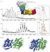
The importance of the membrane for biophysical measurements
- PMID: 33199903
- PMCID: PMC7116504
- DOI: 10.1038/s41589-020-0574-1
The importance of the membrane for biophysical measurements
Abstract
Within cell membranes numerous protein assemblies reside. Among their many functions, these assemblies regulate the movement of molecules between membranes, facilitate signaling into and out of cells, allow movement of cells by cell-matrix attachment, and regulate the electric potential of the membrane. With such critical roles, membrane protein complexes are of considerable interest for human health, yet they pose an enduring challenge for structural biologists because it is difficult to study these protein structures at atomic resolution in in situ environments. To advance structural and functional insights for these protein assemblies, membrane mimetics are typically employed to recapitulate some of the physical and chemical properties of the lipid bilayer membrane. However, extraction from native membranes can sometimes change the structure and lipid-binding properties of these complexes, leading to conflicting results and fueling a drive to study complexes directly from native membranes. Here we consider the co-development of membrane mimetics with technological breakthroughs in both cryo-electron microscopy (cryo-EM) and native mass spectrometry (nMS). Together, these developments are leading to a plethora of high-resolution protein structures, as well as new knowledge of their lipid interactions, from different membrane-like environments.
Figures


Similar articles
-
Fake It 'Till You Make It-The Pursuit of Suitable Membrane Mimetics for Membrane Protein Biophysics.Int J Mol Sci. 2020 Dec 23;22(1):50. doi: 10.3390/ijms22010050. Int J Mol Sci. 2020. PMID: 33374526 Free PMC article. Review.
-
Beyond detergent micelles: The advantages and applications of non-micellar and lipid-based membrane mimetics for solution-state NMR.Prog Nucl Magn Reson Spectrosc. 2019 Oct-Dec;114-115:271-283. doi: 10.1016/j.pnmrs.2019.08.001. Epub 2019 Aug 26. Prog Nucl Magn Reson Spectrosc. 2019. PMID: 31779883 Review.
-
Structures and Interactions of Transmembrane Targets in Native Nanodiscs.SLAS Discov. 2019 Dec;24(10):943-952. doi: 10.1177/2472555219857691. Epub 2019 Jun 26. SLAS Discov. 2019. PMID: 31242812
-
Border controls: Lipids control proteins and proteins control lipids.Biochim Biophys Acta Biomembr. 2017 Apr;1859(4):507-508. doi: 10.1016/j.bbamem.2016.12.016. Epub 2016 Dec 24. Biochim Biophys Acta Biomembr. 2017. PMID: 28024797 No abstract available.
-
Delimiting lipids.Nat Chem Biol. 2020 Dec;16(12):1279. doi: 10.1038/s41589-020-00701-6. Nat Chem Biol. 2020. PMID: 33199910 No abstract available.
Cited by
-
TTYH family members form tetrameric complexes at the cell membrane.Commun Biol. 2022 Aug 30;5(1):886. doi: 10.1038/s42003-022-03862-3. Commun Biol. 2022. PMID: 36042377 Free PMC article.
-
Fake It 'Till You Make It-The Pursuit of Suitable Membrane Mimetics for Membrane Protein Biophysics.Int J Mol Sci. 2020 Dec 23;22(1):50. doi: 10.3390/ijms22010050. Int J Mol Sci. 2020. PMID: 33374526 Free PMC article. Review.
-
In-Cell Fast Photochemical Oxidation Interrogates the Native Structure of Integral Membrane Proteins.Angew Chem Int Ed Engl. 2025 May;64(19):e202424779. doi: 10.1002/anie.202424779. Epub 2025 Mar 9. Angew Chem Int Ed Engl. 2025. PMID: 40033852
-
Selective Nutrient Transport in Bacteria: Multicomponent Transporter Systems Reign Supreme.Front Mol Biosci. 2021 Jun 29;8:699222. doi: 10.3389/fmolb.2021.699222. eCollection 2021. Front Mol Biosci. 2021. PMID: 34268334 Free PMC article. Review.
-
Structural Dynamics of the Slide Helix of Inactive/Closed Conformation of KirBac1.1 in Micelles and Membranes: A Fluorescence Approach.J Membr Biol. 2025 Feb;258(1):97-112. doi: 10.1007/s00232-024-00335-y. Epub 2025 Jan 9. J Membr Biol. 2025. PMID: 39789244 Free PMC article.
References
-
- Callaway E. The revolution will not be crystallized: a new method sweeps through structural biology. Nature. 2015;525:172–4. - PubMed
-
- Murphy BJ, et al. Rotary substates of mitochondrial ATP synthase reveal the basis of flexible F1-Fo coupling. Science. 2019;364 - PubMed
-
- Gu J, et al. Cryo-EM structure of the mammalian ATP synthase tetramer bound with inhibitory protein IF1. Science. 2019;364:1068–1075. - PubMed
Publication types
MeSH terms
Substances
Grants and funding
LinkOut - more resources
Full Text Sources

