Review
. 2020 Dec;16(12):1303-1313.
doi: 10.1038/s41589-020-00678-2. Epub 2020 Nov 16.

Cholesterol access in cellular membranes controls Hedgehog signaling

Affiliations
Review

Cholesterol access in cellular membranes controls Hedgehog signaling

Arun Radhakrishnan et al. Nat Chem Biol. 2020 Dec.

Abstract

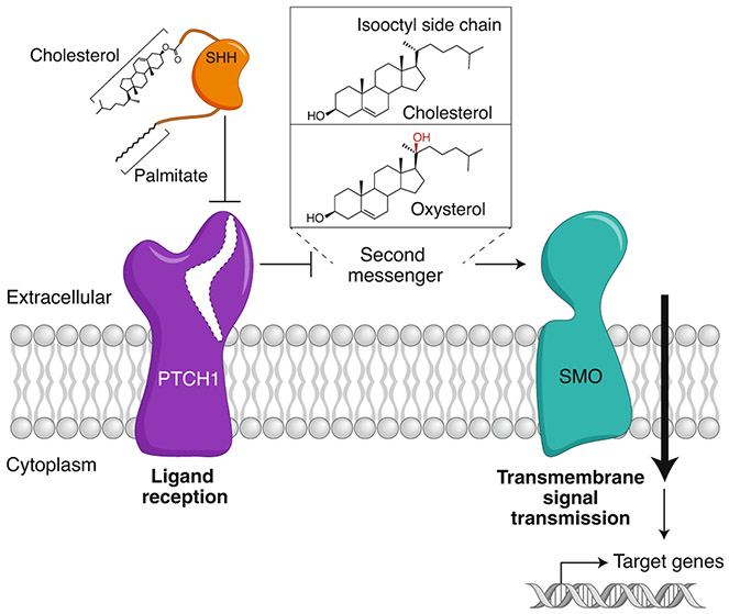
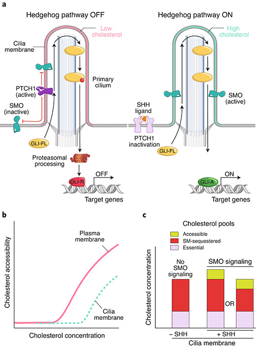
The Hedgehog (Hh) signaling pathway coordinates cell-cell communication in development and regeneration. Defects in this pathway underlie diseases ranging from birth defects to cancer. Hh signals are transmitted across the plasma membrane by two proteins, Patched 1 (PTCH1) and Smoothened (SMO). PTCH1, a transporter-like tumor-suppressor protein, binds to Hh ligands, but SMO, a G-protein-coupled-receptor family oncoprotein, transmits the Hh signal across the membrane. Recent structural, biochemical and cell-biological studies have converged at the surprising model that a specific pool of plasma membrane cholesterol, termed accessible cholesterol, functions as a second messenger that conveys the signal between PTCH1 and SMO. Beyond solving a central puzzle in Hh signaling, these studies are revealing new principles in membrane biology: how proteins respond to and remodel cholesterol accessibility in membranes and how the cholesterol composition of organelle membranes is used to regulate protein function.


Figures

Fig. 1 ∣
Fig. 1 ∣. Hedgehog signal transmission across the plasma membrane.
A double-negative mechanism initiates Hh signaling at the cell surface. PTCH1 inhibits SMO; SHH inhibits PTCH1, allowing SMO activation. Since ligand reception and transmembrane signaling are assigned to different proteins, a second messenger must communicate the signal between PTCH1 and SMO. Candidate second messengers include cholesterol and oxysterols (such as 20(S)-hydroxycholesterol shown here), both of which can bind and activate SMO. Interestingly, cholesterol is also covalently attached to SHH and plays a critical role in its biogenesis from a precursor protein.
Fig. 2 ∣
Fig. 2 ∣. Multiple sterol binding sites in SMO.
a, Schematic of SMO showing the extracellular cysteine-rich domain (CRD), linker domain (LD), transmembrane domain (TMD), third extracellular loop (ECL3) and intracellular domain (ICD). The three structurally and functionally characterized small-molecule binding sites (two in the TMD and one in the CRD) are highlighted. N and C refer to N- and C-termini, respectively. b–e, Cartoon representation of the closed SMO–cholesterol complex (b; PDB ID 5L7D), the SMO–Vismodegib (Vismo) complex in an inhibited conformation (c; PDB ID 5L7I), the SMO–cholesterol–nanobody-SAG (SMO agonist) complex in an activated conformation (d; PDB ID 6O3C) and the SMO–G-protein complex (e, PDB ID 6OT0). Dotted arrows indicate movement compared to that of the closed SMO–cholesterol complex (shown in b). Small molecules are labeled and depicted in stick representation. The V329F mutation that locks SMO in a closed conformation is highlighted as a red sphere in b.
Fig. 3 ∣
Fig. 3 ∣. Structural similarities between PTCH1 and the cholesterol transporter NPC1.
a, Both PTCH1 (top) and NPC1 (bottom) contain two large extracellular domains (ECD1 and ECD2 in PTCH1 and the middle (MLD) and C-terminal (CTD) luminal domains in NPC1) and a 12-pass transmembrane domain (TMD) that includes an evolutionarily conserved sterol-sensing domain (SSD). NPC1 contains an additional N-terminal transmembrane helix (1′) and an extracellular domain (NTD) involved in cholesterol transfer (see b). b,c, Structures of the NPC1–NPC2 complex (b; PDB ID 6W5V) and human PTCH1 (c; PDB ID 6RVD). Yellow surfaces show potential tunnels (calculated with CAVER99) connecting the extracellular/luminal and TMD cholesterol-binding sites. Blue arrows indicate possible transport routes for cholesterol. d,e, Inhibited-state structures of NPC1 (PDB ID 6UOX) and PTCH1 (PDB ID 6RVD). NPC1 is inhibited by itraconazole, which occupies the TMD cholesterol-binding site (d). Binding of SHH to PTCH1 results in a 2:1 PTCH1:SHH complex (e). SHH interacts with PTCH1 mol1 via a high-affinity protein–protein interface involving the SHH-metal binding sites (green and black spheres represent calcium and zinc ions, respectively) and by inserting its N- and C-terminal palmitate and cholesterol modifications into PTCH1 mol2. f,h, Close-up views of the TMD cholesterol-binding sites show that the palmitoyl moiety of SHH overlaps with the cholesterol- and itraconazole-binding sites, suggesting a shared binding site critical for cholesterol transport in both PTCH1 and NPC1.
Fig. 4 ∣
Fig. 4 ∣. Cholesterol accessibility in cellular membranes.
a, Calculated chemical activity (related to the chemical potential) of cholesterol as a function of membrane cholesterol content (expressed as the mole fraction of cholesterol relative to total lipids). Shown are chemical activities for binary membrane mixtures of cholesterol and phospholipid assuming no interactions (blue curve) or a complex containing one cholesterol and two phospholipid molecules (pink curve, derived from a regular solution free energy calculation). At concentrations below the equivalence point, cholesterol is sequestered by phospholipids. Above the equivalence point, cholesterol exceeds the sequestering capacity of phospholipids, and its accessibility sharply rises. b, Three pools of cholesterol in plasma membranes, along with toxin-based probes that can be used to detect and manipulate the sphingomyelin (SM)-sequestered (OlyA) and accessible (ALOD4 or PFO) pools. c, Schematic showing the three cholesterol pools in unperturbed plasma membranes or after cholesterol loading or SM depletion.
Fig. 5 ∣
Fig. 5 ∣. Changes in accessible cholesterol influence Hh signaling in target cells.
a, Accessible cholesterol can be reduced (left) in the plasma membrane by trapping it in the outer leaflet with ALOD4 (Fig. 4b) or by removing it with methyl-β-cyclodextrin (MβCD). Conversely, accessible cholesterol can be increased (right) by depleting sphingomyelin (SM) or by delivering cholesterol to the outer leaflet using MβCD–cholesterol complexes. Due to rapid flip-flop of cholesterol between the leaflets, changes in the outer leaflet are also transmitted to the inner leaflet. b, Changes in total cholesterol, the accessible pool of cholesterol and Hedgehog signaling strength after each of the manipulations shown immediately above in a. c, Conceptual cholesterol activity vs. concentration curves (see Fig. 4a for description) depicting how the manipulations shown in a change total and accessible cholesterol in the plasma membrane. The starting set-point in both panels is point A. ALOD4 (left panel) traps cholesterol (and shifts the curve to the right) without changing total cholesterol abundance (A→B), while cholesterol removal by MβCD reduces total and accessible cholesterol (A→C). Right panel shows two manipulations that increase cholesterol accessibility either by shifting the curve to the left (SM depletion, A→B) or by increasing total cholesterol (A→C).
Fig. 6 ∣
Fig. 6 ∣. Hh signal transmission by ligand-controlled changes in cholesterol accessibility of the ciliary membrane.
a, PTCH1 inhibits SMO (left) by reducing accessible cholesterol in the ciliary membrane (pink). Without SMO activity, full-length GLI proteins (GLI-FL) are proteolytically processed into transcriptional repressors (GLI-R). When PTCH1 is inactivated by SHH (right), accessible cholesterol levels in the ciliary membrane rise (blue), allowing SMO accumulation and activation and ultimately the formation of GLI activators (GLI-A). b, Conceptual cholesterol activity vs. concentration curves (see Fig. 4a) for the plasma membrane and ciliary membrane. The curve for the ciliary membrane is shifted to the right due to its higher sphingomyelin content. c, Schematic showing changes in cholesterol pools in the ciliary membrane in response to PTCH1 inactivation by SHH. Accessible cholesterol in the ciliary membrane rises in response to SHH, driving SMO activation. Two models for how inactivation of PTCH1 by SHH expands the ciliary accessible cholesterol pool are by increasing the total cholesterol in cilia or by converting some of the SM-sequestered cholesterol to an accessible form.
Fig. 7 ∣
Fig. 7 ∣. Two models for the regulation of SMO by PTCH1 at primary cilia.
a, A cartoon representation of cilia is shown at the top, with a rectangle showing a region at the cilia base known as the ‘ciliary pocket’, magnified below. In model 1, PTCH1 uses its transporter function (left) to remove cholesterol from the ciliary membrane, transferring it to an acceptor. Because of the higher abundance of SM in the ciliary membrane, accessible cholesterol drops below the threshold required to activate SMO. Inactivation of PTCH1 (right) allows influx of cholesterol back into the ciliary membrane, driving SMO activity. b, An alternative model in which PTCH1 directly inactivates SMO by accepting cholesterol from its CRD, inspired by how cholesterol bound to the NTD of NPC1 is transferred to the CTD (see Fig. 3b).

Similar articles

Cited by

References

    1. Kong JH, Siebold C & Rohatgi R Biochemical mechanisms of vertebrate hedgehog signaling. Development 146, dev166892 (2019). - PMC - PubMed
    1. Huangfu D et al. Hedgehog signalling in the mouse requires intraflagellar transport proteins. Nature 426, 83–87 (2003). - PubMed
    1. Goetz SC, Ocbina PJR & Anderson KV The primary cilium as a Hedgehog signal transduction machine. Methods Cell Biol. 94, 199–222 (2009). - PMC - PubMed
    1. Wang C et al. Structure of the human smoothened receptor bound to an antitumour agent. Nature 497, 338–343 (2013). - PMC - PubMed
    1. Porter JA, Young KE & Beachy PA Cholesterol modification of Hedgehog signaling proteins in animal development. Science 274, 255–259 (1996). - PubMed

Publication types

MeSH terms