[Preprint]. 2020 Jul 24.
doi: 10.26434/chemrxiv.12682316.

A Multi-Pronged Approach Targeting SARS-CoV-2 Proteins Using Ultra-Large Virtual Screening

Affiliations

A Multi-Pronged Approach Targeting SARS-CoV-2 Proteins Using Ultra-Large Virtual Screening

Christoph Gorgulla et al. ChemRxiv. .

Update in

  • A multi-pronged approach targeting SARS-CoV-2 proteins using ultra-large virtual screening.
    Gorgulla C, Padmanabha Das KM, Leigh KE, Cespugli M, Fischer PD, Wang ZF, Tesseyre G, Pandita S, Shnapir A, Calderaio A, Gechev M, Rose A, Lewis N, Hutcheson C, Yaffe E, Luxenburg R, Herce HD, Durmaz V, Halazonetis TD, Fackeldey K, Patten JJ, Chuprina A, Dziuba I, Plekhova A, Moroz Y, Radchenko D, Tarkhanova O, Yavnyuk I, Gruber C, Yust R, Payne D, Näär AM, Namchuk MN, Davey RA, Wagner G, Kinney J, Arthanari H. Gorgulla C, et al. iScience. 2021 Feb 19;24(2):102021. doi: 10.1016/j.isci.2020.102021. Epub 2021 Jan 5. iScience. 2021. PMID: 33426509 Free PMC article.

Abstract

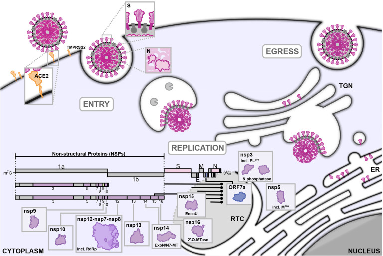
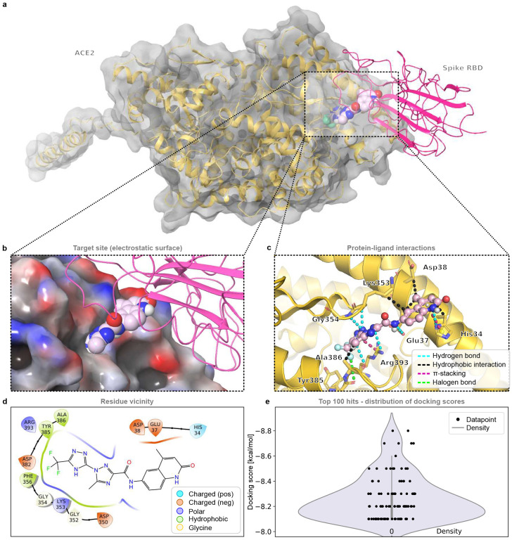
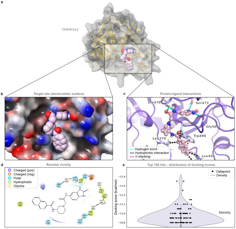
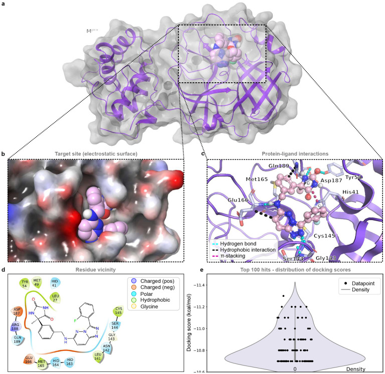
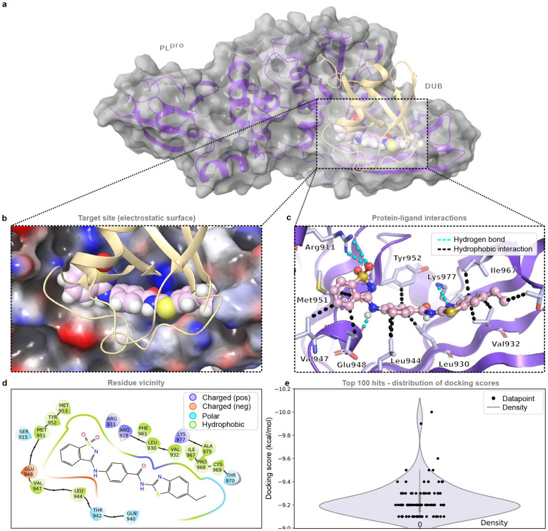
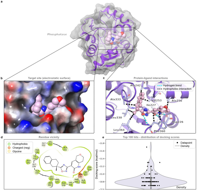
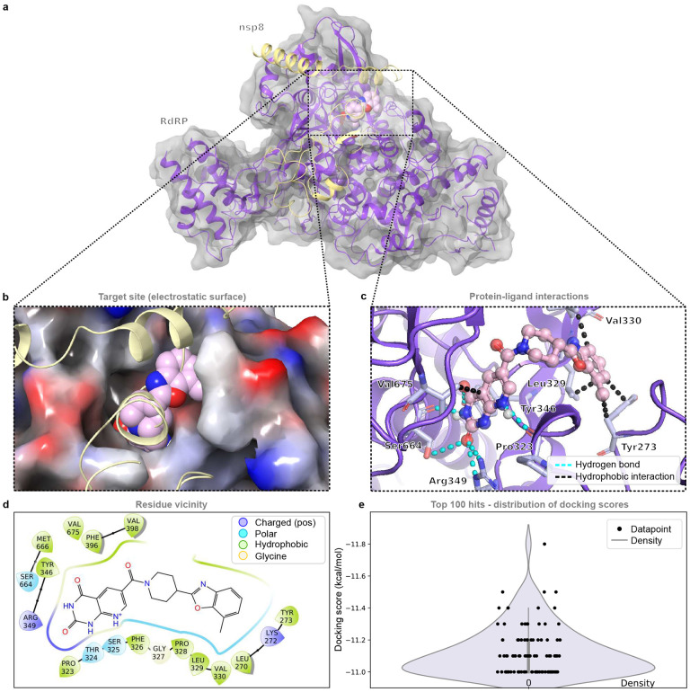
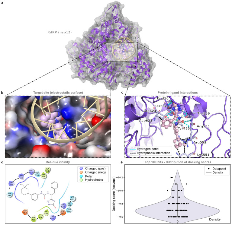
Severe acute respiratory syndrome coronavirus 2 (SARS-CoV-2), previously known as 2019 novel coronavirus (2019-nCoV), has spread rapidly across the globe, creating an unparalleled global health burden and spurring a deepening economic crisis. As of July 7th, 2020, almost seven months into the outbreak, there are no approved vaccines and few treatments available. Developing drugs that target multiple points in the viral life cycle could serve as a strategy to tackle the current as well as future coronavirus pandemics. Here we leverage the power of our recently developed in silico screening platform, VirtualFlow, to identify inhibitors that target SARS-CoV-2. VirtualFlow is able to efficiently harness the power of computing clusters and cloud-based computing platforms to carry out ultra-large scale virtual screens. In this unprecedented structure-based multi-target virtual screening campaign, we have used VirtualFlow to screen an average of approximately 1 billion molecules against each of 40 different target sites on 17 different potential viral and host targets in the cloud. In addition to targeting the active sites of viral enzymes, we also target critical auxiliary sites such as functionally important protein-protein interaction interfaces. This multi-target approach not only increases the likelihood of finding a potent inhibitor, but could also help identify a collection of anti-coronavirus drugs that would retain efficacy in the face of viral mutation. Drugs belonging to different regimen classes could be combined to develop possible combination therapies, and top hits that bind at highly conserved sites would be potential candidates for further development as coronavirus drugs. Here, we present the top 200 in silico hits for each target site. While in-house experimental validation of some of these compounds is currently underway, we want to make this array of potential inhibitor candidates available to researchers worldwide in consideration of the pressing need for fast-tracked drug development.

Keywords: ACE2; COVID-19 data; In silico; MPro; NSP16/NSP10; PLpro inhibitors; RdRP; SARS-CoV -2; SARS-CoV2 Small molecule inhibitor; Spike; TMPRSS2; computational biology; drug discovery; nsp10; nsp13; nsp14-exonuclease; nsp16; nsp3 macrdomain; nsp5; nsp7; nsp8; nsp9; nucleoprotein; virtual screening.


Conflict of interest statement

Alexander Chuprina, Dmytro Radchenko, and Iryna Iavniuk work for Enamine, Kyiv Ukraine. Igor Dziuba works for UkrOrgSyntez Ltd, Kyiv Ukraine. Olga Tarkhanova, Alla Plekhova, and Yurii Moroz work for Chemspace Kyiv, Ukraine. Enamine, UkrOrgSyntez, and Chemspace are companies that are involved in the synthesis and distribution of drug-like compounds. Yurii Moroz is a scientific advisor for Enamine.

Figures

Fig. 1.
Fig. 1.
A schematic of the viral lifecycle of SARS-CoV-2. The genome organization is based on other coronaviruses and published predictions (2, 13). ACE2: angiotensin-converting enzyme 2; TMPRSS2: transmembrane protease, serine 2; RdRp: RNA-dependent RNA polymerase; ExoN: exonuclease; N7-MT N7-methyl transferase; 2’O-MTase: 2’O-methyl transferase; EndoU: uridylate-specific endonuclease; RTC: replication and transcription complex; ER: endoplasmic reticulum; TGN: trans-Golgi network
Fig. 2.
Fig. 2.. The ACE2 receptor (open conformation) and an example compound from the top 0.0001% of screened compounds bound at the spike interaction interface (site1).
a, The target protein ACE2 (gold) in an open conformation bound to the RBD of the spike protein (magenta) and an example compound (light pink) from the virtual screen bound to the spike interaction interface (site 1, around Glu37) (Screen ID: 1). b, The electrostatic surface of the target protein (ACE2) bound to the RBD domain of the spike protein (magenta) and an example compound (light pink). c, An overview of the interactions between the ligand and the receptor structure. d, Receptor residues within 4 Å of the ligand. e, Distribution of the docking scores of the top 100 virtual screening hits.
Fig. 3.
Fig. 3.. TMPRSS2 and an example compound from the top 0.0001% of screened compounds bound at the active site of the serine protease domain.
a, The target protein TMPRSS2 (gold) and an example compound (light pink) from the virtual screen bound at the active site of the serine protease domain (Screen ID: 6). b, Electrostatic surface of the target protein to which an example compound (light pink) is bound. c, An overview of the interactions between the inhibitor and protease structure. d, Residues within 4 Å of the inhibitor. e, Distribution of the docking scores of the top 100 virtual screening hits.
Fig. 4.
Fig. 4.. The RBD of the spike protein and an example compound from the top 0.0001% of screened compounds bound at the ACE2 binding interface.
a, The targeted spike protein (magenta) and an example compound (light pink) from the virtual screen bound to the ACE2 binding interface (Screen ID: 7). b, Electrostatic surface of the target protein to which an example compound (light pink) is bound. c, An overview of the interactions between the inhibitor and the receptor structure. d, Residues within 4 Å of the inhibitor. e, Distribution of the docking scores of the top 100 virtual screening hits.
Fig. 5.
Fig. 5.. The HR1 domain and an example compound from the top 0.0001% of screened compounds bound at the interaction interface to the HR2 domain of the spike protein.
a, The HR1 domain of the targeted spike protein (magenta) and an example compound (light pink) from the virtual screen bound to the HR2 interaction interface. The H2 domain is shown in light gold (Screen ID: 8). b, Electrostatic surface of the target protein to which an example compound (light pink) is bound. c, An overview of the interactions between the inhibitor and the HR1 structure. d, Residues within 4 Å of the inhibitor. e, Distribution of the docking scores of the top 100 virtual screening hits.
Fig. 6.
Fig. 6.. Mpro and an example compound from the top 0.0001% of screened compounds bound at the enzymatic active site
a, Mpro (violet) and an example compound (light pink) from the virtual screen bound to the active site. Here, the crystal structure with PBD ID 6lu7 was used (Screen ID: 16). b, Electrostatic surface of the target protein to which an example compound (light pink) is bound. c, An overview of the interactions between the inhibitor and the protease structure. d, Residues within 4 Å of the inhibitor. e, Distribution of the docking scores of the top 100 virtual screening hits.
Fig. 7.
Fig. 7.. PLpro and an example compound from the top 0.0001% of screened compounds bound at the enzymatic active site.
a, PLpro (violet) and an example compound (light pink) from the virtual screen bound to the active site (Screen ID: 12). b, Electrostatic surface of the target protein to which an example compound (light pink) is bound. c, An overview of the interactions between the inhibitor and the protease structure. d, Residues within 4 Å of the inhibitor. e, Distribution of the docking scores of the top 100 virtual screening hits.
Fig. 8.
Fig. 8.. PLpro and an example compound from the top 0.0001% of screened compounds bound at the DUB binding site.
a, PLpro (violet) and an example compound (light pink) from the virtual screen bound at the DUB binding site (Screen ID: 14). b, Electrostatic surface of the target protein to which an example compound (light pink) is bound. c, An overview of the interactions between the inhibitor and the receptor structure. d, Receptor residues within 4 Å of the inhibitor. e, Distribution of the docking scores of the top 100 virtual screening hits.
Fig. 9.
Fig. 9.. PLpro and an example compound from the top 0.0001% of screened compounds bound at the enzymatic active site (tunnel region).
a, PLpro (violet) and an example compound (light pink) from the virtual screen bound to the active site (tunnel region) (Screen ID: 15). b, Electrostatic surface of the target protein to which an example compound (light pink) is bound. c, An overview of the interactions between the inhibitor and the protease structure. d, Residues within 4 Å of the inhibitor. e, Distribution of the docking scores of the top 100 virtual screening hits.
Fig. 10.
Fig. 10.. The phosphatase (closed conformation) and an example compound from the top 0.0001% of screened compounds bound at the enzymatic active site.
a, The phosphatase (violet), which is part of nsp3, and an example compound (light pink) from the virtual screen bound to the active site (closed conformation) (Screen ID :10). b, Electrostatic surface of the target protein to which an example compound (light pink) is bound. c, An overview of the interactions between the inhibitor and the phosphatase structure. d, Residues within 4 Å of the inhibitor. e, Distribution of the docking scores of the top 100 virtual screening hits.
Fig. 11.
Fig. 11.. nsp9 and an example compound from the top 0.0001% of screened compounds bound at the dimerization interface (site 2).
a, The nsp9 dimer (one monomer in violet, one monomer in gold), where an example compound (light pink) from the virtual screen is bound to the dimerization interface (site 2) of the nsp9 monomer in violet (Screen ID: 24). b, Electrostatic surface of the target protein to which an example compound (light pink) and a second nsp9 monomer (light gold) is bound. c, An overview of the interactions between the inhibitor and the nsp9 structure. d, Residues within 4 Å of the inhibitor. e, Distribution of the docking scores of the top 100 virtual screening hits.
Fig. 12.
Fig. 12.. nsp7 and an example compound from the top 0.0001% of screened compounds bound to the surface).
a, nsp7 (violet), where an example compound (light pink) from the virtual screen is bound at the nsp8 (light gold) interface. In the virtual screen, a blind docking was carried out for each screened compound over the entire surrounding helical surface. This helical surface includes large parts of the nsp8 (light gold, cyan) and nsp12 (lavender) binding interfaces (Screen ID: 20). b, Electrostatic surface of nsp7 to which an example compound (light pink) is bound. Also shown are the protein-protein interaction partners nsp8 (light gold, cyan) and nsp12 (lavender). c, An overview of the interactions between the inhibitor and the nsp7 structure. d, Residues within 4 Å of the inhibitor. e, Distribution of the docking scores of the top 100 virtual screening hits.
Fig. 13.
Fig. 13.. nsp8 and an example compound from the top 0.0001% of screened compounds bound at the nsp7 binding interface.
a, nsp8 (violet) bound to nsp7 (light gold) and an example compound (light pink) (Screen ID: 21). b, Electrostatic surface of the target protein to which an example compound (light pink) and nsp7 (light gold) bound. c, An overview of the interactions between the inhibitor and the nsp8 structure. d, Residues within 4 Å of the inhibitor. e, Distribution of the docking scores of the top 100 virtual screening hits.
Fig. 14.
Fig. 14.. nsp10 and an example compound from the top 0.0001% of screened compounds bound at the protein-protein interaction interface with nsp14 and nsp16.
a, nsp10 (violet) bound to nsp14 (light gold) and an example compound (light pink), at the nsp14/nsp16 binding interface of nsp10 (Screen ID: 25). b, Electrostatic surface of nsp10 to which an example compound (light pink) as well nsp14 (light gold) are bound. c, An overview of the interactions between the inhibitor and the nsp10 structure. d, Residues within 4 Å of the inhibitor. e, Distribution of the docking scores of the top 100 virtual screening hits.
Fig. 15.
Fig. 15.. nsp12 and an example compound from the top 0.0001% of screened compounds bound at the nsp8 binding interface.
a, nsp12 (violet) bound to nsp8 (light gold) and an example compound (light pink) at the nsp8 binding interface of nsp12 . b, Electrostatic surface of nsp12 to which an example compound (light pink) as well as nsp8 (light gold) are bound. c, An overview of the interactions between the inhibitor and the nsp12 structure. d, Residues within 4 Å of the inhibitor. e, Distribution of the docking scores of the top 100 virtual screening hits.
Fig. 16.
Fig. 16.. nsp12 and an example compound from the top 0.0001% of screened compounds bound at site 1 of the the RNA binding interface.
a, nsp12 (violet) bound to RNA (light gold) and an example compound (light pink) at site 1 of the RNA binding interface of nsp12 (Screen ID: 28). b, Electrostatic surface of nsp12 to which an example compound (light pink) is bound at site 1 of the RNA (light gold) binding interface. c, An overview of the interactions between the inhibitor and the nsp12 structure. d, Residues within 4 Å of the inhibitor. e, Distribution of the docking scores of the top 100 virtual screening hits.
Fig. 17.
Fig. 17.. nsp12 and an example compound from the top 0.0001% of screened compounds bound at the nucleotide binding site.
a, nsp12 (violet) bound to RNA (light gold) and an example compound (light pink) at the nucleotide binding site (Screen ID: 30). b, Electrostatic surface of nsp12 to which an example compound (light pink) is bound at the nucleotide binding site. RNA is depicted in light gold. c, An overview of the interactions between the inhibitor and the nsp12 structure. d, Residues within 4 Å of the inhibitor. e, Distribution of the docking scores of the top 100 virtual screening hits.
Fig. 18.
Fig. 18.. The helicase (nsp13) and an example compound from the top 0.0001% of screened compounds bound at the enzymatic active site.
a, The helicase (violet) bound to an example compound (light pink) at the enzymatic active site. A docked DNA strand is shown in light gold (Screen ID: 33). b, Electrostatic surface of the helicase to which an example compound (light pink) is bound. c, An overview of the interactions between the compound and the helicase structure. d, Residues within 4 Å of the inhibitor. e, Distribution of the docking scores of the top 100 virtual screening hits.
Fig. 19.
Fig. 19.. The helicase (nsp13) and an example compound from the top 0.0001% of screened compounds bound at region 1 of the RNA-binding interface.
a, The helicase (violet) bound to an example compound (light pink) at region 1 of the RNA-binding interface. A docked DNA strand is shown in light gold (Screen ID: 34). b, Electrostatic surface of the helicase to which an example compound (light pink) and a docked DNA strand (light gold) are bound. c, An overview of the interactions between the compound and the helicase structure. d, Residues within 4 Å of the inhibitor. e, Distribution of the docking scores of the top 100 virtual screening hits.
Fig. 20.
Fig. 20.. nsp15 and an example compound from the top 0.0001% of screened compounds bound at the active site.
a, nsp15 (violet) bound to an example compound (light pink) at the active site (Screen ID: 39. b, Electrostatic surface of nsp15 to which an example compound (light pink) is bound. c, An overview of the interactions between the compound and the nsp15 structure. d, Residues within 4 Å of the inhibitor. e, Distribution of the docking scores of the top 100 virtual screening hits.
Fig. 21.
Fig. 21.. ORF7a and an example compound from the top 0.0001% of screened compounds bound.
a, ORF7a (violet) bound to an example compound (light pink) at the surface (Screen ID: 9). b, Electrostatic surface of ORF7a to which an example compound (light pink) is bound. c, An overview of the interactions between the compound and the ORF7a structure. d, Residues within 4 Å of the inhibitor. e, Distribution of the docking scores of the top 100 virtual screening hits.
Fig. 22.
Fig. 22.. nsp14 and an example compound from the top 0.0001% of screened compounds bound at the active site of the N7 methyltransferase domain.
a, nsp14 (violet) bound to a example compound (light pink) at the active site of the N7 methyltransferase domain (Screen ID: 38). b, Electrostatic surface of nsp14 to which an example compound (light pink) is bound. c, An overview of the interactions between the compound and the nsp14 structure. d, Residues within 4 Å of the inhibitor. e, Distribution of the docking scores of the top 100 virtual screening hits.
Fig. 23.
Fig. 23.. nsp16 and an example compound from the top 0.0001% of screened compounds bound at the active site.
a, nsp16 (violet) bound to an example compound (light pink) at the active site (Screen ID: 40). b, Electrostatic surface of nsp16 to which an example compound (light pink) is bound. c, An overview of the interactions between the compound and the nsp16 structure. d, Residues within 4 Å of the inhibitor. e, Distribution of the docking scores of the top 100 virtual screening hits.
Fig. 24.
Fig. 24.. N-terminal domain of the nucleoprotein and an example compound from the top 0.0001% of screened compounds bound at the RNA-binding interface.
a, The N-terminal domain (NTD) of nucleoprotein (violet) bound to an example compound (light pink) at the RNA binding interface (Screen ID: 41). b, Electrostatic surface of nucleoprotein to which an example compound (light pink) is bound. c, An overview of the interactions between the compound and the NTD structure. d, Residues within 4 Å of the inhibitor. e, Distribution of the docking scores of the top 100 virtual screening hits.
Fig. 25.
Fig. 25.. C-terminal domain of the nucleoprotein and an example compound from the top 0.0001% of screened compounds bound at the oligomerization interface.
a, The C-terminal domain (CTD) dimer of nucleoprotein (violet) bound to an example compound (light pink) at the oligomerization interface Screen ID: 45). b, Electrostatic surface of the CTD dimer of nucleoprotein to which an example compound (light pink) is bound. c, An overview of the interactions between the compound and the CTD structure. d, Residues within 4 Å of the inhibitor. e, Distribution of the docking scores of the top 100 virtual screening hits.

References

    1. Zhou Peng, Yang Xing-Lou, Wang Xian-Guang, Hu Ben, Zhang Lei, Zhang Wei, Si Hao-Rui, Zhu Yan, Li Bei, Huang Chao-Lin, Chen Hui-Dong, Chen Jing, Luo Yun, Guo Hua, Jiang Ren-Di, Liu Mei-Qin, Chen Ying, Shen Xu-Rui, Wang Xi, Zheng Xiao-Shuang, Zhao Kai, Chen Quan-Jiao, Deng Fei, Liu Lin-Lin, Yan Bing, Zhan Fa-Xian, Wang Yan-Yi, Xiao Geng-Fu, and Shi Zheng-Li. A pneumonia outbreak associated with a new coronavirus of probable bat origin. Nature, 579(7798):270–273, 2020. ISSN 0028–0836 1476–4687. doi: 10.1038/s41586-020-2012-7 - DOI - PMC - PubMed
    1. Wu Fan, Zhao Su, Yu Bin, Chen Yan-Mei, Wang Wen, Song Zhi-Gang, Hu Yi, Tao Zhao-Wu, Tian Jun-Hua, Pei Yuan-Yuan, Yuan Ming-Li, Zhang Yu-Ling, Dai Fa-Hui, Liu Yi, Wang Qi-Min, Zheng Jiao-Jiao, Xu Lin, Holmes Edward C., and Zhang Yong-Zhen. A new coronavirus associated with human respiratory disease in China. Nature, 579(7798):265–269, 2020. ISSN 0028–0836 1476–4687. doi: 10.1038/s41586-020-2008-3 - DOI - PMC - PubMed
    1. Andersen Kristian G., Rambaut Andrew, Lipkin W. Ian, Holmes Edward C., and Garry Robert F.. The proximal origin of SARS-CoV-2. Nature Medicine, 26(4):450–452, April 2020. ISSN 1078–8956. doi: 10.1038/s41591-020-0820-9 - DOI - PMC - PubMed
    1. Zhu Na, Zhang Dingyu, Wang Wenling, Li Xingwang, Yang Bo, Song Jingdong, Zhao Xiang, Huang Baoying, Shi Weifeng, Lu Roujian, Niu Peihua, Zhan Faxian, Ma Xuejun, Wang Dayan, Xu Wenbo, Wu Guizhen, Gao George F., and Tan Wenjie. A novel coronavirus from patients with pneumonia in China, 2019. New England Journal of Medicine, 382(8):727–733, 2020. ISSN 0028–4793 1533–4406. doi: 10.1056/NEJMoa2001017 - DOI - PMC - PubMed
    1. Dong Ensheng, Du Hongru, and Gardner Lauren. An interactive web-based dashboard to track COVID-19 in real time. The Lancet Infectious Diseases, 20(5):533–534, 2020. ISSN 14733099. doi: 10.1016/s1473-3099(20)30120-1 - DOI - PMC - PubMed

Publication types

LinkOut - more resources