. 2021 Mar;36(3):729-739.
doi: 10.1002/mds.28385. Epub 2020 Nov 16.

A Gain-of-Function Variant in Dopamine D2 Receptor and Progressive Chorea and Dystonia Phenotype

Affiliations

A Gain-of-Function Variant in Dopamine D2 Receptor and Progressive Chorea and Dystonia Phenotype

Marlous C M van der Weijden et al. Mov Disord. 2021 Mar.

Abstract

Background: We describe a 4-generation Dutch pedigree with a unique dominantly inherited clinical phenotype of a combined progressive chorea and cervical dystonia carrying a novel heterozygous dopamine D2 receptor (DRD2) variant.

Objectives: The objective of this study was to identify the genetic cause of the disease and to further investigate the functional consequences of the genetic variant.

Methods: After detailed clinical and neurological examination, whole-exome sequencing was performed. Because a novel variant in the DRD2 gene was found as the likely causative gene defect in our pedigree, we sequenced the DRD2 gene in a cohort of 121 Huntington-like cases with unknown genetic cause (Germany). Moreover, functional characterization of the DRD2 variant included arrestin recruitment, G protein activation, and G protein-mediated inhibition of adenylyl cyclase determined in a cell model, and G protein-regulated inward-rectifying potassium channels measured in midbrain slices of mice.

Result: We identified a novel heterozygous variant c.634A > T, p.Ile212Phe in exon 5 of DRD2 that cosegregated with the clinical phenotype. Screening of the German cohort did not reveal additional putative disease-causing variants. We demonstrated that the D2S/L -I212 F receptor exhibited increased agonist potency and constitutive activation of G proteins in human embryonic kidney 239 cells as well as significantly reduced arrestin3 recruitment. We further showed that the D2S -I212 F receptor exhibited aberrant receptor function in mouse midbrain slices.

Conclusions: Our results support an association between the novel p.Ile212Phe variant in DRD2, its modified D2 receptor activity, and the hyperkinetic movement disorder reported in the 4-generation pedigree. © 2020 The Authors. Movement Disorders published by Wiley Periodicals LLC on behalf of International Parkinson and Movement Disorder Society.

Keywords: chorea; dopamine D2 receptor; dystonia; hyperkinetic movement disorder.


Figures

FIG. 1
FIG. 1
Four‐generation pedigree and amino acid sequence homology. (A) Four‐generation pedigree carrying the missense variant c.634A > T, p.Ile212Phe in DRD2. Genetic analysis was performed in the individuals indicated by “DNA.” Proband is indicated by an arrow. Individuals marked by an asterisk underwent whole‐exome sequencing. The variant (c.634A > T) was identified in 5 affected subjects and was absent in 3 unaffected subjects. Male = square, female = circle, sex unknown = hexagon. Filled symbols = affected. Open symbols = unaffected. Sequence alignments showing the amino acid sequence identity of the affected amino acids in (B) multiple species orthologs of DRD2 and (C) among the human dopamine receptor family. The mutated amino acid isoleucine at position 212 is indicated by the vertical box. The region of arrestin‐binding (IYIV) in DRD2 is indicated by the horizontal box. [Color figure can be viewed at wileyonlinelibrary.com]
FIG. 2
FIG. 2
Arrestin recruitment in response to quinpirole. (A,B) Dose‐response curves for arrestin recruitment mediated by D2‐WT, D2‐I212F, and D2‐WT/I212F in response to a 20‐minute stimulation with quinpirole. Results are expressed as a percentage of maximum arrestin recruitment by D2‐WT. (A) Data for D2S. (B) Data for D2L. (C,D) Time course of maximal recruitment of arrestin for each condition. Basal response was subtracted from the maximal quinpirole stimulation response for each indicated time. Maximal recruitment was normalized to its initial response (1‐minute response). There was a significant interaction between genotype and time for maximal recruitment (2‐way analysis of variance: F 12,63 = 3.628, P = 0.0004 for D2S, and F 12,42 = 4.304, P = 0.0002 for D2L). Values plotted are mean ± SD of 4 (A,C) and 3 (B,D) independent experiments performed in quadruplicate. *P < 0.05, **P < 0.01, and ***P < 0.001 compared to D2‐WT and †P < 0.05, ††P < 0.01, and †††P < 0.001 compared to D2‐WT/I212F, Tukey's multiple comparisons test. Emax, maximum response; WT, wild type. [Color figure can be viewed at wileyonlinelibrary.com]
FIG. 3
FIG. 3
Protein activation in response to quinpirole. (A,B) Dose‐response curves for Gαi1 protein activation mediated by D2‐WT, D2‐I212F, and D2‐WT/I212F in response to a 10‐minute stimulation with quinpirole. Results are expressed as a percentage of maximum G protein–activation by D2‐WT. Values plotted represent mean ± SD of 4 independent experiments performed in quadruplicate. (C,D) Dose‐response curves for the inhibition of forskolin‐stimulated cAMP accumulation mediated by D2‐WT, D2‐I212F, and D2‐WT/I212F in response to incubation with quinpirole for 10 minutes in the presence of 10 μM forskolin. Results are expressed as a percentage of maximum cAMP accumulation for each condition. Values plotted are mean ± SD of 4 (C) and 6 (D) independent experiments performed in triplicate. Left and right panels depict data for D2S and D2L, respectively. cAMP, cyclic adenosine monophosphate; Emax, maximum response; FSK, forskolin; WT, wild type. [Color figure can be viewed at wileyonlinelibrary.com]
FIG. 4
FIG. 4
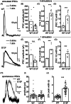
D2S receptor‐GIRK IPSCs in dopamine neurons. (A) Example recordings of electrically stimulated IPSCs from neurons expressing D2S‐WT (5 stimuli at 40 Hz [black] and 1 stimulus [gray]) and D2S‐I212F (5 stimuli at 40 Hz [black] and 1 stimulus [gray]). When 5 stimuli were used to elicit IPSCs, neurons expressing D2S‐WT had (B) larger amplitude IPSCs (Student'st‐test; t = 2.2141, P = 0.043), (C) faster time to peak (Student's t‐test; 5 stimulations, t = 5.1519, P < 0.001), and (D) shorter half‐width compared to IPSCs in neurons expressing D2S‐I212F (N = 7 cells from 3 animals for D2S‐WT, N = 10 cells from 3 animals for D2S‐I212F; Student's t‐test; for 5 stimulations, t = 5.3117, P < 0.001). When 1 stimulus was used to elicit IPSCs, (E) neurons expressing D2S‐WT and D2S‐I212F had IPSCs that did not differ significantly in amplitude (Student's t‐test; t = 1.8838, P = 0.086), but (F) the IPSC time to peak (Student's t‐test; 1 stimulation, t = 4.5137, P < 0.001) and (G) half‐widths were significantly shorter in neurons expressing D2S‐WT compared with D2S‐I212F (N = 5 cells from 2 animals for D2S‐WT, N = 8 cells from 3 animals for D2S‐I212F; Student's t‐test; for 1 stimulation, t = 6.6569, P < 0.001). (H) Averages of spontaneous D2 receptor IPSCs from DA neurons expressing D2‐WT (black) or D2‐I212F (gray). (I) The average spontaneous IPSC amplitudes did not differ between neurons expressing D2S‐WT and D2S‐I212F (N = 61 events from 3 animals for D2S‐WT, N = 50 events from 5 animals for D2S‐I212F; Student's t‐test; t = 0.72164, P = 0.4721). (J) The average spontaneous IPSC width at 20% of peak was significantly longer in DA neurons expressing D2S‐I212F compared with those expressing D2S‐WT (N = 61 events from 3 animals for D2S‐WT, N = 50 events from 5 animals for D2S‐I212F; Student's t‐test; t = 14.341, P < 0.0001). *P < 0.05; **P < 0.01; ***P < 0.001 compared with D2‐WT. IPSCs, inhibitory postsynaptic currents; n.s., not significant; stims, stimulations; WT, wild type.

Comment in

Similar articles

Cited by

References

    1. Korchounov A, Meyer MF, Krasnianski M. Postsynaptic nigrostriatal dopamine receptors and their role in movement regulation. J Neural Trans 2010;117(12):1359–1369. - PMC - PubMed
    1. Gurevich EV, Gainetdinov RR, Gurevich VV. G protein‐coupled receptor kinases as regulators of dopamine receptor functions. Pharmacol Res 2016;111:1–16. - PMC - PubMed
    1. Mishra A, Singh S, Shukla S. Physiological and functional basis of dopamine receptors and their role in neurogenesis: possible implication for Parkinson's disease. J Exp Neurosci 2018;31(12):1179069518779829. - PMC - PubMed
    1. Lüscher C, Slesinger PA. Emerging roles for G protein‐gated inwardly rectifying potassium (GIRK) channels in health and disease. Nat Rev Neurosci 2010;11(5):301–315. - PMC - PubMed
    1. Beaulieu J‐M. Beyond cAMP: the regulation of Akt and GSK3 by dopamine receptors. Front Mol Neurosci 2011;1(4):38. - PMC - PubMed

Publication types

Substances