. 2020 Nov;25(11):116005.
doi: 10.1117/1.JBO.25.11.116005.

Prediction of optimal contrast times post-imaging agent administration to inform personalized fluorescence-guided surgery

Affiliations

Prediction of optimal contrast times post-imaging agent administration to inform personalized fluorescence-guided surgery

Negar Sadeghipour et al. J Biomed Opt. 2020 Nov.

Abstract

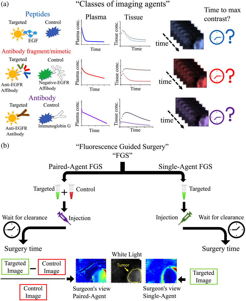
Significance: Fluorescence guidance in cancer surgery (FGS) using molecular-targeted contrast agents is accelerating, yet the influence of individual patients' physiology on the optimal time to perform surgery post-agent-injection is not fully understood.

Aim: Develop a mathematical framework and analytical expressions to estimate patient-specific time-to-maximum contrast after imaging agent administration for single- and paired-agent (coadministration of targeted and control agents) protocols.

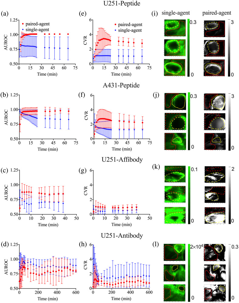
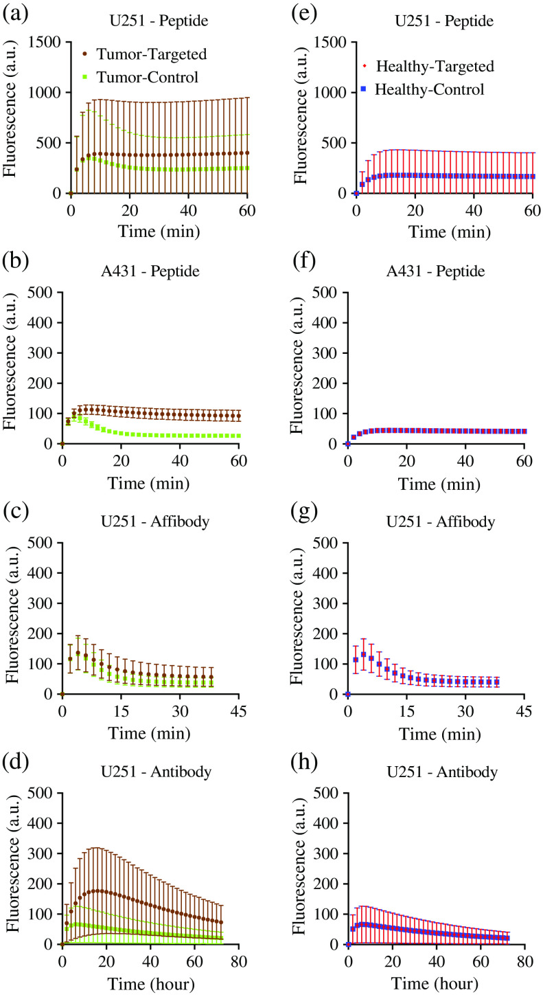
Approach: The framework was validated in mouse subcutaneous xenograft studies for three classes of imaging agents: peptide, antibody mimetic, and antibody. Analytical expressions estimating time-to-maximum-tumor-discrimination potential were evaluated over a range of parameters using the validated framework for human cancer parameters.

Results: Correlations were observed between simulations and matched experiments and metrics of tumor discrimination potential (p < 0.05). Based on human cancer physiology, times-to-maximum contrast for peptide and antibody mimetic agents were <200 min, >15 h for antibodies, on average. The analytical estimates of time-to-maximum tumor discrimination performance exhibited errors of <10 % on average, whereas patient-to-patient variance is expected to be greater than 100%.

Conclusion: We demonstrated that analytical estimates of time-to-maximum contrast in FGS carried out patient-to-patient can outperform the population average time-to-maximum contrast used currently in clinical trials. Such estimates can be made with preoperative DCE-MRI (or similar) and knowledge of the targeted agent's binding affinity.

Keywords: fluorescence-guided surgery; intraoperative visualization; kinetic modeling; optimal time of surgery; paired-agent imaging.


Figures

Fig. 1
Fig. 1
Overview of FGS. (a) Schematic of three classes of protein-based molecular imaging agents that were investigated: peptides, antibody fragment/mimetic, and antibodies (specifically here: targeted: IRDye® 800CW EGF, control: IRDye® 700DX), medium sized antibody fragment/mimetic (targeted: IRDye® 800CW Anti-EGFR affibody, control: IRDye® 680RD negative control affibody®), and larger sized antibodies (targeted: IRDye® 800CW Cetuximab, control: IRDye® 700DX IgG). The plasma and tissue clearance and kinetics of these imaging agents differ, and consequently time-to-maximum contrast can differ for each of these groups. The goal of this article was to develop and test a mathematical framework for estimated timing of optimal surgical contrast enabled by different imaging agent properties and different clinically relevant physiological conditions. (b) Two molecular imaging protocols in FGS are discussed: SA and PA imaging. In SA imaging, a targeted imaging agent is injected and there is a delay between injection and the operation to let the unbound imaging agent washout from the body. In PA imaging, a control imaging agent is mixed with the targeted imaging agent and the cocktail is injected. After a delay, the imaging agents can reach an equilibrium level at which improved tumor contrast and a quantitative estimate of target concentrations are achievable, despite residual unbound imaging agents.
Fig. 2
Fig. 2
Targeted and control imaging agents in three different imaging agent classes: peptides, affibodies, and antibodies. Mouse human cancer xenograft (subcutaneous thigh tumor models) fluorescent imaging dynamics from the peptide-based imaging agent (targeted: IRDye® 800CW EGF, control: IRDye® 700DX) studies are presented from the moderate EGFR-expressing cell line (human glioblastoma; U251); the high EGFR-expressing cell line (human epidermoid, A431); the affibody-based imaging agent study in U251 xenografts (targeted: IRDye® 800CW anti-EGFR affibody, control: IRDye® 680RD negative control affibody®); the antibody-based imaging agent study in U251 xenografts (targeted: IRDye® 800CW Cetuximab, control: IRDye® 700DX IgG). (a)–(d) The mean fluorescence measured for the targeted and control imaging agents in the tumors in each group (errors are SD between animals). (e)–(h) The same information in a proportionally sized region-of-interest in the muscle surrounding the tumors in each case.
Fig. 3
Fig. 3
Comparison of the PA and SA FGS protocols. The AUROCs and the CVRs measured from PA (red data) and SA (blue data) analyses of the experimental results as a function of time post-imaging-agent injection. The mean of the AUROCs measured in each group (errors are SD between animals) for (a) the U251-peptide group, (b) the A431-peptide group, (c) the U251-affibody group, and (d) the U251-antibody group. The CVR measured in each group (errors are SD between animals) for (e) the U251-peptide group, (f) the A431-peptide group, (g) the U251-affibody group, and (h) the U251-antibody group. The third column depicts maps of targeted imaging agent fluorescence the SA (targeted; green) and the PA binding potentials (BPratio; grayscale) for three randomly selected mice in each of (i) the U251-peptide group, (j) the A431-peptide group, (k) the U251-affibody group, and (l) the U251-antibody group. The yellow dashed circles depict the location of the tumors and the red dashed circles show the normal muscle tissue surrounding the tumor that were selected as representative of “background.” The ROIs were selected based on the white-light images (not shown).
Fig. 4
Fig. 4
Simulation results for three different classes of imaging agents: peptides, affibodies and antibodies. Simulated fluorescence signal are presented for mouse human cancer xenograft (subcutaneous thigh tumor models) fluorescent imaging dynamics from the peptide-based imaging agent (targeted: IRDye® 800CW EGF, control: IRDye® 700DX) from the moderate EGFR-expressing cell line (human glioblastoma; U251); the high EGFR-expressing cell line (human epidermoid, A431); the affibody-based imaging agent study in U251 xenografts (targeted: IRDye® 800CW anti-EGFR affibody, control: IRDye® 680RD negative control affibody®); and the antibody-based imaging agent study in U251 xenografts (targeted: IRDye® 800CW Cetuximab, control: IRDye® 700DX IgG). (a)–(d) The mean fluorescence measured for the targeted and control imaging agents in the tumors in each group (errors are SD between animals). (e)–(h) The same information in a proportionally sized region-of-interest in the muscle surrounding the tumors in each case.
Fig. 5
Fig. 5
Simulation results for three different classes of imaging agents: peptides, affibodies and antibodies. The mean±SD of AUROC (blue = SA, red = PA) for (a) the U251-peptide, (b) the A431-peptide, (c) the U251-affibody and (d) the U251-antibody simulated groups. The mean±SD of CVR (blue = SA, red = PA) for (e) the U251-peptide, (f) the A431-peptide, (g) the U251-affibody, and (h) the U251-antibody simulated groups.
Fig. 6
Fig. 6
Comparison between experimental and estimated simulation results. Correlations between estimated and simulated areas under the receiver operating characteristic curve (AUROCs) for (a) SA and (b) PA imaging protocols. CVRs for (c) SA and (d) PA imaging agent protocols. The correlations are tested at the frame of maximum value.
Fig. 7
Fig. 7
Predicting time-to-maximum tumor contrast (CVR Tmax) after imaging agent injection based on human cancer parameters. Correlations between true simulated values and estimations by the analytical solutions of CVR Tmax for (a) SA and (b) PA imaging techniques. (c) Correlations between true simulated values of CVR Tmax and estimations at 98% CVR before and after CVR Tmax for SA and PA imaging techniques. Time Diff refers to the difference in time between the pre 98% of maximum CVR threshold pre-Tmax (circle data) or post-Tmax (solid dots) and the CVR Tmax. Red data refer to the SA simulations and blue data to the PA simulations. The input parameters were perturbed to cover a range of parameters from small peptides to large antibodies with different binding kinetics. (d) Predicted CVR Tmax,SA based on blood extravasation rate constants of the targeted imaging agent in the tumor and “normal”/background tissues, respectively (K1), the corresponding tissue-to-blood efflux rate constants (k2), and the tumor binding potential (BP). (e) Predicted CVR Tmax,PA based on tissue-to-blood efflux rate constant in the tumor (k2) and the tumor binding potential (BP).

Similar articles

Cited by

References

    1. Keles G. E., Anderson B., Berger M. S., “The effect of extent of resection on time to tumor progression and survival in patients with glioblastoma multiforme of the cerebral hemisphere,” Surg. Neurol. 52, 371–379 (1999).SGNRAI10.1016/S0090-3019(99)00103-2 - DOI - PubMed
    1. McGrath P. C., et al. , “Improved survival following complete excision of retroperitoneal sarcomas,” Ann. Surg. 200, 200 (1984).10.1097/00000658-198408000-00014 - DOI - PMC - PubMed
    1. Schulick R. D., Brennan M. F., “Long-term survival after complete resection and repeat resection in patients with adrenocortical carcinoma,” Ann. Surg. Oncol. 6, 719–726 (1999).10.1007/s10434-999-0719-7 - DOI - PubMed
    1. Hoogstins C. E., et al. , “A novel tumor-specific agent for intraoperative near-infrared fluorescence imaging: a translational study in healthy volunteers and patients with ovarian cancer,” Clin. Cancer Res. 22, 2929–2938 (2016).10.1158/1078-0432.CCR-15-2640 - DOI - PubMed
    1. Rosenthal E. L., et al. , “Safety and tumor specificity of cetuximab-IRDye800 for surgical navigation in head and neck cancer,” Clin. Cancer Res. 21, 3658–3666 (2015).10.1158/1078-0432.CCR-14-3284 - DOI - PMC - PubMed

Publication types