. 2020;64(10-11-12):485-494.
doi: 10.1387/ijdb.200114jw.

Precise control of ion channel and gap junction expression is required for patterning of the regenerating axolotl limb

Affiliations

Precise control of ion channel and gap junction expression is required for patterning of the regenerating axolotl limb

Konstantinos Sousounis et al. Int J Dev Biol. 2020.

Abstract

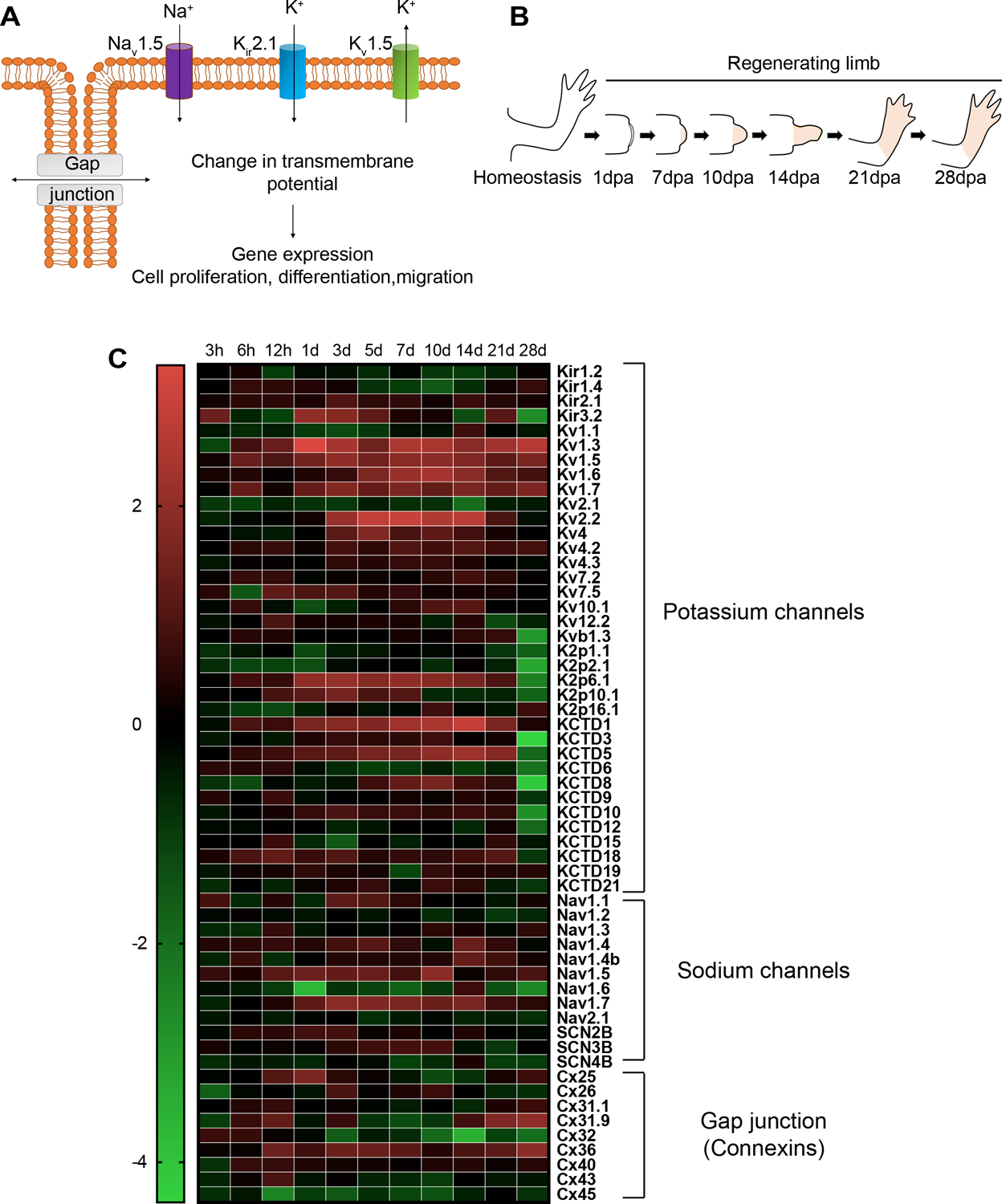
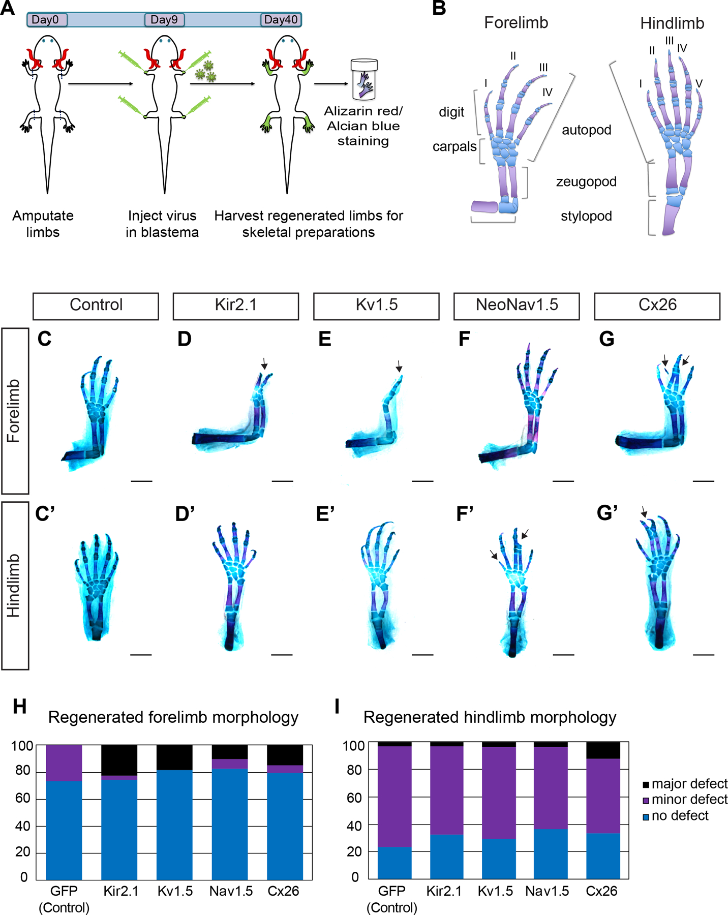
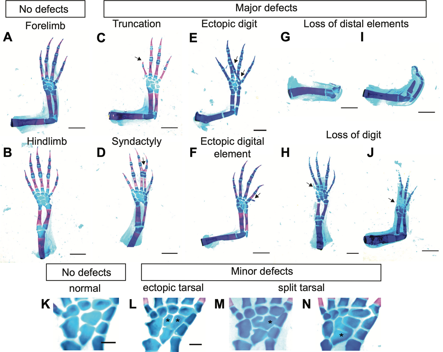
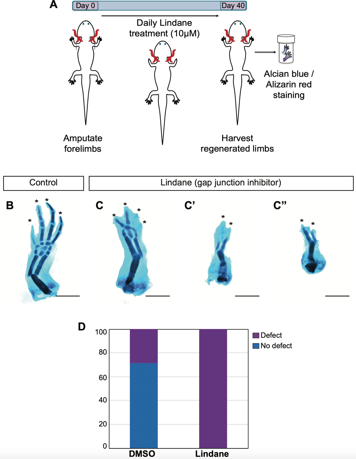
Axolotls and other salamanders have the capacity to regenerate lost tissue after an amputation or injury. Growth and morphogenesis are coordinated within cell groups in many contexts by the interplay of transcriptional networks and biophysical properties such as ion flows and voltage gradients. It is not, however, known whether regulators of a cell's ionic state are involved in limb patterning at later stages of regeneration. Here we manipulated expression and activities of ion channels and gap junctions in vivo, in axolotl limb blastema cells. Limb amputations followed by retroviral infections were performed to drive expression of a human gap junction protein Connexin 26 (Cx26), potassium (Kir2.1-Y242F and Kv1.5) and sodium (NeoNav1.5) ion channel proteins along with EGFP control. Skeletal preparation revealed that overexpressing Cx26 caused syndactyly, while overexpression of ion channel proteins resulted in digit loss and structural abnormalities compared to EGFP expressing control limbs. Additionally, we showed that exposing limbs to the gap junction inhibitor lindane during the regeneration process caused digit loss. Our data reveal that manipulating native ion channel and gap junction function in blastema cells results in patterning defects involving the number and structure of the regenerated digits. Gap junctions and ion channels have been shown to mediate ion flows that control the endogenous voltage gradients which are tightly associated with the regulation of gene expression, cell cycle progression, migration, and other cellular behaviors. Therefore, we postulate that mis-expression of these channels may have disturbed this regulation causing uncoordinated cell behavior which results in morphological defects.


Conflict of interest statement

The authors declare no conflicts of interest.

Figures

Fig. 1.
Fig. 1.. Ion channel and gap junction isoforms are transcribed in regenerating forelimb tissues.
(A) Schematic representation of sodium (Na+), potassium (K+) ion channels and gap junctions located on the cell membrane mediating ion exchange to induce transmembrane potential to activate downstream cellular activities. (B) Cartoon showing the stages of forelimb regeneration. (C) Heatmap of the log2 ratios ofTPMs (transcripts per million) of transcripts encoding potassium channels, sodium channels, and gap junction forming protein connexins at different time points during forelimb regeneration relative to homeostasis (0 h).
Fig. 2.
Fig. 2.. Ion channel and gap junction overexpression causes limb morphology defects in regenerated fore and hindlimbs.
(A) Cartoon illustrating the experimental outline. Axolotl fore and hindlimbs were amputated at day 0. 9 days post amputation (Day9) retrovirus carrying ion channel constructs Kir2.1, Kv1.5, NeoNav1.5 and Connexin 26 (Cx26) were injected into blastemas. 40 days post amputation (Day40) all four limbs were harvested and processed with alcian blue (cartilage) and alizarin red (bone) stain for visualization of skeletal morphology. (B) Cartoon illustrating the fore and hindlimb skeletal elements (cartilage in blue; bone in magenta). Forelimb possess 4 digits and 8 carpals and hindlimb possess 5 digits and 9 tarsals. (C-G’) Representative regenerated fore (C-G) and hindlimb (C’-G’) skeletal morphologies of EGFP control (C-C’), Kir2.1(D-D’), Kv1.5 (E-E’), NeoNav1.5 (F-F’) and Connexin 26 (G-G’) expressing limbs. Overexpression of ion channel and gap junction protein resulted in major defects elicited as digit (D) and distal element loss (E), digit truncation (F’) and syndactyly (G, G’). (H-I) Bar graph depicting the quantification of morphological defects observed in ion channel and gap junction overexpressing regenerated limbs. (Scoring details can be found in Table1). Scale bar, 1mm.
Fig. 3.
Fig. 3.. Representative alcian blue/ alizarin red images depicting skeletal morphology defects used in scoring.
(A,B) Representative fore and hindlimbs with no defect. Representative images of major defects, truncation (C), syndactyly (D), ectopic digit formation (E), ectopic digital element formation (F), loss of distal elements (G-I) digit loss (H-J). Scale bar, 2 mm. (K-N) Representative images of central elements (K) normal central elements present in hindlimb with 9 tarsals. (L) ectopic tarsal, (M,N) split tarsal. Scale bar, 0.5 mm.
Fig. 4.
Fig. 4.. The gap junction blocker lindane causes severe skeletal morphological defects in regenerating forelimbs.
(A) Cartoon illustrating the experimental timeline. Axolotl forelimbs were amputated at day 0. Lindane at a final concentration of 10 mM is added daily into axolotl housing water until the limbs are harvested 40 days after amputation. Harvested limbs are processed for skeletal morphology evaluation. (B-C”) Representative skeletal images of DMSO (B) and lindane (C,C”) treated control and gap junction communication blocked forelimbs. (D) Bar graph depicting the scoring of the defects observed in DMSO treated control (4 out of 14 limb showed defect) versus lindane treated forelimbs (all 14 forelimbs examined showed defect). Scale bar, 1 mm.

Similar articles

Cited by

References

    1. ADAMS DS, MASI A & LEVIN M (2007). H+ pump-dependent changes in membrane voltage are an early mechanism necessary and sufficient to induce Xenopus tail regeneration. Development, 134: 1323–35. - PubMed
    1. ADAMS DS, TSENG AS & LEVIN M (2013). Light-activation of the Archaerhodopsin H(+)-pump reverses age-dependent loss of vertebrate regeneration: sparking system-level controls in vivo. Biology open, 2: 306–13. - PMC - PubMed
    1. ADAMS DS, UZEL SG, AKAGI J, WLODKOWIC D, ANDREEVA V, YELICK PC, DEVITT-LEE A, PARE JF & LEVIN M (2016). Bioelectric signalling via potassium channels: a mechanism for craniofacial dysmorphogenesis in KCNJ2-associated Andersen-Tawil Syndrome. J Physiol, 594: 3245–3270. - PMC - PubMed
    1. BATES E (2015). Ion Channels in Development and Cancer. Annu Rev Cell Dev Biol, 31: 231–47. - PubMed
    1. BEANE WS, MOROKUMA J, LEMIRE JM & LEVIN M (2013). Bioelectric signaling regulates head and organ size during planarian regeneration. Development, 140: 313–22. - PMC - PubMed

Publication types