. 2020 Dec 7;219(12):e202009128.
doi: 10.1083/jcb.202009128.

STING induces LC3B lipidation onto single-membrane vesicles via the V-ATPase and ATG16L1-WD40 domain

Affiliations

STING induces LC3B lipidation onto single-membrane vesicles via the V-ATPase and ATG16L1-WD40 domain

Tara D Fischer et al. J Cell Biol. .

Abstract

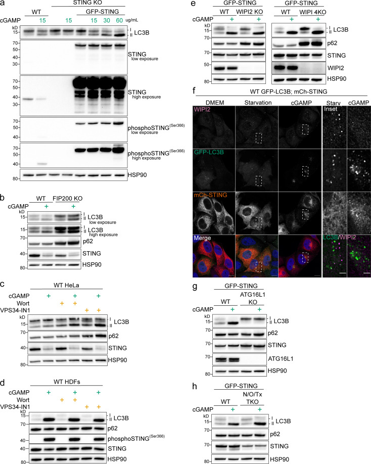
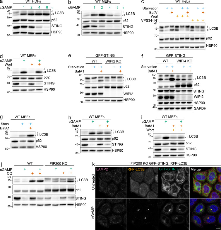
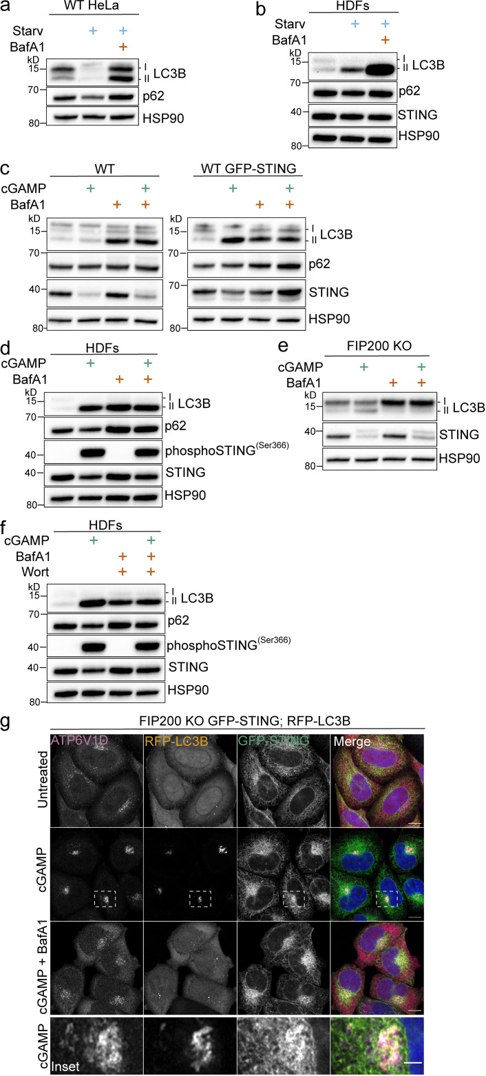
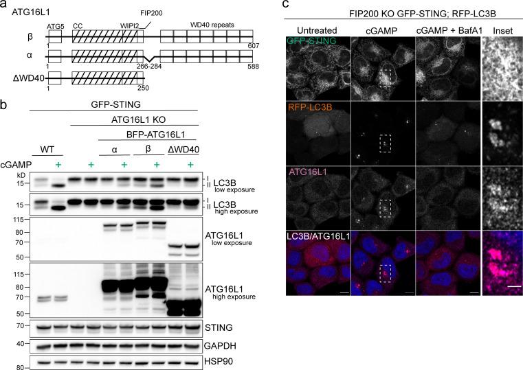
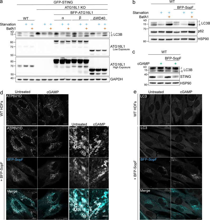
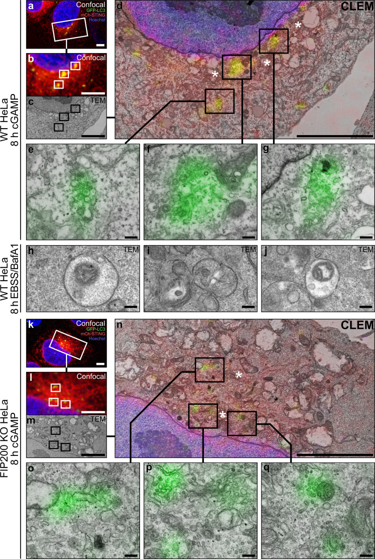
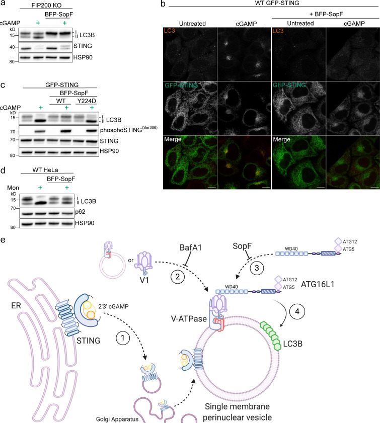
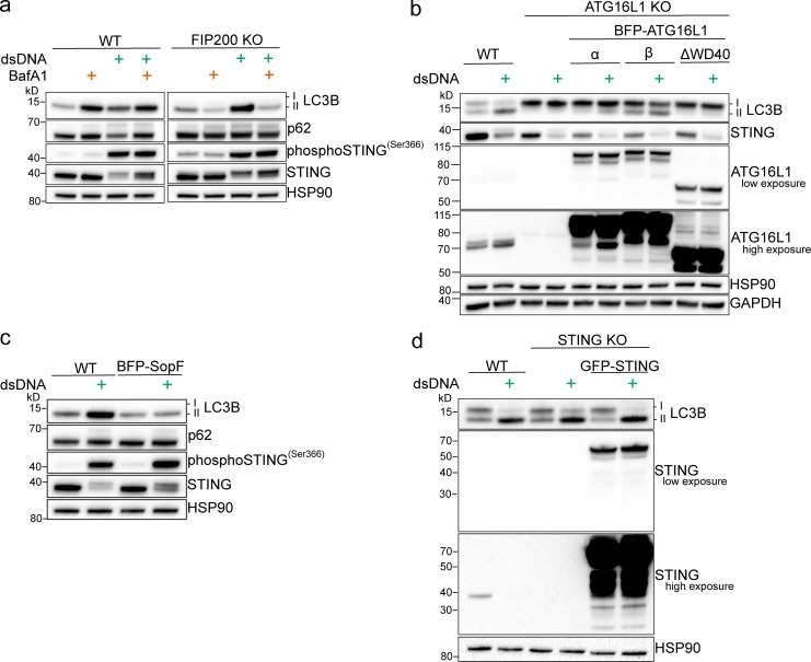
Following the detection of cytosolic double-stranded DNA from viral or bacterial infection in mammalian cells, cyclic dinucleotide activation of STING induces interferon β expression to initiate innate immune defenses. STING activation also induces LC3B lipidation, a classical but equivocal marker of autophagy, that promotes a cell-autonomous antiviral response that arose before evolution of the interferon pathway. We report that STING activation induces LC3B lipidation onto single-membrane perinuclear vesicles mediated by ATG16L1 via its WD40 domain, bypassing the requirement of canonical upstream autophagy machinery. This process is blocked by bafilomycin A1 that binds and inhibits the vacuolar ATPase (V-ATPase) and by SopF, a bacterial effector that catalytically modifies the V-ATPase to inhibit LC3B lipidation via ATG16L1. These results indicate that activation of the cGAS-STING pathway induces V-ATPase-dependent LC3B lipidation that may mediate cell-autonomous host defense, an unanticipated mechanism that is distinct from LC3B lipidation onto double-membrane autophagosomes.


Figures

Figure 1.
Figure 1.
Characterization of autophagy machinery required for STING-dependent LC3B lipidation. (a) WT, STING KO, and STING KO HeLa cells stably expressing GFP-STING were treated with 15 µg/ml or the indicated concentrations of cGAMP for 8 h in normal DMEM. (b) WT and FIP200 KO HeLa cells were treated with 15 µg/ml cGAMP for 8 h in normal DMEM (all the following experiments were treated the same unless specified). (c) WT HeLa cells were treated with cGAMP alone or with cGAMP and wortmannin (200 nM) or VPS34-IN1 (300 nM) for 8 h. (d) Primary HDFs were treated with 60 µg/ml cGAMP and wortmannin or VPS34-IN1 at the same concentrations as in c for 4 h. (e) WT, WIPI2 KO, and WIPI 4KO HeLa cells stably expressing GFP-STING were treated with 60 µg/ml cGAMP. (f) Representative Airyscan-processed confocal imaging of WT HeLa cells stably expressing GFP-LC3B (green) and mCh-STING (red) were treated with 60 µg/ml cGAMP or starvation media (HBSS with Ca2+ and Mg2+) for 8 h before immunofluorescent labeling of endogenous WIPI2 (magenta). Scale bar, 10 µm, 2 µm (inset). (g and h) WT, ATG16L1 KO, and NDP52 (N), optineurin (O), and TAX1BP1 (Tx) triple KO (N/O/Tx TKO) HeLa cells stably expressing GFP-STING were treated with 60 µg/ml cGAMP. Each Western blotting experiment was independently replicated three times. Starv, starvation; Wort, wortmannin.
Figure S1.
Figure S1.
Experiments supplementary to Figs. 1 and 2. (a and b) Primary HDFs (a) and primary MEFs (b) were treated with 60 μg/ml cGAMP, and cell lysates were collected at the indicated time points. (c) WT HeLa cells were incubated in starvation media (HBSS with Ca2+ and Mg2+) alone and with 100 nM BafA1 and with or without wortmannin (200 nM) or VPS34-IN1 (300 nM) for 4 h. All the following experiments were treated the same unless specified. (d) MEFs were treated with 60 μg/ml cGAMP alone and with wortmannin for 2 h. All the following experiments using MEFs use the same conditions. (e and f) WT, WIPI2 KO (e), and WIPI 4KO (f) HeLa cells stably expressing GFP-STING were incubated in starvation media alone and with BafA1 for 4 h. (g) MEFs were incubated in starvation media alone and with BafA1 for 4 h. (h and i) MEFs were incubated with cGAMP alone and with BafA1 (h) or with BafA1 and wortmannin (i). (j) WT and FIP200 KO HeLa cells were incubated with 15 µg/ml cGAMP alone and with 20 μM chloroquine (CQ) for 8 h. (k) Representative Airyscan-processed confocal imaging of FIP200 KO HeLa cells with stable expression of RFP-LC3B and GFP-STING treated with cGAMP (60 µg/ml) for 8 h, fixed, and immunostained for endogenous LAMP2. Scale bar, 10 µm. Western blotting experiments were independently replicated two (b, c, e, and g) or three (a, d, f, h, i, and j) times. Starv, starvation; Wort, wortmannin.
Figure 2.
Figure 2.
cGAMP induces LC3B lipidation and redistribution of the V1 complex in the perinuclear region that is sensitive to pharmacological inhibition of the V-ATPase. (a and b) WT HeLa cells and primary HDFs were incubated in starvation media (HBSS with Ca2+ and Mg2+) alone and with 100 nM BafA1 for 8 h (a) and 4 h (b). (c) WT HeLa cells and WT HeLa cells with stable expression of GFP-STING were incubated with 15 µg/ml (WT) or 60 µg/ml (WT GFP-STING) cGAMP alone and with 100 nM BafA1 for 8 h. All the following experiments were treated the same unless specified. (d) HDFs were incubated with 60 µg/ml cGAMP alone and with BafA1 for 4 h. (e) FIP200 KO HeLa cells were incubated with cGAMP alone and with BafA1. (f) HDFs were incubated with 60 µg/ml cGAMP alone, with BafA1 and 200 nM wortmannin (Wort) alone, and with cGAMP, BafA1, and Wort for 4 h. (g) Representative Airyscan-processed confocal imaging of FIP200 KO HeLa cells with stable expression of RFP-LC3B and GFP-STING treated with cGAMP (60 µg/ml) alone and with BafA1, fixed, and immunostained for endogenous ATP6V1D. Scale bar, 10 µm, 2 µm (inset). Each Western blotting experiment was independently replicated three times. Starv, starvation.
Figure 3.
Figure 3.
STING activation induced LC3B lipidation is mediated by the WD40 domain of ATG16L1. (a) Schematic representation of domains in the ATG16L1 isoforms (α and β) and ΔWD40 assessed in d. CC, coiled-coil. WIPI2 and FIP200 indicate interacting regions. (b) WT and ATG16L1 KO HeLa cells stably expressing GFP-STING and BFP-ATG16L1-α, BFP-ATG16L1-β, or ATG16L1-ΔWD40 were treated with 60 µg/ml cGAMP for 8 h. (c) Representative Airyscan-processed confocal imaging of FIP200 KO HeLa cells with stable expression of RFP-LC3B and GFP-STING treated with cGAMP (60 µg/ml) alone and with 100 nM BafA1 for 8 h, fixed, and immunostained for endogenous ATG16L1. Scale bar, 10 µm, 2 µm (inset). Each Western blotting experiment was independently replicated three times.
Figure S2.
Figure S2.
Experiments supplementary to Figs. 3 and 5. (a) WT and ATG16L KO HeLa cells with stable expression of GFP-STING and BFP-ATG16L-α, BFP-ATG16L-β, or ATG16L-ΔWD40 were incubated in starvation media (HBSS with Ca2+ and Mg2+) alone and with 100 nM BafA1 for 8 h. All the following experiments use the same conditions unless specified. (b and c) WT HeLa cells and WT HeLa cells with stable expression of BFP-SopF were incubated in starvation media alone and with BafA1 for 4 h (b) and treated with 15 µg/ml cGAMP for 8 h (c). Western blotting experiments were independently replicated three times. (d and e) Representative Airyscan confocal imaging of primary HDFs and HDFs with stable expression of BFP-SopF were treated with 60 μg/ml cGAMP for 4 h, fixed, and immunostained for endogenous ATP6V1D (d) or LC3 (e). Scale bar, 10 µm.
Figure 4.
Figure 4.
GFP-LC3B is targeted to perinuclear single-membrane vesicles upon STING activation by cGAMP. (a) Confocal image data (deconvolved; maximum projected) acquired from WT HeLa cells expressing GFP-LC3B (green) and mCh-STING (red), and stained with Hoechst 33342 (blue) after 8 h cGAMP (60 µg/ml) incubation, before correlative imaging of the indicated region of interest (white inset frame). (b–d) Spatial alignment between the (b) optical and (c) electron micrographs acquired within the region indicated in a, (d) with direct overlay between imaging modalities. (e–g) Correlative micrographs of GFP-LC3B–positive structures detected from d. (h–j) Representative electron micrographs of starvation induced autophagosomal structures in WT HeLa cells after 8 h incubation with Earle's Balanced Salt Solution in the presence of BafA1 (100 nM). (k) Confocal image data (deconvolved; maximum projected) acquired from FIP200 KO HeLa cells expressing GFP-LC3B and mCh-STING, stained with Hoechst 33342 after 8 h cGAMP (60 µg/ml) incubation. (l–n) Spatial alignment between (l) optical and (m) electron micrographs acquired at region indicated in k, (n) displayed with direct overlay between micrographs. (o–q) Selected examples of GFP-LC3B–positive structures detected from n. Portions of the Golgi apparatus indicated by asterisk. Scale bars, a–d and k–n, 5 µm; e–j and o–q, 200 nm. EBSS, Earle's Balanced Salt Solution.
Figure 5.
Figure 5.
The V-ATPase targeting bacterial effector, SopF, blocks STING-mediated LC3B lipidation and perinuclear foci formation. (a) FIP200 KO HeLa cells and FIP200 KO HeLa cells with stable expression of mammalian codon optimized BFP-SopF were incubated with 15 µg/ml cGAMP for 8 h. (b) Representative Airyscan confocal imaging of WT HeLa cells with stable expression of GFP-STING alone and with GFP-STING and BFP-SopF were treated with 60 µg/ml cGAMP for 8 h, fixed, and immunostained for endogenous LC3. Scale bar, 10 µm. (c) WT HeLa cells with stable expression of GFP-STING alone and with either stable expression of BFP-SopF or BFP-SopF-Y224D were incubated with 60 µg/ml cGAMP for 8 h. (d) WT HeLa cells and WT HeLa cells with stable expression of BFP-SopF were incubated with 100 μM monensin for 1 h. Western blotting experiments were independently replicated three times. (e) Model of VAIL onto single-membrane perinuclear vesicles induced by cGAMP activation of STING created with BioRender.com. (1) cGAMP-activated STING translocates from the ER to the Golgi apparatus to colocalize at or around single-membrane perinuclear vesicles. (2) The V1 complex docks to resident V0 domains in perinuclear vesicles, or vesicles with assembled V-ATPases redistribute to a denser formation in the perinuclear region. This process is blocked by BafA1. (3) ATG16L1 is recruited to interact with the V-ATPase via its WD40 domain (as reported by Xu et al., 2019). (4) LC3B is conjugated to phosphatidylethanolamine on single-membrane perinuclear vesicles by the ATG16L1-ATG5-12 complex. This process is inhibited by BafA1 and SopF. Mon, monensin.
Figure S3.
Figure S3.
VAIL is induced by transfection of poly(dA:dT) independently of STING. (a) WT and FIP200 KO HeLa cells were transfected with 4 μg/ml poly(dA:dT) (dsDNA) and Lipofectamine 2000 at (1:1.5 μg DNA: μl Lipofectamine) alone and with BafA1 (100 nM) for 4 h. All the following experiments were treated the same unless specified. (b) WT and ATG16L KO HeLa cells with stable expression of GFP-STING and BFP-ATG16L-α, BFP-ATG16L-β, or ATG16L-ΔWD40 were transfected with dsDNA. (c) WT HeLa cells and WT HeLa cells with stable expression of BFP-SopF were transfected with dsDNA. (d) WT, STING KO, and STING KO HeLa cells stably expressing GFP-STING were transfected with dsDNA.

References

    1. Ahn, J., and Barber G.N.. 2019. STING signaling and host defense against microbial infection. Exp. Mol. Med. 51:1–10. 10.1038/s12276-019-0333-0 - DOI - PMC - PubMed
    1. Bajagic, M., Archna A., Büsing P., and Scrima A.. 2017. Structure of the WD40-domain of human ATG16L1. Protein Sci. 26:1828–1837. 10.1002/pro.3222 - DOI - PMC - PubMed
    1. Chen, Q., Sun L., and Chen Z.J.. 2016. Regulation and function of the cGAS-STING pathway of cytosolic DNA sensing. Nat. Immunol. 17:1142–1149. 10.1038/ni.3558 - DOI - PubMed
    1. Choy, A., Dancourt J., Mugo B., O’Connor T.J., Isberg R.R., Melia T.J., and Roy C.R.. 2012. The Legionella effector RavZ inhibits host autophagy through irreversible Atg8 deconjugation. Science. 338:1072–1076. 10.1126/science.1227026 - DOI - PMC - PubMed
    1. Cotter, K., Stransky L., McGuire C., and Forgac M.. 2015. Recent Insights into the Structure, Regulation, and Function of the V-ATPases. Trends Biochem. Sci. 40:611–622. 10.1016/j.tibs.2015.08.005 - DOI - PMC - PubMed

Publication types

MeSH terms